WO2005047458A2 - Modified recombinant vaccina viruses and other microorganisms, uses thereof - Google Patents
Modified recombinant vaccina viruses and other microorganisms, uses thereof Download PDFInfo
- Publication number
- WO2005047458A2 WO2005047458A2 PCT/US2004/019866 US2004019866W WO2005047458A2 WO 2005047458 A2 WO2005047458 A2 WO 2005047458A2 US 2004019866 W US2004019866 W US 2004019866W WO 2005047458 A2 WO2005047458 A2 WO 2005047458A2
- Authority
- WO
- WIPO (PCT)
- Prior art keywords
- microorganism
- cells
- gene
- tumor
- tissues
- Prior art date
Links
- 244000005700 microbiome Species 0.000 title claims abstract description 244
- 241000700605 Viruses Species 0.000 title claims description 95
- 210000004027 cell Anatomy 0.000 claims abstract description 203
- 108090000623 proteins and genes Proteins 0.000 claims abstract description 185
- 206010028980 Neoplasm Diseases 0.000 claims abstract description 132
- 238000000034 method Methods 0.000 claims abstract description 122
- 102000004169 proteins and genes Human genes 0.000 claims abstract description 88
- 241000700618 Vaccinia virus Species 0.000 claims abstract description 43
- 210000000056 organ Anatomy 0.000 claims abstract description 41
- 238000002560 therapeutic procedure Methods 0.000 claims abstract description 31
- 238000004519 manufacturing process Methods 0.000 claims abstract description 15
- 230000001413 cellular effect Effects 0.000 claims abstract description 10
- 210000000170 cell membrane Anatomy 0.000 claims abstract description 9
- 238000001415 gene therapy Methods 0.000 claims abstract description 7
- 231100000053 low toxicity Toxicity 0.000 claims abstract description 5
- 102000048850 Neoplasm Genes Human genes 0.000 claims abstract description 3
- 108700019961 Neoplasm Genes Proteins 0.000 claims abstract description 3
- 150000007523 nucleic acids Chemical class 0.000 claims description 93
- 102000039446 nucleic acids Human genes 0.000 claims description 91
- 108020004707 nucleic acids Proteins 0.000 claims description 91
- 150000001875 compounds Chemical class 0.000 claims description 84
- 210000004881 tumor cell Anatomy 0.000 claims description 80
- 241001465754 Metazoa Species 0.000 claims description 53
- MJNIWUJSIGSWKK-UHFFFAOYSA-N Riboflavine 2',3',4',5'-tetrabutanoate Chemical compound CCCC(=O)OCC(OC(=O)CCC)C(OC(=O)CCC)C(OC(=O)CCC)CN1C2=CC(C)=C(C)C=C2N=C2C1=NC(=O)NC2=O MJNIWUJSIGSWKK-UHFFFAOYSA-N 0.000 claims description 51
- 108020004414 DNA Proteins 0.000 claims description 48
- 230000028993 immune response Effects 0.000 claims description 47
- 239000000427 antigen Substances 0.000 claims description 42
- 108091007433 antigens Proteins 0.000 claims description 42
- 102000036639 antigens Human genes 0.000 claims description 42
- 206010046865 Vaccinia virus infection Diseases 0.000 claims description 37
- 108090000765 processed proteins & peptides Proteins 0.000 claims description 37
- 208000007089 vaccinia Diseases 0.000 claims description 37
- 230000001225 therapeutic effect Effects 0.000 claims description 36
- 108060001084 Luciferase Proteins 0.000 claims description 34
- 102000004196 processed proteins & peptides Human genes 0.000 claims description 34
- 101150056927 F3 gene Proteins 0.000 claims description 33
- 238000006243 chemical reaction Methods 0.000 claims description 33
- 230000014509 gene expression Effects 0.000 claims description 33
- 238000005415 bioluminescence Methods 0.000 claims description 31
- 230000029918 bioluminescence Effects 0.000 claims description 31
- 229920001184 polypeptide Polymers 0.000 claims description 30
- 239000000203 mixture Substances 0.000 claims description 28
- 230000002238 attenuated effect Effects 0.000 claims description 27
- 241000894006 Bacteria Species 0.000 claims description 22
- 239000005089 Luciferase Substances 0.000 claims description 22
- 230000004048 modification Effects 0.000 claims description 21
- 238000012986 modification Methods 0.000 claims description 21
- 241000282414 Homo sapiens Species 0.000 claims description 16
- 238000003780 insertion Methods 0.000 claims description 16
- 230000037431 insertion Effects 0.000 claims description 16
- 230000001105 regulatory effect Effects 0.000 claims description 14
- 230000012010 growth Effects 0.000 claims description 13
- 239000008194 pharmaceutical composition Substances 0.000 claims description 13
- 231100000331 toxic Toxicity 0.000 claims description 13
- 230000002588 toxic effect Effects 0.000 claims description 13
- 210000003527 eukaryotic cell Anatomy 0.000 claims description 12
- 238000012544 monitoring process Methods 0.000 claims description 12
- 230000007918 pathogenicity Effects 0.000 claims description 12
- 101150039660 HA gene Proteins 0.000 claims description 11
- 101150003725 TK gene Proteins 0.000 claims description 11
- 210000004962 mammalian cell Anatomy 0.000 claims description 11
- 238000012360 testing method Methods 0.000 claims description 11
- 241000588724 Escherichia coli Species 0.000 claims description 10
- 239000003814 drug Substances 0.000 claims description 10
- 230000008030 elimination Effects 0.000 claims description 10
- 238000003379 elimination reaction Methods 0.000 claims description 10
- 230000001939 inductive effect Effects 0.000 claims description 9
- 230000001580 bacterial effect Effects 0.000 claims description 8
- 239000012634 fragment Substances 0.000 claims description 8
- 238000003306 harvesting Methods 0.000 claims description 8
- 239000000126 substance Substances 0.000 claims description 8
- 230000000259 anti-tumor effect Effects 0.000 claims description 6
- 230000006907 apoptotic process Effects 0.000 claims description 6
- 230000001086 cytosolic effect Effects 0.000 claims description 6
- 229940002612 prodrug Drugs 0.000 claims description 6
- 239000000651 prodrug Substances 0.000 claims description 6
- 210000002966 serum Anatomy 0.000 claims description 6
- 210000001550 testis Anatomy 0.000 claims description 6
- 230000007423 decrease Effects 0.000 claims description 5
- 108091006047 fluorescent proteins Proteins 0.000 claims description 5
- 102000034287 fluorescent proteins Human genes 0.000 claims description 5
- 239000013612 plasmid Substances 0.000 claims description 5
- 108700008625 Reporter Genes Proteins 0.000 claims description 4
- 241000607598 Vibrio Species 0.000 claims description 4
- 210000004369 blood Anatomy 0.000 claims description 4
- 239000008280 blood Substances 0.000 claims description 4
- 230000006037 cell lysis Effects 0.000 claims description 4
- 230000002401 inhibitory effect Effects 0.000 claims description 4
- 231100000252 nontoxic Toxicity 0.000 claims description 4
- 210000001236 prokaryotic cell Anatomy 0.000 claims description 4
- ZCCUUQDIBDJBTK-UHFFFAOYSA-N psoralen Chemical compound C1=C2OC(=O)C=CC2=CC2=C1OC=C2 ZCCUUQDIBDJBTK-UHFFFAOYSA-N 0.000 claims description 4
- 230000009467 reduction Effects 0.000 claims description 4
- 238000007910 systemic administration Methods 0.000 claims description 4
- 229960005486 vaccine Drugs 0.000 claims description 4
- 102000053602 DNA Human genes 0.000 claims description 3
- 241000607142 Salmonella Species 0.000 claims description 3
- 230000002708 enhancing effect Effects 0.000 claims description 3
- 238000009472 formulation Methods 0.000 claims description 3
- 210000002865 immune cell Anatomy 0.000 claims description 3
- 230000003000 nontoxic effect Effects 0.000 claims description 3
- 210000001672 ovary Anatomy 0.000 claims description 3
- 238000002360 preparation method Methods 0.000 claims description 3
- 210000000130 stem cell Anatomy 0.000 claims description 3
- VXGRJERITKFWPL-UHFFFAOYSA-N 4',5'-Dihydropsoralen Natural products C1=C2OC(=O)C=CC2=CC2=C1OCC2 VXGRJERITKFWPL-UHFFFAOYSA-N 0.000 claims description 2
- 238000001990 intravenous administration Methods 0.000 claims description 2
- 241000607626 Vibrio cholerae Species 0.000 claims 1
- 241000933945 bacterium 42 Species 0.000 claims 1
- 229940118696 vibrio cholerae Drugs 0.000 claims 1
- 210000001519 tissue Anatomy 0.000 abstract description 114
- 230000002062 proliferating effect Effects 0.000 abstract description 8
- 238000002255 vaccination Methods 0.000 abstract description 5
- -1 tumor antigens Chemical class 0.000 description 383
- 239000000047 product Substances 0.000 description 75
- 208000037265 diseases, disorders, signs and symptoms Diseases 0.000 description 40
- 108091032973 (ribonucleotides)n+m Proteins 0.000 description 31
- 230000001988 toxicity Effects 0.000 description 28
- 231100000419 toxicity Toxicity 0.000 description 28
- 201000010099 disease Diseases 0.000 description 27
- 230000030833 cell death Effects 0.000 description 24
- 230000000694 effects Effects 0.000 description 23
- 239000000758 substrate Substances 0.000 description 22
- 230000002829 reductive effect Effects 0.000 description 21
- 239000013598 vector Substances 0.000 description 21
- 229940024606 amino acid Drugs 0.000 description 20
- 150000001413 amino acids Chemical class 0.000 description 20
- 230000035508 accumulation Effects 0.000 description 19
- 238000009825 accumulation Methods 0.000 description 19
- 238000011282 treatment Methods 0.000 description 19
- 230000000890 antigenic effect Effects 0.000 description 16
- IGXWBGJHJZYPQS-SSDOTTSWSA-N D-Luciferin Chemical compound OC(=O)[C@H]1CSC(C=2SC3=CC=C(O)C=C3N=2)=N1 IGXWBGJHJZYPQS-SSDOTTSWSA-N 0.000 description 15
- 125000003729 nucleotide group Chemical group 0.000 description 15
- 230000010076 replication Effects 0.000 description 15
- CYCGRDQQIOGCKX-UHFFFAOYSA-N Dehydro-luciferin Natural products OC(=O)C1=CSC(C=2SC3=CC(O)=CC=C3N=2)=N1 CYCGRDQQIOGCKX-UHFFFAOYSA-N 0.000 description 14
- BJGNCJDXODQBOB-UHFFFAOYSA-N Fivefly Luciferin Natural products OC(=O)C1CSC(C=2SC3=CC(O)=CC=C3N=2)=N1 BJGNCJDXODQBOB-UHFFFAOYSA-N 0.000 description 14
- DDWFXDSYGUXRAY-UHFFFAOYSA-N Luciferin Natural products CCc1c(C)c(CC2NC(=O)C(=C2C=C)C)[nH]c1Cc3[nH]c4C(=C5/NC(CC(=O)O)C(C)C5CC(=O)O)CC(=O)c4c3C DDWFXDSYGUXRAY-UHFFFAOYSA-N 0.000 description 14
- 210000000987 immune system Anatomy 0.000 description 14
- 208000015181 infectious disease Diseases 0.000 description 14
- 239000002773 nucleotide Substances 0.000 description 14
- 102000005962 receptors Human genes 0.000 description 14
- 108020003175 receptors Proteins 0.000 description 14
- 239000000243 solution Substances 0.000 description 14
- 208000035475 disorder Diseases 0.000 description 13
- 231100000617 superantigen Toxicity 0.000 description 12
- 230000027455 binding Effects 0.000 description 11
- 102000004190 Enzymes Human genes 0.000 description 10
- 108090000790 Enzymes Proteins 0.000 description 10
- 201000011510 cancer Diseases 0.000 description 10
- 239000003795 chemical substances by application Substances 0.000 description 10
- 229940088598 enzyme Drugs 0.000 description 10
- 238000001727 in vivo Methods 0.000 description 10
- 230000001613 neoplastic effect Effects 0.000 description 10
- 230000004044 response Effects 0.000 description 10
- 208000024891 symptom Diseases 0.000 description 10
- 206010027476 Metastases Diseases 0.000 description 9
- 108020004459 Small interfering RNA Proteins 0.000 description 9
- 230000003612 virological effect Effects 0.000 description 9
- 108020004440 Thymidine kinase Proteins 0.000 description 8
- 239000003153 chemical reaction reagent Substances 0.000 description 8
- 238000003745 diagnosis Methods 0.000 description 8
- 239000002157 polynucleotide Substances 0.000 description 8
- 102000040430 polynucleotide Human genes 0.000 description 8
- 108091033319 polynucleotide Proteins 0.000 description 8
- 239000000523 sample Substances 0.000 description 8
- 238000013518 transcription Methods 0.000 description 8
- 230000035897 transcription Effects 0.000 description 8
- 108090000695 Cytokines Proteins 0.000 description 7
- 102000004127 Cytokines Human genes 0.000 description 7
- 108010021625 Immunoglobulin Fragments Proteins 0.000 description 7
- 102000008394 Immunoglobulin Fragments Human genes 0.000 description 7
- 241000699666 Mus <mouse, genus> Species 0.000 description 7
- 102000006601 Thymidine Kinase Human genes 0.000 description 7
- 125000003275 alpha amino acid group Chemical group 0.000 description 7
- 238000009396 hybridization Methods 0.000 description 7
- 238000003384 imaging method Methods 0.000 description 7
- 231100000765 toxin Toxicity 0.000 description 7
- 239000003053 toxin Substances 0.000 description 7
- 108700012359 toxins Proteins 0.000 description 7
- 241000699670 Mus sp. Species 0.000 description 6
- 108091028043 Nucleic acid sequence Proteins 0.000 description 6
- 208000037065 Subacute sclerosing leukoencephalitis Diseases 0.000 description 6
- 206010042297 Subacute sclerosing panencephalitis Diseases 0.000 description 6
- 230000000692 anti-sense effect Effects 0.000 description 6
- 230000004071 biological effect Effects 0.000 description 6
- 238000012217 deletion Methods 0.000 description 6
- 230000037430 deletion Effects 0.000 description 6
- 229940079593 drug Drugs 0.000 description 6
- 239000003446 ligand Substances 0.000 description 6
- 230000001404 mediated effect Effects 0.000 description 6
- 238000006467 substitution reaction Methods 0.000 description 6
- 239000003981 vehicle Substances 0.000 description 6
- 241000972773 Aulopiformes Species 0.000 description 5
- 108700039691 Genetic Promoter Regions Proteins 0.000 description 5
- 108060003951 Immunoglobulin Proteins 0.000 description 5
- 108010052090 Renilla Luciferases Proteins 0.000 description 5
- 239000012190 activator Substances 0.000 description 5
- 239000002246 antineoplastic agent Substances 0.000 description 5
- 230000015572 biosynthetic process Effects 0.000 description 5
- 230000000295 complement effect Effects 0.000 description 5
- 238000002591 computed tomography Methods 0.000 description 5
- 230000006870 function Effects 0.000 description 5
- 230000002068 genetic effect Effects 0.000 description 5
- 102000018358 immunoglobulin Human genes 0.000 description 5
- 230000005764 inhibitory process Effects 0.000 description 5
- 201000001441 melanoma Diseases 0.000 description 5
- 230000009401 metastasis Effects 0.000 description 5
- 230000003647 oxidation Effects 0.000 description 5
- 238000007254 oxidation reaction Methods 0.000 description 5
- 235000019515 salmon Nutrition 0.000 description 5
- 230000014616 translation Effects 0.000 description 5
- 108090000363 Bacterial Luciferases Proteins 0.000 description 4
- 108090000626 DNA-directed RNA polymerases Proteins 0.000 description 4
- 102000004163 DNA-directed RNA polymerases Human genes 0.000 description 4
- 101100192384 Escherichia coli (strain K12) manY gene Proteins 0.000 description 4
- 101150000435 GSS gene Proteins 0.000 description 4
- 241000282412 Homo Species 0.000 description 4
- 102000006830 Luminescent Proteins Human genes 0.000 description 4
- 108010047357 Luminescent Proteins Proteins 0.000 description 4
- 101000686985 Mouse mammary tumor virus (strain C3H) Protein PR73 Proteins 0.000 description 4
- 241000699660 Mus musculus Species 0.000 description 4
- 101100180429 Mus musculus Klk1b3 gene Proteins 0.000 description 4
- YHIPILPTUVMWQT-UHFFFAOYSA-N Oplophorus luciferin Chemical compound C1=CC(O)=CC=C1CC(C(N1C=C(N2)C=3C=CC(O)=CC=3)=O)=NC1=C2CC1=CC=CC=C1 YHIPILPTUVMWQT-UHFFFAOYSA-N 0.000 description 4
- 101150001670 PRKCG gene Proteins 0.000 description 4
- 241000242739 Renilla Species 0.000 description 4
- FAPWRFPIFSIZLT-UHFFFAOYSA-M Sodium chloride Chemical compound [Na+].[Cl-] FAPWRFPIFSIZLT-UHFFFAOYSA-M 0.000 description 4
- 230000000875 corresponding effect Effects 0.000 description 4
- 230000009089 cytolysis Effects 0.000 description 4
- KSMVZQYAVGTKIV-UHFFFAOYSA-N decanal Chemical compound CCCCCCCCCC=O KSMVZQYAVGTKIV-UHFFFAOYSA-N 0.000 description 4
- FVTCRASFADXXNN-SCRDCRAPSA-N flavin mononucleotide Chemical compound OP(=O)(O)OC[C@@H](O)[C@@H](O)[C@@H](O)CN1C=2C=C(C)C(C)=CC=2N=C2C1=NC(=O)NC2=O FVTCRASFADXXNN-SCRDCRAPSA-N 0.000 description 4
- 230000002163 immunogen Effects 0.000 description 4
- 230000005847 immunogenicity Effects 0.000 description 4
- 238000000338 in vitro Methods 0.000 description 4
- 101150066555 lacZ gene Proteins 0.000 description 4
- 230000002101 lytic effect Effects 0.000 description 4
- 239000000463 material Substances 0.000 description 4
- 230000007246 mechanism Effects 0.000 description 4
- 230000035772 mutation Effects 0.000 description 4
- 238000011580 nude mouse model Methods 0.000 description 4
- 230000009870 specific binding Effects 0.000 description 4
- 238000013519 translation Methods 0.000 description 4
- 239000013603 viral vector Substances 0.000 description 4
- QKNYBSVHEMOAJP-UHFFFAOYSA-N 2-amino-2-(hydroxymethyl)propane-1,3-diol;hydron;chloride Chemical compound Cl.OCC(N)(CO)CO QKNYBSVHEMOAJP-UHFFFAOYSA-N 0.000 description 3
- RZVAJINKPMORJF-UHFFFAOYSA-N Acetaminophen Chemical compound CC(=O)NC1=CC=C(O)C=C1 RZVAJINKPMORJF-UHFFFAOYSA-N 0.000 description 3
- 102100026189 Beta-galactosidase Human genes 0.000 description 3
- 108020004705 Codon Proteins 0.000 description 3
- 101100028689 Drosophila melanogaster rho-7 gene Proteins 0.000 description 3
- KCXVZYZYPLLWCC-UHFFFAOYSA-N EDTA Chemical compound OC(=O)CN(CC(O)=O)CCN(CC(O)=O)CC(O)=O KCXVZYZYPLLWCC-UHFFFAOYSA-N 0.000 description 3
- 101150019331 FGF2 gene Proteins 0.000 description 3
- YTNIXZGTHTVJBW-SCRDCRAPSA-L FMNH2(2-) Chemical compound [O-]P(=O)([O-])OC[C@@H](O)[C@@H](O)[C@@H](O)CN1C=2C=C(C)C(C)=CC=2NC2=C1NC(=O)NC2=O YTNIXZGTHTVJBW-SCRDCRAPSA-L 0.000 description 3
- 108010043121 Green Fluorescent Proteins Proteins 0.000 description 3
- 101150073396 LTA gene Proteins 0.000 description 3
- 241000124008 Mammalia Species 0.000 description 3
- 101150063838 Myo1a gene Proteins 0.000 description 3
- 101150114487 NPPB gene Proteins 0.000 description 3
- 101150073096 NRAS gene Proteins 0.000 description 3
- 206010029113 Neovascularisation Diseases 0.000 description 3
- 108090000417 Oxygenases Proteins 0.000 description 3
- 102000004020 Oxygenases Human genes 0.000 description 3
- 101150020891 PRKCA gene Proteins 0.000 description 3
- 239000004365 Protease Substances 0.000 description 3
- 101710150451 Protein Bel-1 Proteins 0.000 description 3
- 206010037660 Pyrexia Diseases 0.000 description 3
- 108091030071 RNAI Proteins 0.000 description 3
- 101150073296 SELL gene Proteins 0.000 description 3
- FKNQFGJONOIPTF-UHFFFAOYSA-N Sodium cation Chemical compound [Na+] FKNQFGJONOIPTF-UHFFFAOYSA-N 0.000 description 3
- 101150033527 TNF gene Proteins 0.000 description 3
- 102000016663 Vascular Endothelial Growth Factor Receptor-3 Human genes 0.000 description 3
- 206010052428 Wound Diseases 0.000 description 3
- 208000027418 Wounds and injury Diseases 0.000 description 3
- 230000002159 abnormal effect Effects 0.000 description 3
- 101150019302 alkA gene Proteins 0.000 description 3
- 230000033115 angiogenesis Effects 0.000 description 3
- 230000001640 apoptogenic effect Effects 0.000 description 3
- QVGXLLKOCUKJST-UHFFFAOYSA-N atomic oxygen Chemical compound [O] QVGXLLKOCUKJST-UHFFFAOYSA-N 0.000 description 3
- 238000000376 autoradiography Methods 0.000 description 3
- 108010005774 beta-Galactosidase Proteins 0.000 description 3
- 239000000872 buffer Substances 0.000 description 3
- 230000000981 bystander Effects 0.000 description 3
- 230000022534 cell killing Effects 0.000 description 3
- 101150048956 coaA gene Proteins 0.000 description 3
- 230000003247 decreasing effect Effects 0.000 description 3
- 238000013461 design Methods 0.000 description 3
- 231100000655 enterotoxin Toxicity 0.000 description 3
- 230000002255 enzymatic effect Effects 0.000 description 3
- 239000013604 expression vector Substances 0.000 description 3
- FVTCRASFADXXNN-UHFFFAOYSA-N flavin mononucleotide Natural products OP(=O)(O)OCC(O)C(O)C(O)CN1C=2C=C(C)C(C)=CC=2N=C2C1=NC(=O)NC2=O FVTCRASFADXXNN-UHFFFAOYSA-N 0.000 description 3
- 229940013640 flavin mononucleotide Drugs 0.000 description 3
- 239000011768 flavin mononucleotide Substances 0.000 description 3
- 230000009368 gene silencing by RNA Effects 0.000 description 3
- 239000005090 green fluorescent protein Substances 0.000 description 3
- 101150006844 groES gene Proteins 0.000 description 3
- 230000036541 health Effects 0.000 description 3
- 101150081485 hypA gene Proteins 0.000 description 3
- 230000032832 immune response to tumor cell Effects 0.000 description 3
- 230000001965 increasing effect Effects 0.000 description 3
- 239000003112 inhibitor Substances 0.000 description 3
- 238000002347 injection Methods 0.000 description 3
- 239000007924 injection Substances 0.000 description 3
- 150000002632 lipids Chemical class 0.000 description 3
- 238000004020 luminiscence type Methods 0.000 description 3
- 231100000219 mutagenic Toxicity 0.000 description 3
- 230000003505 mutagenic effect Effects 0.000 description 3
- 229910052760 oxygen Inorganic materials 0.000 description 3
- 239000001301 oxygen Substances 0.000 description 3
- 230000001717 pathogenic effect Effects 0.000 description 3
- 101150093025 pepA gene Proteins 0.000 description 3
- 101150064613 pepN gene Proteins 0.000 description 3
- BASFCYQUMIYNBI-UHFFFAOYSA-N platinum Chemical compound [Pt] BASFCYQUMIYNBI-UHFFFAOYSA-N 0.000 description 3
- 230000008569 process Effects 0.000 description 3
- 238000000746 purification Methods 0.000 description 3
- 230000006798 recombination Effects 0.000 description 3
- 238000005215 recombination Methods 0.000 description 3
- 101150073450 ribE gene Proteins 0.000 description 3
- 235000019231 riboflavin-5'-phosphate Nutrition 0.000 description 3
- 101150114996 sdhd gene Proteins 0.000 description 3
- 229910001415 sodium ion Inorganic materials 0.000 description 3
- 241000894007 species Species 0.000 description 3
- 230000004083 survival effect Effects 0.000 description 3
- 238000003786 synthesis reaction Methods 0.000 description 3
- 101150014006 thrA gene Proteins 0.000 description 3
- 238000005406 washing Methods 0.000 description 3
- 102000040650 (ribonucleotides)n+m Human genes 0.000 description 2
- 101150071927 AANAT gene Proteins 0.000 description 2
- 101150048692 ABCB11 gene Proteins 0.000 description 2
- 101150060184 ACHE gene Proteins 0.000 description 2
- 101150067539 AMBP gene Proteins 0.000 description 2
- 101150036753 ASMT gene Proteins 0.000 description 2
- 241000242764 Aequorea victoria Species 0.000 description 2
- 108010000239 Aequorin Proteins 0.000 description 2
- 101100449747 Aneurinibacillus migulanus gsp gene Proteins 0.000 description 2
- 101150035467 BDNF gene Proteins 0.000 description 2
- 101100399280 Bacillus subtilis (strain 168) licH gene Proteins 0.000 description 2
- 101100096476 Bacillus subtilis (strain 168) splB gene Proteins 0.000 description 2
- 101100192284 Bacillus subtilis (strain 168) spoVC gene Proteins 0.000 description 2
- 101100259583 Bacillus subtilis (strain 168) tyrS2 gene Proteins 0.000 description 2
- 101100478849 Bifidobacterium adolescentis (strain ATCC 15703 / DSM 20083 / NCTC 11814 / E194a) sucP gene Proteins 0.000 description 2
- 101150020334 C18L gene Proteins 0.000 description 2
- 101150084923 C19L gene Proteins 0.000 description 2
- 101150008415 CALCA gene Proteins 0.000 description 2
- 101150012716 CDK1 gene Proteins 0.000 description 2
- 108010083123 CDX2 Transcription Factor Proteins 0.000 description 2
- 102000006277 CDX2 Transcription Factor Human genes 0.000 description 2
- 101150115442 CKM gene Proteins 0.000 description 2
- 101150050712 CRK gene Proteins 0.000 description 2
- 101150053778 CSF1R gene Proteins 0.000 description 2
- 101100004988 Caenorhabditis elegans cdh-3 gene Proteins 0.000 description 2
- 101100393838 Caenorhabditis elegans gst-2 gene Proteins 0.000 description 2
- 101100140896 Caenorhabditis elegans rgs-2 gene Proteins 0.000 description 2
- 102000004646 Calcium-Calmodulin-Dependent Protein Kinase Type 4 Human genes 0.000 description 2
- 101800001318 Capsid protein VP4 Proteins 0.000 description 2
- 101100273639 Carassius auratus ccna1 gene Proteins 0.000 description 2
- 102000014914 Carrier Proteins Human genes 0.000 description 2
- 101150027751 Casr gene Proteins 0.000 description 2
- 241000282994 Cervidae Species 0.000 description 2
- 101150029603 Ckb gene Proteins 0.000 description 2
- 101150017724 Crhr1 gene Proteins 0.000 description 2
- 101150084548 Cubn gene Proteins 0.000 description 2
- 241000035538 Cypridina Species 0.000 description 2
- 241000701022 Cytomegalovirus Species 0.000 description 2
- 101150050688 DGKA gene Proteins 0.000 description 2
- 101150040913 DUT gene Proteins 0.000 description 2
- 101100063500 Danio rerio dlx2a gene Proteins 0.000 description 2
- 101100077761 Danio rerio mpp7 gene Proteins 0.000 description 2
- 101100026470 Danio rerio nipsnap2 gene Proteins 0.000 description 2
- 101100411708 Danio rerio rarga gene Proteins 0.000 description 2
- 101100202237 Danio rerio rxrab gene Proteins 0.000 description 2
- 101100202242 Danio rerio rxrba gene Proteins 0.000 description 2
- 101100309320 Danio rerio rxrga gene Proteins 0.000 description 2
- 101100152865 Danio rerio thraa gene Proteins 0.000 description 2
- 101100284004 Dictyostelium discoideum stkA gene Proteins 0.000 description 2
- MYMOFIZGZYHOMD-UHFFFAOYSA-N Dioxygen Chemical group O=O MYMOFIZGZYHOMD-UHFFFAOYSA-N 0.000 description 2
- 101150068576 Dpys gene Proteins 0.000 description 2
- 101100421425 Drosophila melanogaster Sply gene Proteins 0.000 description 2
- 108060006698 EGF receptor Proteins 0.000 description 2
- 101150059401 EGR2 gene Proteins 0.000 description 2
- 101150064205 ESR1 gene Proteins 0.000 description 2
- 101150110799 ETFA gene Proteins 0.000 description 2
- 101150046595 ETFB gene Proteins 0.000 description 2
- 241000196324 Embryophyta Species 0.000 description 2
- 241000701832 Enterobacteria phage T3 Species 0.000 description 2
- 102000010911 Enzyme Precursors Human genes 0.000 description 2
- 108010062466 Enzyme Precursors Proteins 0.000 description 2
- 101000964041 Escherichia coli (strain K12) 6-phosphogluconolactonase Proteins 0.000 description 2
- 101100108660 Escherichia coli (strain K12) alpA gene Proteins 0.000 description 2
- 101100398755 Escherichia coli (strain K12) ldcC gene Proteins 0.000 description 2
- 101150071111 FADD gene Proteins 0.000 description 2
- 101150036586 FES gene Proteins 0.000 description 2
- 101150092822 FGF5 gene Proteins 0.000 description 2
- 101150082429 FGFR4 gene Proteins 0.000 description 2
- 101150030274 FSHB gene Proteins 0.000 description 2
- 241000282326 Felis catus Species 0.000 description 2
- 108090000331 Firefly luciferases Proteins 0.000 description 2
- ZHNUHDYFZUAESO-UHFFFAOYSA-N Formamide Chemical compound NC=O ZHNUHDYFZUAESO-UHFFFAOYSA-N 0.000 description 2
- 101150023900 G6PC1 gene Proteins 0.000 description 2
- 101150093612 GAP43 gene Proteins 0.000 description 2
- 101150005894 GCLC gene Proteins 0.000 description 2
- 101150099894 GDHA gene Proteins 0.000 description 2
- 101150011391 GFER gene Proteins 0.000 description 2
- 101150110792 GNRHR gene Proteins 0.000 description 2
- 101150006929 GRIK2 gene Proteins 0.000 description 2
- 101150072276 Gabrb3 gene Proteins 0.000 description 2
- 102100039556 Galectin-4 Human genes 0.000 description 2
- 101150030440 Glrx gene Proteins 0.000 description 2
- 102000053187 Glucuronidase Human genes 0.000 description 2
- 108010060309 Glucuronidase Proteins 0.000 description 2
- 101150007061 Gnai2 gene Proteins 0.000 description 2
- 101150087426 Gnal gene Proteins 0.000 description 2
- 101150050733 Gnas gene Proteins 0.000 description 2
- 102000004144 Green Fluorescent Proteins Human genes 0.000 description 2
- 102100034192 Guanine nucleotide exchange factor MSS4 Human genes 0.000 description 2
- 101150055539 HADH gene Proteins 0.000 description 2
- 101150061822 HBEGF gene Proteins 0.000 description 2
- 108010050568 HLA-DM antigens Proteins 0.000 description 2
- 101710154606 Hemagglutinin Proteins 0.000 description 2
- 241000836430 Hilda Species 0.000 description 2
- 101000608765 Homo sapiens Galectin-4 Proteins 0.000 description 2
- 101001134268 Homo sapiens Guanine nucleotide exchange factor MSS4 Proteins 0.000 description 2
- 101000842302 Homo sapiens Protein-cysteine N-palmitoyltransferase HHAT Proteins 0.000 description 2
- 101001123986 Homo sapiens Protein-serine O-palmitoleoyltransferase porcupine Proteins 0.000 description 2
- 101000637726 Homo sapiens Toll/interleukin-1 receptor domain-containing adapter protein Proteins 0.000 description 2
- 101150005343 INHA gene Proteins 0.000 description 2
- 101150046085 INHBA gene Proteins 0.000 description 2
- 101150110522 INHBB gene Proteins 0.000 description 2
- 101150026109 INSR gene Proteins 0.000 description 2
- 101150002416 Igf2 gene Proteins 0.000 description 2
- 102000004372 Insulin-like growth factor binding protein 2 Human genes 0.000 description 2
- 102000004374 Insulin-like growth factor binding protein 3 Human genes 0.000 description 2
- 101150026829 JUNB gene Proteins 0.000 description 2
- 101150038414 LAP gene Proteins 0.000 description 2
- 101150046735 LEPR gene Proteins 0.000 description 2
- 101150063827 LEPROT gene Proteins 0.000 description 2
- 101150084825 LGALSL gene Proteins 0.000 description 2
- 101150071228 Lifr gene Proteins 0.000 description 2
- 101150051213 MAOA gene Proteins 0.000 description 2
- 101150022024 MYCN gene Proteins 0.000 description 2
- 101150104791 MYOC gene Proteins 0.000 description 2
- 101150005607 Mdk gene Proteins 0.000 description 2
- 101100000438 Mus musculus Acacb gene Proteins 0.000 description 2
- 101100490488 Mus musculus Add3 gene Proteins 0.000 description 2
- 101100439101 Mus musculus Cebpa gene Proteins 0.000 description 2
- 101100126960 Mus musculus Cmpk1 gene Proteins 0.000 description 2
- 101100496858 Mus musculus Colec12 gene Proteins 0.000 description 2
- 101100449533 Mus musculus Cxcl1 gene Proteins 0.000 description 2
- 101100485273 Mus musculus Ercc4 gene Proteins 0.000 description 2
- 101100333722 Mus musculus Ercc5 gene Proteins 0.000 description 2
- 101100446506 Mus musculus Fgf3 gene Proteins 0.000 description 2
- 101100335081 Mus musculus Flt3 gene Proteins 0.000 description 2
- 101100447665 Mus musculus Gas2 gene Proteins 0.000 description 2
- 101100175316 Mus musculus Gdf5 gene Proteins 0.000 description 2
- 101100286588 Mus musculus Igfl gene Proteins 0.000 description 2
- 101100020591 Mus musculus Lamc1 gene Proteins 0.000 description 2
- 101100153533 Mus musculus Ltbr gene Proteins 0.000 description 2
- 101100514855 Mus musculus Mtx1 gene Proteins 0.000 description 2
- 101100027996 Mus musculus Omg gene Proteins 0.000 description 2
- 101100242031 Mus musculus Pdha2 gene Proteins 0.000 description 2
- 101100083442 Mus musculus Pitx1 gene Proteins 0.000 description 2
- 101100137157 Mus musculus Pou5f1 gene Proteins 0.000 description 2
- 101100198353 Mus musculus Rnasel gene Proteins 0.000 description 2
- 101100043623 Mus musculus Ss18 gene Proteins 0.000 description 2
- 101100425747 Mus musculus Tnfrsf17 gene Proteins 0.000 description 2
- 101100317378 Mus musculus Wnt3 gene Proteins 0.000 description 2
- 101100268066 Mus musculus Zap70 gene Proteins 0.000 description 2
- 101100449767 Musca domestica Gst2 gene Proteins 0.000 description 2
- 101150090410 NEFL gene Proteins 0.000 description 2
- 101150064037 NGF gene Proteins 0.000 description 2
- 101150050438 NPPA gene Proteins 0.000 description 2
- 101150056901 NPPC gene Proteins 0.000 description 2
- 101150036143 NTF3 gene Proteins 0.000 description 2
- 101150091206 Nfkbia gene Proteins 0.000 description 2
- 101150026055 Ngfr gene Proteins 0.000 description 2
- 108700026244 Open Reading Frames Proteins 0.000 description 2
- 241000283973 Oryctolagus cuniculus Species 0.000 description 2
- 101710093908 Outer capsid protein VP4 Proteins 0.000 description 2
- 101710135467 Outer capsid protein sigma-1 Proteins 0.000 description 2
- 101150095020 Oxct1 gene Proteins 0.000 description 2
- 101150077266 PARL gene Proteins 0.000 description 2
- 101150116046 PCBD1 gene Proteins 0.000 description 2
- 101150045055 PCSK2 gene Proteins 0.000 description 2
- 101150057744 PDGFA gene Proteins 0.000 description 2
- 101150117945 PDGFB gene Proteins 0.000 description 2
- 101150038994 PDGFRA gene Proteins 0.000 description 2
- 101150062285 PGF gene Proteins 0.000 description 2
- 102100035591 POU domain, class 2, transcription factor 2 Human genes 0.000 description 2
- 101710084411 POU domain, class 2, transcription factor 2 Proteins 0.000 description 2
- 101150056612 PPIA gene Proteins 0.000 description 2
- 102100040853 PRKC apoptosis WT1 regulator protein Human genes 0.000 description 2
- 101710162991 PRKC apoptosis WT1 regulator protein Proteins 0.000 description 2
- 101150096038 PTH1R gene Proteins 0.000 description 2
- 101150071808 PTHLH gene Proteins 0.000 description 2
- 108091005804 Peptidases Proteins 0.000 description 2
- 102000035195 Peptidases Human genes 0.000 description 2
- 108091093037 Peptide nucleic acid Proteins 0.000 description 2
- 101150093295 Pla2g4a gene Proteins 0.000 description 2
- 108010051742 Platelet-Derived Growth Factor beta Receptor Proteins 0.000 description 2
- 102000018967 Platelet-Derived Growth Factor beta Receptor Human genes 0.000 description 2
- 101150005409 Pmch gene Proteins 0.000 description 2
- 101150015730 Prlr gene Proteins 0.000 description 2
- 101710101148 Probable 6-oxopurine nucleoside phosphorylase Proteins 0.000 description 2
- 101710176177 Protein A56 Proteins 0.000 description 2
- 108010076504 Protein Sorting Signals Proteins 0.000 description 2
- 241000589516 Pseudomonas Species 0.000 description 2
- 101100084022 Pseudomonas aeruginosa (strain ATCC 15692 / DSM 22644 / CIP 104116 / JCM 14847 / LMG 12228 / 1C / PRS 101 / PAO1) lapA gene Proteins 0.000 description 2
- 102000030764 Purine-nucleoside phosphorylase Human genes 0.000 description 2
- 101150052439 Qdpr gene Proteins 0.000 description 2
- 101150105130 RORB gene Proteins 0.000 description 2
- 101150097436 RXRB gene Proteins 0.000 description 2
- 101150066717 Rara gene Proteins 0.000 description 2
- 101100065098 Rattus norvegicus Egr4 gene Proteins 0.000 description 2
- 101100508762 Rattus norvegicus Impa1 gene Proteins 0.000 description 2
- 101100288126 Rattus norvegicus Klk3 gene Proteins 0.000 description 2
- 101100453992 Rattus norvegicus Ngfg gene Proteins 0.000 description 2
- 108020004511 Recombinant DNA Proteins 0.000 description 2
- 101150093156 Rras gene Proteins 0.000 description 2
- 101150029512 SCG2 gene Proteins 0.000 description 2
- 101150099060 SGPL1 gene Proteins 0.000 description 2
- 101150034022 SHBG gene Proteins 0.000 description 2
- 101100109098 Saccharolobus solfataricus (strain ATCC 35092 / DSM 1617 / JCM 11322 / P2) ape2 gene Proteins 0.000 description 2
- 240000004808 Saccharomyces cerevisiae Species 0.000 description 2
- 235000014680 Saccharomyces cerevisiae Nutrition 0.000 description 2
- 108091081021 Sense strand Proteins 0.000 description 2
- 238000012300 Sequence Analysis Methods 0.000 description 2
- 241000194017 Streptococcus Species 0.000 description 2
- 101100083037 Streptococcus mutans serotype c (strain ATCC 700610 / UA159) act gene Proteins 0.000 description 2
- 210000001744 T-lymphocyte Anatomy 0.000 description 2
- 101150108167 TAC1 gene Proteins 0.000 description 2
- 101150064021 TDO2 gene Proteins 0.000 description 2
- 101150109894 TGFA gene Proteins 0.000 description 2
- 101150024271 TKT gene Proteins 0.000 description 2
- 101150054439 TRHR gene Proteins 0.000 description 2
- 101150082269 TSHB gene Proteins 0.000 description 2
- 101150098159 TSHR gene Proteins 0.000 description 2
- 101100111814 Thermococcus kodakarensis (strain ATCC BAA-918 / JCM 12380 / KOD1) suhB gene Proteins 0.000 description 2
- ATJFFYVFTNAWJD-UHFFFAOYSA-N Tin Chemical compound [Sn] ATJFFYVFTNAWJD-UHFFFAOYSA-N 0.000 description 2
- 101150074062 Tnfsf11 gene Proteins 0.000 description 2
- 108091023040 Transcription factor Proteins 0.000 description 2
- 108020004566 Transfer RNA Proteins 0.000 description 2
- 101100502056 Vaccinia virus (strain Copenhagen) F14L gene Proteins 0.000 description 2
- 101100226407 Vaccinia virus (strain Copenhagen) F15L gene Proteins 0.000 description 2
- 101100502058 Vaccinia virus (strain Western Reserve) VACWR053 gene Proteins 0.000 description 2
- 101100226409 Vaccinia virus (strain Western Reserve) VACWR054 gene Proteins 0.000 description 2
- 241000238584 Vargula Species 0.000 description 2
- 108010053100 Vascular Endothelial Growth Factor Receptor-3 Proteins 0.000 description 2
- 101150091393 Vegfb gene Proteins 0.000 description 2
- 101150111878 Vegfd gene Proteins 0.000 description 2
- 102000052556 Wnt-2 Human genes 0.000 description 2
- 102000052549 Wnt-3 Human genes 0.000 description 2
- 108700020985 Wnt-3 Proteins 0.000 description 2
- 101100323865 Xenopus laevis arg1 gene Proteins 0.000 description 2
- 101150070497 accC gene Proteins 0.000 description 2
- 101150008263 accD gene Proteins 0.000 description 2
- 101150113917 acnA gene Proteins 0.000 description 2
- 101150045155 adk gene Proteins 0.000 description 2
- 101150024831 ahpC gene Proteins 0.000 description 2
- 101150061301 ahpF gene Proteins 0.000 description 2
- 150000001299 aldehydes Chemical class 0.000 description 2
- 230000004075 alteration Effects 0.000 description 2
- 230000001668 ameliorated effect Effects 0.000 description 2
- 101150039027 ampH gene Proteins 0.000 description 2
- 239000012491 analyte Substances 0.000 description 2
- 238000004458 analytical method Methods 0.000 description 2
- 229940034982 antineoplastic agent Drugs 0.000 description 2
- 230000005975 antitumor immune response Effects 0.000 description 2
- 238000013459 approach Methods 0.000 description 2
- 101150037081 aroA gene Proteins 0.000 description 2
- 101150042732 aroC gene Proteins 0.000 description 2
- 101150040872 aroE gene Proteins 0.000 description 2
- 101150111036 arsB gene Proteins 0.000 description 2
- 101150014732 asnS gene Proteins 0.000 description 2
- 101150024707 astC gene Proteins 0.000 description 2
- 101150088806 atpA gene Proteins 0.000 description 2
- 108091008324 binding proteins Proteins 0.000 description 2
- 239000013060 biological fluid Substances 0.000 description 2
- 210000004556 brain Anatomy 0.000 description 2
- 102220347073 c.42C>A Human genes 0.000 description 2
- 101150057051 caiC gene Proteins 0.000 description 2
- 101150059448 cdk7 gene Proteins 0.000 description 2
- 230000008859 change Effects 0.000 description 2
- 101150087654 chrnd gene Proteins 0.000 description 2
- 101150035484 cobT gene Proteins 0.000 description 2
- 238000011961 computed axial tomography Methods 0.000 description 2
- 238000004590 computer program Methods 0.000 description 2
- 101150085553 cpb-2 gene Proteins 0.000 description 2
- 101150000386 cpfC gene Proteins 0.000 description 2
- 101150110403 cspA gene Proteins 0.000 description 2
- 101150049887 cspB gene Proteins 0.000 description 2
- 101150041068 cspJ gene Proteins 0.000 description 2
- 101150084890 cstA gene Proteins 0.000 description 2
- 101150029709 cysM gene Proteins 0.000 description 2
- 101150049467 dadX gene Proteins 0.000 description 2
- 230000006378 damage Effects 0.000 description 2
- 101150109673 dbh gene Proteins 0.000 description 2
- 101150060629 def gene Proteins 0.000 description 2
- 230000002950 deficient Effects 0.000 description 2
- 101150018266 degP gene Proteins 0.000 description 2
- 238000001514 detection method Methods 0.000 description 2
- 230000029087 digestion Effects 0.000 description 2
- 229910001882 dioxygen Inorganic materials 0.000 description 2
- 238000009826 distribution Methods 0.000 description 2
- 101150003155 dnaG gene Proteins 0.000 description 2
- 101150007302 dntt gene Proteins 0.000 description 2
- 239000012636 effector Substances 0.000 description 2
- 101150064931 elavl4 gene Proteins 0.000 description 2
- 238000000295 emission spectrum Methods 0.000 description 2
- 239000003623 enhancer Substances 0.000 description 2
- 101150031187 fba gene Proteins 0.000 description 2
- 101150009993 fdhD gene Proteins 0.000 description 2
- YSFTYXKQUONNFY-NQXPEFQPSA-N fgf2 Chemical compound C([C@@H](C(=O)N[C@@H](CCCCN)C(=O)N[C@@H](CC(O)=O)C(=O)N1CCC[C@H]1C(=O)N[C@@H](CCCCN)C(=O)N[C@@H](CCCNC(N)=N)C(=O)N[C@@H](CC(C)C)C(=O)N[C@@H](CC=1C=CC(O)=CC=1)C(O)=O)NC(=O)[C@H](CC=1N=CNC=1)NC(=O)CNC(=O)[C@H]1N(CCC1)C(=O)[C@H]1N(CCC1)C(=O)[C@H](CC=1C=CC=CC=1)NC(=O)[C@H](C)NC(=O)CNC(=O)[C@H](CO)NC(=O)CNC(=O)CNC(=O)[C@H](CC(O)=O)NC(=O)[C@H](CCC(O)=O)NC(=O)[C@H]1N(CCC1)C(=O)[C@H](CC(C)C)NC(=O)[C@H](C)NC(=O)[C@H]1NCCC1)C1=CC=CC=C1 YSFTYXKQUONNFY-NQXPEFQPSA-N 0.000 description 2
- 239000012530 fluid Substances 0.000 description 2
- 101150064107 fosB gene Proteins 0.000 description 2
- 101150002100 ftsK gene Proteins 0.000 description 2
- 230000004927 fusion Effects 0.000 description 2
- IRSCQMHQWWYFCW-UHFFFAOYSA-N ganciclovir Chemical compound O=C1NC(N)=NC2=C1N=CN2COC(CO)CO IRSCQMHQWWYFCW-UHFFFAOYSA-N 0.000 description 2
- 229960002963 ganciclovir Drugs 0.000 description 2
- 101150073818 gap gene Proteins 0.000 description 2
- 101150046612 gatD gene Proteins 0.000 description 2
- 101150035294 gdx gene Proteins 0.000 description 2
- 239000000499 gel Substances 0.000 description 2
- 101150117187 glmS gene Proteins 0.000 description 2
- 101150103988 gltX gene Proteins 0.000 description 2
- 101150071168 glyQS gene Proteins 0.000 description 2
- 150000004676 glycans Chemical class 0.000 description 2
- 101150106565 gmd gene Proteins 0.000 description 2
- 101150084612 gpmA gene Proteins 0.000 description 2
- 101150104722 gpmI gene Proteins 0.000 description 2
- 101150077981 groEL gene Proteins 0.000 description 2
- 239000003102 growth factor Substances 0.000 description 2
- 101150002138 grxA gene Proteins 0.000 description 2
- 101150066370 hipA gene Proteins 0.000 description 2
- 101150056694 hisC gene Proteins 0.000 description 2
- 229940088597 hormone Drugs 0.000 description 2
- 239000005556 hormone Substances 0.000 description 2
- 230000005745 host immune response Effects 0.000 description 2
- 101150059304 hup gene Proteins 0.000 description 2
- 101150071737 igfbp2 gene Proteins 0.000 description 2
- 101150117117 ihfB gene Proteins 0.000 description 2
- 101150077793 ilvH gene Proteins 0.000 description 2
- SEOVTRFCIGRIMH-UHFFFAOYSA-N indole-3-acetic acid Chemical compound C1=CC=C2C(CC(=O)O)=CNC2=C1 SEOVTRFCIGRIMH-UHFFFAOYSA-N 0.000 description 2
- 208000027866 inflammatory disease Diseases 0.000 description 2
- NOESYZHRGYRDHS-UHFFFAOYSA-N insulin Chemical compound N1C(=O)C(NC(=O)C(CCC(N)=O)NC(=O)C(CCC(O)=O)NC(=O)C(C(C)C)NC(=O)C(NC(=O)CN)C(C)CC)CSSCC(C(NC(CO)C(=O)NC(CC(C)C)C(=O)NC(CC=2C=CC(O)=CC=2)C(=O)NC(CCC(N)=O)C(=O)NC(CC(C)C)C(=O)NC(CCC(O)=O)C(=O)NC(CC(N)=O)C(=O)NC(CC=2C=CC(O)=CC=2)C(=O)NC(CSSCC(NC(=O)C(C(C)C)NC(=O)C(CC(C)C)NC(=O)C(CC=2C=CC(O)=CC=2)NC(=O)C(CC(C)C)NC(=O)C(C)NC(=O)C(CCC(O)=O)NC(=O)C(C(C)C)NC(=O)C(CC(C)C)NC(=O)C(CC=2NC=NC=2)NC(=O)C(CO)NC(=O)CNC2=O)C(=O)NCC(=O)NC(CCC(O)=O)C(=O)NC(CCCNC(N)=N)C(=O)NCC(=O)NC(CC=3C=CC=CC=3)C(=O)NC(CC=3C=CC=CC=3)C(=O)NC(CC=3C=CC(O)=CC=3)C(=O)NC(C(C)O)C(=O)N3C(CCC3)C(=O)NC(CCCCN)C(=O)NC(C)C(O)=O)C(=O)NC(CC(N)=O)C(O)=O)=O)NC(=O)C(C(C)CC)NC(=O)C(CO)NC(=O)C(C(C)O)NC(=O)C1CSSCC2NC(=O)C(CC(C)C)NC(=O)C(NC(=O)C(CCC(N)=O)NC(=O)C(CC(N)=O)NC(=O)C(NC(=O)C(N)CC=1C=CC=CC=1)C(C)C)CC1=CN=CN1 NOESYZHRGYRDHS-UHFFFAOYSA-N 0.000 description 2
- 239000000543 intermediate Substances 0.000 description 2
- 230000002147 killing effect Effects 0.000 description 2
- 101150015333 kup gene Proteins 0.000 description 2
- 101150041530 ldha gene Proteins 0.000 description 2
- 101150025049 leuB gene Proteins 0.000 description 2
- 101150091094 lipA gene Proteins 0.000 description 2
- 239000002502 liposome Substances 0.000 description 2
- 101150077407 livF gene Proteins 0.000 description 2
- 101150020767 livG gene Proteins 0.000 description 2
- 210000004185 liver Anatomy 0.000 description 2
- 101150052914 lolA gene Proteins 0.000 description 2
- 101150009359 lptD gene Proteins 0.000 description 2
- 210000001165 lymph node Anatomy 0.000 description 2
- 101150110767 lysS gene Proteins 0.000 description 2
- 101150029821 lysX gene Proteins 0.000 description 2
- 101150094164 lysY gene Proteins 0.000 description 2
- 101150039489 lysZ gene Proteins 0.000 description 2
- 238000002595 magnetic resonance imaging Methods 0.000 description 2
- 101150019727 malQ gene Proteins 0.000 description 2
- 230000003211 malignant effect Effects 0.000 description 2
- 101150032120 manC gene Proteins 0.000 description 2
- 101150088250 matK gene Proteins 0.000 description 2
- 239000011159 matrix material Substances 0.000 description 2
- 101150070711 mcm2 gene Proteins 0.000 description 2
- 239000012528 membrane Substances 0.000 description 2
- 210000004379 membrane Anatomy 0.000 description 2
- 101150017274 menF gene Proteins 0.000 description 2
- 108020004999 messenger RNA Proteins 0.000 description 2
- 230000002503 metabolic effect Effects 0.000 description 2
- 239000002207 metabolite Substances 0.000 description 2
- 230000001394 metastastic effect Effects 0.000 description 2
- 206010061289 metastatic neoplasm Diseases 0.000 description 2
- 239000000693 micelle Substances 0.000 description 2
- 108700005457 microfibrillar Proteins 0.000 description 2
- 101150055009 mnmE gene Proteins 0.000 description 2
- 101150051171 mtgA gene Proteins 0.000 description 2
- 101150012186 mtr gene Proteins 0.000 description 2
- 208000005340 mucopolysaccharidosis III Diseases 0.000 description 2
- 238000002703 mutagenesis Methods 0.000 description 2
- 231100000350 mutagenesis Toxicity 0.000 description 2
- 101150103941 nuoBCD gene Proteins 0.000 description 2
- 101150029123 osa gene Proteins 0.000 description 2
- 239000002245 particle Substances 0.000 description 2
- 239000006072 paste Substances 0.000 description 2
- 101150049339 pflA gene Proteins 0.000 description 2
- 101150111581 pflB gene Proteins 0.000 description 2
- 101150053253 pgi gene Proteins 0.000 description 2
- 101150105758 phnN gene Proteins 0.000 description 2
- 101150109000 pik3ip1 gene Proteins 0.000 description 2
- 101150005648 polB gene Proteins 0.000 description 2
- 229920001282 polysaccharide Polymers 0.000 description 2
- 239000005017 polysaccharide Substances 0.000 description 2
- 238000002600 positron emission tomography Methods 0.000 description 2
- 230000035755 proliferation Effects 0.000 description 2
- 230000001737 promoting effect Effects 0.000 description 2
- 235000019419 proteases Nutrition 0.000 description 2
- 101150053856 psmb9 gene Proteins 0.000 description 2
- 101150031139 pth gene Proteins 0.000 description 2
- 101150045242 ptsH gene Proteins 0.000 description 2
- 101150002764 purA gene Proteins 0.000 description 2
- 101150015622 pyk gene Proteins 0.000 description 2
- 101150116440 pyrF gene Proteins 0.000 description 2
- 101150079808 radA gene Proteins 0.000 description 2
- 230000005855 radiation Effects 0.000 description 2
- 101150033993 recR gene Proteins 0.000 description 2
- 238000011160 research Methods 0.000 description 2
- 101150080850 rhlB gene Proteins 0.000 description 2
- 229940101201 ringl Drugs 0.000 description 2
- 102220228926 rs1064793592 Human genes 0.000 description 2
- 150000003839 salts Chemical class 0.000 description 2
- 101150047761 sdhA gene Proteins 0.000 description 2
- 101150108347 sdhB gene Proteins 0.000 description 2
- 101150006137 sir gene Proteins 0.000 description 2
- 101150018269 sodB gene Proteins 0.000 description 2
- 239000011780 sodium chloride Substances 0.000 description 2
- 238000010561 standard procedure Methods 0.000 description 2
- 230000000638 stimulation Effects 0.000 description 2
- 101150032811 suhB gene Proteins 0.000 description 2
- 230000002459 sustained effect Effects 0.000 description 2
- 101150106193 tal gene Proteins 0.000 description 2
- 239000011269 tar Substances 0.000 description 2
- 238000011285 therapeutic regimen Methods 0.000 description 2
- 101150084216 thiB gene Proteins 0.000 description 2
- 101150054688 thiM gene Proteins 0.000 description 2
- 238000004809 thin layer chromatography Methods 0.000 description 2
- 101150072448 thrB gene Proteins 0.000 description 2
- 101150031434 tnp2 gene Proteins 0.000 description 2
- 238000003325 tomography Methods 0.000 description 2
- 230000005026 transcription initiation Effects 0.000 description 2
- 230000002103 transcriptional effect Effects 0.000 description 2
- 101150118060 trxA gene Proteins 0.000 description 2
- 101150062288 ubiB gene Proteins 0.000 description 2
- 210000003932 urinary bladder Anatomy 0.000 description 2
- 210000005166 vasculature Anatomy 0.000 description 2
- 101150024760 xpf gene Proteins 0.000 description 2
- YURDCJXYOLERLO-LCYFTJDESA-N (2E)-5-methyl-2-phenylhex-2-enal Chemical compound CC(C)C\C=C(\C=O)C1=CC=CC=C1 YURDCJXYOLERLO-LCYFTJDESA-N 0.000 description 1
- ZPEZUAAEBBHXBT-WCCKRBBISA-N (2s)-2-amino-3-methylbutanoic acid;2-amino-3-methylbutanoic acid Chemical compound CC(C)C(N)C(O)=O.CC(C)[C@H](N)C(O)=O ZPEZUAAEBBHXBT-WCCKRBBISA-N 0.000 description 1
- LPJXPACOXRZCCP-VIFPVBQESA-N (2s)-2-benzamidopentanedioic acid Chemical compound OC(=O)CC[C@@H](C(O)=O)NC(=O)C1=CC=CC=C1 LPJXPACOXRZCCP-VIFPVBQESA-N 0.000 description 1
- KZPYGQFFRCFCPP-UHFFFAOYSA-N 1,1'-bis(diphenylphosphino)ferrocene Chemical compound [Fe+2].C1=CC=C[C-]1P(C=1C=CC=CC=1)C1=CC=CC=C1.C1=CC=C[C-]1P(C=1C=CC=CC=1)C1=CC=CC=C1 KZPYGQFFRCFCPP-UHFFFAOYSA-N 0.000 description 1
- LQRURPBJFLYJBO-UHFFFAOYSA-N 1,1-bis(2-chloroethyl)-3-(4-hydroxyphenyl)urea Chemical compound OC1=CC=C(NC(=O)N(CCCl)CCCl)C=C1 LQRURPBJFLYJBO-UHFFFAOYSA-N 0.000 description 1
- BDJZCCWUSOZUQG-UHFFFAOYSA-N 2,4-dichloro-1-fluorobenzene Chemical compound FC1=CC=C(Cl)C=C1Cl BDJZCCWUSOZUQG-UHFFFAOYSA-N 0.000 description 1
- CDSZITPHFYDYIK-UHFFFAOYSA-N 2-[[ethyl(2-methylpropoxy)phosphinothioyl]sulfanylmethyl]isoindole-1,3-dione Chemical compound C1=CC=C2C(=O)N(CSP(=S)(OCC(C)C)CC)C(=O)C2=C1 CDSZITPHFYDYIK-UHFFFAOYSA-N 0.000 description 1
- 125000001340 2-chloroethyl group Chemical group [H]C([H])(Cl)C([H])([H])* 0.000 description 1
- 108010050421 23S rRNA m1G745 methyltransferase Proteins 0.000 description 1
- 108010067083 3 beta-hydroxysteroid dehydrogenase type II Proteins 0.000 description 1
- ALYNCZNDIQEVRV-UHFFFAOYSA-N 4-aminobenzoic acid Chemical compound NC1=CC=C(C(O)=O)C=C1 ALYNCZNDIQEVRV-UHFFFAOYSA-N 0.000 description 1
- BCJVBDBJSMFBRW-UHFFFAOYSA-N 4-diphenylphosphanylbutyl(diphenyl)phosphane Chemical compound C=1C=CC=CC=1P(C=1C=CC=CC=1)CCCCP(C=1C=CC=CC=1)C1=CC=CC=C1 BCJVBDBJSMFBRW-UHFFFAOYSA-N 0.000 description 1
- SYMHUEFSSMBHJA-UHFFFAOYSA-N 6-methylpurine Chemical compound CC1=NC=NC2=C1NC=N2 SYMHUEFSSMBHJA-UHFFFAOYSA-N 0.000 description 1
- BSFODEXXVBBYOC-UHFFFAOYSA-N 8-[4-(dimethylamino)butan-2-ylamino]quinolin-6-ol Chemical compound C1=CN=C2C(NC(CCN(C)C)C)=CC(O)=CC2=C1 BSFODEXXVBBYOC-UHFFFAOYSA-N 0.000 description 1
- NYHBQMYGNKIUIF-FJFJXFQQSA-N 9-beta-D-arabinofuranosylguanine Chemical compound C12=NC(N)=NC(O)=C2N=CN1[C@@H]1O[C@H](CO)[C@@H](O)[C@@H]1O NYHBQMYGNKIUIF-FJFJXFQQSA-N 0.000 description 1
- 101150011812 AADAC gene Proteins 0.000 description 1
- 101150013604 ACADS gene Proteins 0.000 description 1
- 101150065145 ACADSB gene Proteins 0.000 description 1
- 101150080366 ACADVL gene Proteins 0.000 description 1
- 101150077457 ACOX1 gene Proteins 0.000 description 1
- 101150065296 ACP2 gene Proteins 0.000 description 1
- 101150004974 ACP3 gene Proteins 0.000 description 1
- 101150078509 ADH2 gene Proteins 0.000 description 1
- 101150026777 ADH5 gene Proteins 0.000 description 1
- 101150033193 ADRA2B gene Proteins 0.000 description 1
- 101150033809 ADRB2 gene Proteins 0.000 description 1
- 101150090657 ADRB3 gene Proteins 0.000 description 1
- 101150116940 AGPS gene Proteins 0.000 description 1
- 101150079978 AGRN gene Proteins 0.000 description 1
- 101150106303 AHSG gene Proteins 0.000 description 1
- 101150082527 ALAD gene Proteins 0.000 description 1
- 101150058750 ALB gene Proteins 0.000 description 1
- 101150014309 ALCAM gene Proteins 0.000 description 1
- 101150090289 ALDOB gene Proteins 0.000 description 1
- 101150035093 AMPD gene Proteins 0.000 description 1
- 101150103672 AMT gene Proteins 0.000 description 1
- 101150058497 ANPEP gene Proteins 0.000 description 1
- 101150037123 APOE gene Proteins 0.000 description 1
- 101150094949 APRT gene Proteins 0.000 description 1
- 101150019464 ARAF gene Proteins 0.000 description 1
- 101150032765 ARNTL gene Proteins 0.000 description 1
- 101150099190 ARR3 gene Proteins 0.000 description 1
- 101150082201 ASIP gene Proteins 0.000 description 1
- 101150007902 ASPA gene Proteins 0.000 description 1
- 101150081035 ATCAY gene Proteins 0.000 description 1
- 101150009360 ATF4 gene Proteins 0.000 description 1
- 101150020662 ATP5F1B gene Proteins 0.000 description 1
- 101150095401 AURKA gene Proteins 0.000 description 1
- 101150067361 Aars1 gene Proteins 0.000 description 1
- 101150058502 Acaca gene Proteins 0.000 description 1
- 101150054866 Acadl gene Proteins 0.000 description 1
- 101150113336 Acadm gene Proteins 0.000 description 1
- 241000023308 Acca Species 0.000 description 1
- 101100016215 Acetivibrio thermocellus (strain ATCC 27405 / DSM 1237 / JCM 9322 / NBRC 103400 / NCIMB 10682 / NRRL B-4536 / VPI 7372) celF gene Proteins 0.000 description 1
- 101100220718 Acetivibrio thermocellus (strain ATCC 27405 / DSM 1237 / JCM 9322 / NBRC 103400 / NCIMB 10682 / NRRL B-4536 / VPI 7372) cipA gene Proteins 0.000 description 1
- 101100235004 Acetobacterium woodii (strain ATCC 29683 / DSM 1030 / JCM 2381 / KCTC 1655 / WB1) lctD gene Proteins 0.000 description 1
- 101100061328 Acinetobacter baumannii (strain ATCC 19606 / DSM 30007 / JCM 6841 / CCUG 19606 / CIP 70.34 / NBRC 109757 / NCIMB 12457 / NCTC 12156 / 81) craA gene Proteins 0.000 description 1
- 101100295756 Acinetobacter baumannii (strain ATCC 19606 / DSM 30007 / JCM 6841 / CCUG 19606 / CIP 70.34 / NBRC 109757 / NCIMB 12457 / NCTC 12156 / 81) omp38 gene Proteins 0.000 description 1
- 101100536799 Acinetobacter baylyi (strain ATCC 33305 / BD413 / ADP1) tgnE gene Proteins 0.000 description 1
- 101150099236 Acly gene Proteins 0.000 description 1
- 101150077153 Acsm3 gene Proteins 0.000 description 1
- 101150020966 Acta2 gene Proteins 0.000 description 1
- MROJXXOCABQVEF-UHFFFAOYSA-N Actarit Chemical compound CC(=O)NC1=CC=C(CC(O)=O)C=C1 MROJXXOCABQVEF-UHFFFAOYSA-N 0.000 description 1
- 101100148256 Actinobacillus pleuropneumoniae apxIB gene Proteins 0.000 description 1
- 101100148259 Actinobacillus pleuropneumoniae apxIIA gene Proteins 0.000 description 1
- 101150097677 Adhr gene Proteins 0.000 description 1
- 241001103870 Adia Species 0.000 description 1
- 241000009881 Aega Species 0.000 description 1
- 101100511537 Aeropyrum pernix (strain ATCC 700893 / DSM 11879 / JCM 9820 / NBRC 100138 / K1) lplA gene Proteins 0.000 description 1
- 241001385733 Aesculus indica Species 0.000 description 1
- 101150016886 Afdn gene Proteins 0.000 description 1
- 229920001817 Agar Polymers 0.000 description 1
- 235000001674 Agaricus brunnescens Nutrition 0.000 description 1
- 102100036794 Agouti-related protein Human genes 0.000 description 1
- 101150003270 Agxt gene Proteins 0.000 description 1
- 101150100721 Alas2 gene Proteins 0.000 description 1
- 101150037140 Aldoa gene Proteins 0.000 description 1
- 101100163490 Alkalihalobacillus halodurans (strain ATCC BAA-125 / DSM 18197 / FERM 7344 / JCM 9153 / C-125) aroA1 gene Proteins 0.000 description 1
- 101100175632 Alkalihalobacillus halodurans (strain ATCC BAA-125 / DSM 18197 / FERM 7344 / JCM 9153 / C-125) glcK gene Proteins 0.000 description 1
- 101100015910 Alkalihalobacillus halodurans (strain ATCC BAA-125 / DSM 18197 / FERM 7344 / JCM 9153 / C-125) hemL2 gene Proteins 0.000 description 1
- 101100438526 Alkalimonas amylolytica caxA gene Proteins 0.000 description 1
- 101100256840 Allochromatium vinosum (strain ATCC 17899 / DSM 180 / NBRC 103801 / NCIMB 10441 / D) sgpB gene Proteins 0.000 description 1
- 101150050490 Alox5 gene Proteins 0.000 description 1
- 101710095306 Alpha-crystallin Proteins 0.000 description 1
- 101150014183 Alpl gene Proteins 0.000 description 1
- 101150061900 Ambn gene Proteins 0.000 description 1
- 101150068340 Amelx gene Proteins 0.000 description 1
- 102400000068 Angiostatin Human genes 0.000 description 1
- 108010079709 Angiostatins Proteins 0.000 description 1
- 101150026842 Anp gene Proteins 0.000 description 1
- 108020000948 Antisense Oligonucleotides Proteins 0.000 description 1
- 101150103359 Apeh gene Proteins 0.000 description 1
- 241001550224 Apha Species 0.000 description 1
- 101150102415 Apob gene Proteins 0.000 description 1
- 101150104773 Apoh gene Proteins 0.000 description 1
- 102100021569 Apoptosis regulator Bcl-2 Human genes 0.000 description 1
- 101100323184 Aquifex aeolicus (strain VF5) amt gene Proteins 0.000 description 1
- 101100168799 Aquifex aeolicus (strain VF5) csp gene Proteins 0.000 description 1
- 101100281124 Aquifex aeolicus (strain VF5) flaA gene Proteins 0.000 description 1
- 101100234243 Aquifex aeolicus (strain VF5) kdtA gene Proteins 0.000 description 1
- 101100456369 Aquifex aeolicus (strain VF5) mdh1 gene Proteins 0.000 description 1
- 101100019907 Arabidopsis thaliana KDSA2 gene Proteins 0.000 description 1
- 241000180579 Arca Species 0.000 description 1
- 101100278439 Archaeoglobus fulgidus (strain ATCC 49558 / DSM 4304 / JCM 9628 / NBRC 100126 / VC-16) pol gene Proteins 0.000 description 1
- 101100049748 Archaeoglobus fulgidus (strain ATCC 49558 / DSM 4304 / JCM 9628 / NBRC 100126 / VC-16) wtpA gene Proteins 0.000 description 1
- 101100049754 Archaeoglobus fulgidus (strain ATCC 49558 / DSM 4304 / JCM 9628 / NBRC 100126 / VC-16) wtpB gene Proteins 0.000 description 1
- 101100049760 Archaeoglobus fulgidus (strain ATCC 49558 / DSM 4304 / JCM 9628 / NBRC 100126 / VC-16) wtpC gene Proteins 0.000 description 1
- 241000370685 Arge Species 0.000 description 1
- 101150033550 Arhgdia gene Proteins 0.000 description 1
- 241001167018 Aroa Species 0.000 description 1
- 241000238421 Arthropoda Species 0.000 description 1
- 101150017103 Asgr2 gene Proteins 0.000 description 1
- 101100460671 Aspergillus flavus (strain ATCC 200026 / FGSC A1120 / IAM 13836 / NRRL 3357 / JCM 12722 / SRRC 167) norA gene Proteins 0.000 description 1
- 101100107238 Aspergillus flavus (strain ATCC 200026 / FGSC A1120 / IAM 13836 / NRRL 3357 / JCM 12722 / SRRC 167) znf27 gene Proteins 0.000 description 1
- 101100437173 Aspergillus niger (strain ATCC 1015 / CBS 113.46 / FGSC A1144 / LSHB Ac4 / NCTC 3858a / NRRL 328 / USDA 3528.7) azaA gene Proteins 0.000 description 1
- 101100437174 Aspergillus niger (strain ATCC 1015 / CBS 113.46 / FGSC A1144 / LSHB Ac4 / NCTC 3858a / NRRL 328 / USDA 3528.7) azaB gene Proteins 0.000 description 1
- 101100028193 Aspergillus oryzae (strain ATCC 42149 / RIB 40) alp1 gene Proteins 0.000 description 1
- 101100345994 Aspergillus oryzae (strain ATCC 42149 / RIB 40) mns1B gene Proteins 0.000 description 1
- 101100500652 Aspergillus oryzae (strain ATCC 42149 / RIB 40) tef1 gene Proteins 0.000 description 1
- 101100425074 Aspergillus oryzae (strain ATCC 42149 / RIB 40) thiA gene Proteins 0.000 description 1
- 101100162212 Aspergillus parasiticus (strain ATCC 56775 / NRRL 5862 / SRRC 143 / SU-1) aflO gene Proteins 0.000 description 1
- 101100215658 Aspergillus parasiticus (strain ATCC 56775 / NRRL 5862 / SRRC 143 / SU-1) aflY gene Proteins 0.000 description 1
- 101100538446 Aspergillus terreus (strain NIH 2624 / FGSC A1156) trt2 gene Proteins 0.000 description 1
- 102000007370 Ataxin2 Human genes 0.000 description 1
- 108010032951 Ataxin2 Proteins 0.000 description 1
- 208000031212 Autoimmune polyendocrinopathy Diseases 0.000 description 1
- 208000017354 Autosomal recessive polycystic kidney disease Diseases 0.000 description 1
- 208000034320 Autosomal recessive spastic ataxia of Charlevoix-Saguenay Diseases 0.000 description 1
- 101100450654 Azorhizobium caulinodans (strain ATCC 43989 / DSM 5975 / JCM 20966 / LMG 6465 / NBRC 14845 / NCIMB 13405 / ORS 571) hfq gene Proteins 0.000 description 1
- 101100400594 Azotobacter chroococcum mcd 1 hupL gene Proteins 0.000 description 1
- 101100508000 Azotobacter chroococcum mcd 1 hypB gene Proteins 0.000 description 1
- 101100457845 Azotobacter vinelandii modA gene Proteins 0.000 description 1
- 101100184638 Azotobacter vinelandii modB gene Proteins 0.000 description 1
- 101100184647 Azotobacter vinelandii modC1 gene Proteins 0.000 description 1
- 101100514057 Azotobacter vinelandii modE gene Proteins 0.000 description 1
- 101710165244 B-cell CLL/lymphoma 9 protein Proteins 0.000 description 1
- 102100032481 B-cell CLL/lymphoma 9 protein Human genes 0.000 description 1
- 102100025218 B-cell differentiation antigen CD72 Human genes 0.000 description 1
- 101710129514 B-cell differentiation antigen CD72 Proteins 0.000 description 1
- 101150042108 B4galnt1 gene Proteins 0.000 description 1
- 101150022891 BCKDHA gene Proteins 0.000 description 1
- 101150077604 BLMH gene Proteins 0.000 description 1
- 101150061927 BMP2 gene Proteins 0.000 description 1
- 108700020463 BRCA1 Proteins 0.000 description 1
- 101150072950 BRCA1 gene Proteins 0.000 description 1
- 101150034980 BRDT gene Proteins 0.000 description 1
- 101100288630 Bacillus amyloliquefaciens sipS2 gene Proteins 0.000 description 1
- 101100012355 Bacillus anthracis fabH1 gene Proteins 0.000 description 1
- 101100510836 Bacillus caldolyticus lepB gene Proteins 0.000 description 1
- 101100442929 Bacillus licheniformis (strain ATCC 14580 / DSM 13 / JCM 2505 / CCUG 7422 / NBRC 12200 / NCIMB 9375 / NCTC 10341 / NRRL NRS-1264 / Gibson 46) deoC2 gene Proteins 0.000 description 1
- 101100510837 Bacillus licheniformis lepB gene Proteins 0.000 description 1
- 101100138645 Bacillus licheniformis pelA gene Proteins 0.000 description 1
- 101100260134 Bacillus megaterium (strain ATCC 14581 / DSM 32 / JCM 2506 / NBRC 15308 / NCIMB 9376 / NCTC 10342 / NRRL B-14308 / VKM B-512) teg gene Proteins 0.000 description 1
- 101100427060 Bacillus spizizenii (strain ATCC 23059 / NRRL B-14472 / W23) thyA1 gene Proteins 0.000 description 1
- 101100378010 Bacillus subtilis (strain 168) accC1 gene Proteins 0.000 description 1
- 101100322122 Bacillus subtilis (strain 168) accC2 gene Proteins 0.000 description 1
- 101100000756 Bacillus subtilis (strain 168) acpA gene Proteins 0.000 description 1
- 101100162670 Bacillus subtilis (strain 168) amyE gene Proteins 0.000 description 1
- 101100002068 Bacillus subtilis (strain 168) araR gene Proteins 0.000 description 1
- 101100216993 Bacillus subtilis (strain 168) aroD gene Proteins 0.000 description 1
- 101100381793 Bacillus subtilis (strain 168) bioK gene Proteins 0.000 description 1
- 101100462570 Bacillus subtilis (strain 168) bsdB gene Proteins 0.000 description 1
- 101100488070 Bacillus subtilis (strain 168) bsdC gene Proteins 0.000 description 1
- 101100059000 Bacillus subtilis (strain 168) capA gene Proteins 0.000 description 1
- 101100438857 Bacillus subtilis (strain 168) ccpA gene Proteins 0.000 description 1
- 101100113088 Bacillus subtilis (strain 168) cgoX gene Proteins 0.000 description 1
- 101100167120 Bacillus subtilis (strain 168) cheY gene Proteins 0.000 description 1
- 101100007857 Bacillus subtilis (strain 168) cspB gene Proteins 0.000 description 1
- 101100008469 Bacillus subtilis (strain 168) cysE gene Proteins 0.000 description 1
- 101100170098 Bacillus subtilis (strain 168) defA gene Proteins 0.000 description 1
- 101100170444 Bacillus subtilis (strain 168) dhbA gene Proteins 0.000 description 1
- 101100170447 Bacillus subtilis (strain 168) dhbE gene Proteins 0.000 description 1
- 101100010253 Bacillus subtilis (strain 168) dnaN gene Proteins 0.000 description 1
- 101100444239 Bacillus subtilis (strain 168) dxs gene Proteins 0.000 description 1
- 101100226150 Bacillus subtilis (strain 168) estA gene Proteins 0.000 description 1
- 101100226155 Bacillus subtilis (strain 168) estB gene Proteins 0.000 description 1
- 101100012357 Bacillus subtilis (strain 168) fabHA gene Proteins 0.000 description 1
- 101100434663 Bacillus subtilis (strain 168) fbaA gene Proteins 0.000 description 1
- 101100066782 Bacillus subtilis (strain 168) flgG gene Proteins 0.000 description 1
- 101100281245 Bacillus subtilis (strain 168) fliY gene Proteins 0.000 description 1
- 101100281246 Bacillus subtilis (strain 168) fliZ gene Proteins 0.000 description 1
- 101100013238 Bacillus subtilis (strain 168) folB gene Proteins 0.000 description 1
- 101100287449 Bacillus subtilis (strain 168) fruK gene Proteins 0.000 description 1
- 101100325906 Bacillus subtilis (strain 168) ganA gene Proteins 0.000 description 1
- 101100176689 Bacillus subtilis (strain 168) hemL gene Proteins 0.000 description 1
- 101100339280 Bacillus subtilis (strain 168) hisK gene Proteins 0.000 description 1
- 101100394813 Bacillus subtilis (strain 168) hprT gene Proteins 0.000 description 1
- 101100451792 Bacillus subtilis (strain 168) htrB gene Proteins 0.000 description 1
- 101100508888 Bacillus subtilis (strain 168) iolJ gene Proteins 0.000 description 1
- 101100397224 Bacillus subtilis (strain 168) isp gene Proteins 0.000 description 1
- 101100354311 Bacillus subtilis (strain 168) licB gene Proteins 0.000 description 1
- 101100354312 Bacillus subtilis (strain 168) licC gene Proteins 0.000 description 1
- 101100343501 Bacillus subtilis (strain 168) lipO gene Proteins 0.000 description 1
- 101100455080 Bacillus subtilis (strain 168) lmrB gene Proteins 0.000 description 1
- 101100455969 Bacillus subtilis (strain 168) mdxK gene Proteins 0.000 description 1
- 101100290837 Bacillus subtilis (strain 168) metAA gene Proteins 0.000 description 1
- 101100076641 Bacillus subtilis (strain 168) metE gene Proteins 0.000 description 1
- 101100130094 Bacillus subtilis (strain 168) metK gene Proteins 0.000 description 1
- 101100239088 Bacillus subtilis (strain 168) murAA gene Proteins 0.000 description 1
- 101100131847 Bacillus subtilis (strain 168) murAB gene Proteins 0.000 description 1
- 101100404144 Bacillus subtilis (strain 168) nasD gene Proteins 0.000 description 1
- 101100404147 Bacillus subtilis (strain 168) nasE gene Proteins 0.000 description 1
- 101100079887 Bacillus subtilis (strain 168) nfrA1 gene Proteins 0.000 description 1
- 101100162669 Bacillus subtilis (strain 168) nrgA gene Proteins 0.000 description 1
- 101100351124 Bacillus subtilis (strain 168) pckA gene Proteins 0.000 description 1
- 101100242035 Bacillus subtilis (strain 168) pdhA gene Proteins 0.000 description 1
- 101100350224 Bacillus subtilis (strain 168) pdhB gene Proteins 0.000 description 1
- 101100084110 Bacillus subtilis (strain 168) pps gene Proteins 0.000 description 1
- 101100355997 Bacillus subtilis (strain 168) recA gene Proteins 0.000 description 1
- 101100203377 Bacillus subtilis (strain 168) slp gene Proteins 0.000 description 1
- 101100366267 Bacillus subtilis (strain 168) spoIIIE gene Proteins 0.000 description 1
- 101100481176 Bacillus subtilis (strain 168) thiE gene Proteins 0.000 description 1
- 101100207422 Bacillus subtilis (strain 168) treA gene Proteins 0.000 description 1
- 101100299636 Bacillus subtilis (strain 168) treP gene Proteins 0.000 description 1
- 101100489185 Bacillus subtilis (strain 168) yugH gene Proteins 0.000 description 1
- 101100238763 Bacillus subtilis hsdRM gene Proteins 0.000 description 1
- 101100275685 Bacillus thuringiensis cry2Ad gene Proteins 0.000 description 1
- 101100539529 Bacillus thuringiensis subsp. konkukian (strain 97-27) uppP2 gene Proteins 0.000 description 1
- 101100155460 Bacillus thuringiensis subsp. konkukian (strain 97-27) uppP3 gene Proteins 0.000 description 1
- 101100155465 Bacillus thuringiensis subsp. konkukian (strain 97-27) uppP4 gene Proteins 0.000 description 1
- 101100096227 Bacteroides fragilis (strain 638R) argF' gene Proteins 0.000 description 1
- 101100011678 Bacteroides fragilis (strain YCH46) eno gene Proteins 0.000 description 1
- 102100021677 Baculoviral IAP repeat-containing protein 2 Human genes 0.000 description 1
- 241001160050 Bat mastadenovirus Species 0.000 description 1
- 101150084992 Bckdhb gene Proteins 0.000 description 1
- 101150040844 Bin1 gene Proteins 0.000 description 1
- 101150023512 Blvra gene Proteins 0.000 description 1
- 101150072730 Bmp6 gene Proteins 0.000 description 1
- 101100446530 Bordetella pertussis (strain Tohama I / ATCC BAA-589 / NCTC 13251) fhaE gene Proteins 0.000 description 1
- 241000283690 Bos taurus Species 0.000 description 1
- 101100098884 Botryotinia fuckeliana tubA gene Proteins 0.000 description 1
- 101100438796 Bradyrhizobium diazoefficiens (strain JCM 10833 / BCRC 13528 / IAM 13628 / NBRC 14792 / USDA 110) cycW gene Proteins 0.000 description 1
- 101100495087 Bradyrhizobium diazoefficiens (strain JCM 10833 / BCRC 13528 / IAM 13628 / NBRC 14792 / USDA 110) cycX gene Proteins 0.000 description 1
- 101100441812 Bradyrhizobium diazoefficiens (strain JCM 10833 / BCRC 13528 / IAM 13628 / NBRC 14792 / USDA 110) cycY gene Proteins 0.000 description 1
- 101100495076 Bradyrhizobium diazoefficiens (strain JCM 10833 / BCRC 13528 / IAM 13628 / NBRC 14792 / USDA 110) cycZ gene Proteins 0.000 description 1
- 101100169896 Bradyrhizobium diazoefficiens (strain JCM 10833 / BCRC 13528 / IAM 13628 / NBRC 14792 / USDA 110) dctA1 gene Proteins 0.000 description 1
- 101100070731 Bradyrhizobium diazoefficiens (strain JCM 10833 / BCRC 13528 / IAM 13628 / NBRC 14792 / USDA 110) hisE2 gene Proteins 0.000 description 1
- 101100232322 Bradyrhizobium diazoefficiens (strain JCM 10833 / BCRC 13528 / IAM 13628 / NBRC 14792 / USDA 110) hypA2 gene Proteins 0.000 description 1
- 101100396130 Bradyrhizobium diazoefficiens (strain JCM 10833 / BCRC 13528 / IAM 13628 / NBRC 14792 / USDA 110) hypD1 gene Proteins 0.000 description 1
- 101100018292 Bradyrhizobium diazoefficiens (strain JCM 10833 / BCRC 13528 / IAM 13628 / NBRC 14792 / USDA 110) hypD2 gene Proteins 0.000 description 1
- 102100025401 Breast cancer type 1 susceptibility protein Human genes 0.000 description 1
- 101100343729 Buchnera aphidicola subsp. Rhopalosiphum padi leuL gene Proteins 0.000 description 1
- 101100233616 Burkholderia pseudomallei (strain K96243) ispH1 gene Proteins 0.000 description 1
- 101100180240 Burkholderia pseudomallei (strain K96243) ispH2 gene Proteins 0.000 description 1
- 102100031102 C-C motif chemokine 4 Human genes 0.000 description 1
- 208000010482 CADASIL Diseases 0.000 description 1
- 101150111590 CALCB gene Proteins 0.000 description 1
- 101150006300 CALCR gene Proteins 0.000 description 1
- 101150014715 CAP2 gene Proteins 0.000 description 1
- 101150115132 CAPZB gene Proteins 0.000 description 1
- 101150081010 CCKAR gene Proteins 0.000 description 1
- 101150105054 CCKBR gene Proteins 0.000 description 1
- 101150049479 CCNC gene Proteins 0.000 description 1
- 102100032937 CD40 ligand Human genes 0.000 description 1
- 101150035324 CDK9 gene Proteins 0.000 description 1
- 101150045267 CEA gene Proteins 0.000 description 1
- 101150020640 CFL2 gene Proteins 0.000 description 1
- 101150075558 CHGA gene Proteins 0.000 description 1
- 101150020786 CHGB gene Proteins 0.000 description 1
- 101150006084 CHKB gene Proteins 0.000 description 1
- 101150060249 CHRM3 gene Proteins 0.000 description 1
- 101150005883 CHRM4 gene Proteins 0.000 description 1
- 101150013062 CHRNA1 gene Proteins 0.000 description 1
- 101150038588 CHRNB1 gene Proteins 0.000 description 1
- 101150018040 CHRNE gene Proteins 0.000 description 1
- 101150071384 CIRBP gene Proteins 0.000 description 1
- 101150043532 CISH gene Proteins 0.000 description 1
- 101150047589 CLCNKA gene Proteins 0.000 description 1
- 101150115520 CLCNKB gene Proteins 0.000 description 1
- 101150017419 CLTB gene Proteins 0.000 description 1
- 101150113197 CMAH gene Proteins 0.000 description 1
- 101150030072 CNTF gene Proteins 0.000 description 1
- 101150028349 COLQ gene Proteins 0.000 description 1
- 102100022618 COMM domain-containing protein 6 Human genes 0.000 description 1
- 101150106671 COMT gene Proteins 0.000 description 1
- 101150116606 COPS3 gene Proteins 0.000 description 1
- 101150029901 CPD gene Proteins 0.000 description 1
- 101150104923 CPOX gene Proteins 0.000 description 1
- 101150110330 CRAT gene Proteins 0.000 description 1
- 101150092493 CREA gene Proteins 0.000 description 1
- 101150025621 CRIP1 gene Proteins 0.000 description 1
- 101150000551 CRYAA gene Proteins 0.000 description 1
- 101150063057 CRYAB gene Proteins 0.000 description 1
- 101150093380 CRYGB gene Proteins 0.000 description 1
- 101150053424 CRYGC gene Proteins 0.000 description 1
- 101150003886 CRYZ gene Proteins 0.000 description 1
- 101150018129 CSF2 gene Proteins 0.000 description 1
- 101150069031 CSN2 gene Proteins 0.000 description 1
- 101150070727 CSNK2B gene Proteins 0.000 description 1
- 101150022676 CSTB gene Proteins 0.000 description 1
- 101150071000 CTNNA2 gene Proteins 0.000 description 1
- 101150035856 CTSB gene Proteins 0.000 description 1
- 101150085973 CTSD gene Proteins 0.000 description 1
- 101150074911 CTSH gene Proteins 0.000 description 1
- 101150011252 CTSK gene Proteins 0.000 description 1
- 101150064066 CTSL gene Proteins 0.000 description 1
- 101150044903 CTSS gene Proteins 0.000 description 1
- 101150029590 CTSZ gene Proteins 0.000 description 1
- 101150009724 CYBA gene Proteins 0.000 description 1
- 101150065528 CYP51 gene Proteins 0.000 description 1
- 101100378191 Caenorhabditis elegans aco-2 gene Proteins 0.000 description 1
- 101100108091 Caenorhabditis elegans acox-1.2 gene Proteins 0.000 description 1
- 101100108092 Caenorhabditis elegans acox-1.3 gene Proteins 0.000 description 1
- 101100108095 Caenorhabditis elegans acox-3 gene Proteins 0.000 description 1
- 101100162366 Caenorhabditis elegans akt-2 gene Proteins 0.000 description 1
- 101100380241 Caenorhabditis elegans arx-2 gene Proteins 0.000 description 1
- 101100325829 Caenorhabditis elegans bcl-7 gene Proteins 0.000 description 1
- 101100392078 Caenorhabditis elegans cat-4 gene Proteins 0.000 description 1
- 101100005789 Caenorhabditis elegans cdk-4 gene Proteins 0.000 description 1
- 101100220553 Caenorhabditis elegans chd-3 gene Proteins 0.000 description 1
- 101100113692 Caenorhabditis elegans clk-2 gene Proteins 0.000 description 1
- 101100441878 Caenorhabditis elegans cyn-3 gene Proteins 0.000 description 1
- 101100064707 Caenorhabditis elegans eef-2 gene Proteins 0.000 description 1
- 101100491817 Caenorhabditis elegans evl-20 gene Proteins 0.000 description 1
- 101100012465 Caenorhabditis elegans fce-2 gene Proteins 0.000 description 1
- 101100067450 Caenorhabditis elegans fut-2 gene Proteins 0.000 description 1
- 101100067452 Caenorhabditis elegans fut-3 gene Proteins 0.000 description 1
- 101100067453 Caenorhabditis elegans fut-5 gene Proteins 0.000 description 1
- 101100067454 Caenorhabditis elegans fut-6 gene Proteins 0.000 description 1
- 101100013695 Caenorhabditis elegans fut-8 gene Proteins 0.000 description 1
- 101100447571 Caenorhabditis elegans gad-3 gene Proteins 0.000 description 1
- 101100058513 Caenorhabditis elegans glo-2 gene Proteins 0.000 description 1
- 101100122788 Caenorhabditis elegans gpx-2 gene Proteins 0.000 description 1
- 101100122801 Caenorhabditis elegans gpx-3 gene Proteins 0.000 description 1
- 101100393844 Caenorhabditis elegans gst-3 gene Proteins 0.000 description 1
- 101100393846 Caenorhabditis elegans gst-4 gene Proteins 0.000 description 1
- 101100393847 Caenorhabditis elegans gst-5 gene Proteins 0.000 description 1
- 101100017018 Caenorhabditis elegans him-14 gene Proteins 0.000 description 1
- 101100395863 Caenorhabditis elegans hst-2 gene Proteins 0.000 description 1
- 101100508408 Caenorhabditis elegans ifa-3 gene Proteins 0.000 description 1
- 101100508409 Caenorhabditis elegans ifa-4 gene Proteins 0.000 description 1
- 101100179596 Caenorhabditis elegans ins-3 gene Proteins 0.000 description 1
- 101100341170 Caenorhabditis elegans irg-7 gene Proteins 0.000 description 1
- 101100073846 Caenorhabditis elegans klc-2 gene Proteins 0.000 description 1
- 101100020739 Caenorhabditis elegans lect-2 gene Proteins 0.000 description 1
- 101100208731 Caenorhabditis elegans math-33 gene Proteins 0.000 description 1
- 101100508407 Caenorhabditis elegans mua-6 gene Proteins 0.000 description 1
- 101100296182 Caenorhabditis elegans paf-2 gene Proteins 0.000 description 1
- 101100135641 Caenorhabditis elegans par-3 gene Proteins 0.000 description 1
- 101100489581 Caenorhabditis elegans par-5 gene Proteins 0.000 description 1
- 101100263742 Caenorhabditis elegans pdcd-2 gene Proteins 0.000 description 1
- 101100297345 Caenorhabditis elegans pgl-2 gene Proteins 0.000 description 1
- 101100245139 Caenorhabditis elegans prdh-1 gene Proteins 0.000 description 1
- 101100410050 Caenorhabditis elegans psmd-9 gene Proteins 0.000 description 1
- 101100139845 Caenorhabditis elegans rac-2 gene Proteins 0.000 description 1
- 101100033032 Caenorhabditis elegans rbbp-5 gene Proteins 0.000 description 1
- 101100301808 Caenorhabditis elegans rgs-3 gene Proteins 0.000 description 1
- 101100301821 Caenorhabditis elegans rgs-5 gene Proteins 0.000 description 1
- 101100305639 Caenorhabditis elegans rnf-5 gene Proteins 0.000 description 1
- 101100256304 Caenorhabditis elegans sdc-2 gene Proteins 0.000 description 1
- 101100510617 Caenorhabditis elegans sel-8 gene Proteins 0.000 description 1
- 101100478890 Caenorhabditis elegans smo-1 gene Proteins 0.000 description 1
- 101100203566 Caenorhabditis elegans sod-3 gene Proteins 0.000 description 1
- 101100421423 Caenorhabditis elegans spl-1 gene Proteins 0.000 description 1
- 101100310920 Caenorhabditis elegans sra-2 gene Proteins 0.000 description 1
- 101100043727 Caenorhabditis elegans syx-2 gene Proteins 0.000 description 1
- 101100422644 Caenorhabditis elegans syx-5 gene Proteins 0.000 description 1
- 101100152579 Caenorhabditis elegans tbx-2 gene Proteins 0.000 description 1
- 101100501256 Caenorhabditis elegans tceb-3 gene Proteins 0.000 description 1
- 101100536546 Caenorhabditis elegans tcl-2 gene Proteins 0.000 description 1
- 101100314454 Caenorhabditis elegans tra-1 gene Proteins 0.000 description 1
- 101100539153 Caenorhabditis elegans ubc-7 gene Proteins 0.000 description 1
- 108010003613 Calcium-Calmodulin-Dependent Protein Kinase Type 4 Proteins 0.000 description 1
- 101100173127 Caldanaerobacter subterraneus subsp. tengcongensis (strain DSM 15242 / JCM 11007 / NBRC 100824 / MB4) fabZ gene Proteins 0.000 description 1
- 101100175237 Caldanaerobacter subterraneus subsp. tengcongensis (strain DSM 15242 / JCM 11007 / NBRC 100824 / MB4) gcvPB gene Proteins 0.000 description 1
- 101100351592 Caldanaerobacter subterraneus subsp. tengcongensis (strain DSM 15242 / JCM 11007 / NBRC 100824 / MB4) pepT gene Proteins 0.000 description 1
- 241000189662 Calla Species 0.000 description 1
- 101150117824 Calr gene Proteins 0.000 description 1
- 101150069920 Camk2a gene Proteins 0.000 description 1
- 101150100821 Camk2b gene Proteins 0.000 description 1
- 101150093868 Camk4 gene Proteins 0.000 description 1
- 101150109517 Camlg gene Proteins 0.000 description 1
- 101100335500 Campylobacter jejuni subsp. jejuni serotype O:2 (strain ATCC 700819 / NCTC 11168) ftn gene Proteins 0.000 description 1
- 101100339117 Campylobacter jejuni subsp. jejuni serotype O:2 (strain ATCC 700819 / NCTC 11168) hisF1 gene Proteins 0.000 description 1
- 101100485230 Campylobacter jejuni subsp. jejuni serotype O:2 (strain ATCC 700819 / NCTC 11168) xerH gene Proteins 0.000 description 1
- 241000282472 Canis lupus familiaris Species 0.000 description 1
- 101100507655 Canis lupus familiaris HSPA1 gene Proteins 0.000 description 1
- 101150051353 Canx gene Proteins 0.000 description 1
- 101150107838 Capg gene Proteins 0.000 description 1
- 241000283707 Capra Species 0.000 description 1
- 101100069071 Carassius auratus gnrh2a gene Proteins 0.000 description 1
- 102000013392 Carboxylesterase Human genes 0.000 description 1
- 108010051152 Carboxylesterase Proteins 0.000 description 1
- 244000201986 Cassia tora Species 0.000 description 1
- 101100323857 Caulobacter vibrioides (strain ATCC 19089 / CB15) argD1 gene Proteins 0.000 description 1
- 101100173925 Caulobacter vibrioides (strain ATCC 19089 / CB15) fljL gene Proteins 0.000 description 1
- 101100072558 Caulobacter vibrioides (strain ATCC 19089 / CB15) ilvR gene Proteins 0.000 description 1
- 101150047856 Cav2 gene Proteins 0.000 description 1
- 101150091877 Ccn2 gene Proteins 0.000 description 1
- 101150056334 Ccne1 gene Proteins 0.000 description 1
- 101150004064 Ccnh gene Proteins 0.000 description 1
- 101150053721 Cdk5 gene Proteins 0.000 description 1
- 101150090188 Cdk8 gene Proteins 0.000 description 1
- 101150061453 Cebpa gene Proteins 0.000 description 1
- 101150004620 Cebpb gene Proteins 0.000 description 1
- 101150023376 Cebpd gene Proteins 0.000 description 1
- 101150085837 Cebpe gene Proteins 0.000 description 1
- 101150053833 Cenpa gene Proteins 0.000 description 1
- 241000282693 Cercopithecidae Species 0.000 description 1
- 208000033221 Cerebral autosomal dominant arteriopathy with subcortical infarcts and leukoencephalopathy Diseases 0.000 description 1
- 208000033935 Cerebral autosomal dominant arteriopathy-subcortical infarcts-leukoencephalopathy Diseases 0.000 description 1
- 101100278586 Cereibacter sphaeroides dmsA gene Proteins 0.000 description 1
- 201000009744 Charcot-Marie-Tooth disease X-linked recessive 2 Diseases 0.000 description 1
- 201000003679 Charlevoix-Saguenay spastic ataxia Diseases 0.000 description 1
- 102000019034 Chemokines Human genes 0.000 description 1
- 108010012236 Chemokines Proteins 0.000 description 1
- 101100499417 Chlamydia pneumoniae dnaA1 gene Proteins 0.000 description 1
- 101100227595 Chlamydia pneumoniae folKP gene Proteins 0.000 description 1
- 101100121991 Chlamydia pneumoniae glmM gene Proteins 0.000 description 1
- 101100383746 Chlamydomonas reinhardtii chlL gene Proteins 0.000 description 1
- 101100059802 Chlamydophila caviae (strain ATCC VR-813 / DSM 19441 / GPIC) groEL1 gene Proteins 0.000 description 1
- 101100409047 Chlorobaculum tepidum (strain ATCC 49652 / DSM 12025 / NBRC 103806 / TLS) ppk2 gene Proteins 0.000 description 1
- 101150012960 Chrm2 gene Proteins 0.000 description 1
- 101150064250 Chrna4 gene Proteins 0.000 description 1
- 101150018569 Chrna7 gene Proteins 0.000 description 1
- 101150090987 Chrnb4 gene Proteins 0.000 description 1
- 241001112695 Clostridiales Species 0.000 description 1
- 101100018293 Clostridioides difficile pflE gene Proteins 0.000 description 1
- 101100490145 Clostridium perfringens (strain 13 / Type A) ackA2 gene Proteins 0.000 description 1
- 101100163308 Clostridium perfringens (strain 13 / Type A) argR1 gene Proteins 0.000 description 1
- 101100423897 Clostridium perfringens (strain 13 / Type A) glnS gene Proteins 0.000 description 1
- 101100450422 Clostridium perfringens (strain 13 / Type A) hemC gene Proteins 0.000 description 1
- 101100365080 Clostridium perfringens (strain 13 / Type A) scpB gene Proteins 0.000 description 1
- 101100538459 Clostridium perfringens (strain 13 / Type A) truA1 gene Proteins 0.000 description 1
- 101100100749 Clostridium perfringens (strain 13 / Type A) truA2 gene Proteins 0.000 description 1
- 101100185881 Clostridium tetani (strain Massachusetts / E88) mutS2 gene Proteins 0.000 description 1
- 101150075848 Clta gene Proteins 0.000 description 1
- 101150017672 Cntfr gene Proteins 0.000 description 1
- 241000254173 Coleoptera Species 0.000 description 1
- 102100031162 Collagen alpha-1(XVIII) chain Human genes 0.000 description 1
- 101150065984 Comp gene Proteins 0.000 description 1
- 241000239250 Copepoda Species 0.000 description 1
- 241000689227 Cora <basidiomycete fungus> Species 0.000 description 1
- MFYSYFVPBJMHGN-UHFFFAOYSA-N Cortisone Natural products O=C1CCC2(C)C3C(=O)CC(C)(C(CC4)(O)C(=O)CO)C4C3CCC2=C1 MFYSYFVPBJMHGN-UHFFFAOYSA-N 0.000 description 1
- 101100054574 Corynebacterium diphtheriae (strain ATCC 700971 / NCTC 13129 / Biotype gravis) acn gene Proteins 0.000 description 1
- 101100134884 Corynebacterium glutamicum (strain ATCC 13032 / DSM 20300 / BCRC 11384 / JCM 1318 / LMG 3730 / NCIMB 10025) aceF gene Proteins 0.000 description 1
- 101100435903 Corynebacterium glutamicum (strain ATCC 13032 / DSM 20300 / BCRC 11384 / JCM 1318 / LMG 3730 / NCIMB 10025) aroG gene Proteins 0.000 description 1
- 101100236536 Corynebacterium glutamicum (strain ATCC 13032 / DSM 20300 / BCRC 11384 / JCM 1318 / LMG 3730 / NCIMB 10025) glcB gene Proteins 0.000 description 1
- 101100239133 Corynebacterium glutamicum (strain ATCC 13032 / DSM 20300 / BCRC 11384 / JCM 1318 / LMG 3730 / NCIMB 10025) murB1 gene Proteins 0.000 description 1
- 101100263205 Coxiella burnetii (strain RSA 493 / Nine Mile phase I) uspA2 gene Proteins 0.000 description 1
- 101150097853 Crebbp gene Proteins 0.000 description 1
- 101150029544 Crem gene Proteins 0.000 description 1
- 101150068030 Crhbp gene Proteins 0.000 description 1
- 101150118364 Crkl gene Proteins 0.000 description 1
- 101150033025 Cryga gene Proteins 0.000 description 1
- 101150114072 Crygd gene Proteins 0.000 description 1
- 101150080484 Crygs gene Proteins 0.000 description 1
- 101150058070 Crym gene Proteins 0.000 description 1
- 101150092569 Ctsc gene Proteins 0.000 description 1
- 101150023635 Ctse gene Proteins 0.000 description 1
- 101150022161 Ctsg gene Proteins 0.000 description 1
- 101100018311 Cupriavidus necator (strain ATCC 17699 / DSM 428 / KCTC 22496 / NCIMB 10442 / H16 / Stanier 337) hypF1 gene Proteins 0.000 description 1
- 101100138542 Cupriavidus necator (strain ATCC 17699 / DSM 428 / KCTC 22496 / NCIMB 10442 / H16 / Stanier 337) phbH gene Proteins 0.000 description 1
- 101150059484 CycT gene Proteins 0.000 description 1
- 101150024941 Cyp21 gene Proteins 0.000 description 1
- 101150104085 Cyp24a1 gene Proteins 0.000 description 1
- 241000252233 Cyprinus carpio Species 0.000 description 1
- 206010011732 Cyst Diseases 0.000 description 1
- 201000003883 Cystic fibrosis Diseases 0.000 description 1
- UHDGCWIWMRVCDJ-CCXZUQQUSA-N Cytarabine Chemical compound O=C1N=C(N)C=CN1[C@H]1[C@@H](O)[C@H](O)[C@@H](CO)O1 UHDGCWIWMRVCDJ-CCXZUQQUSA-N 0.000 description 1
- 102000002269 Cytochrome P-450 CYP2C9 Human genes 0.000 description 1
- 108010000543 Cytochrome P-450 CYP2C9 Proteins 0.000 description 1
- 108700021993 Cytochrome P-450 CYP2J2 Proteins 0.000 description 1
- 102000004328 Cytochrome P-450 CYP3A Human genes 0.000 description 1
- 108010081668 Cytochrome P-450 CYP3A Proteins 0.000 description 1
- 108010015742 Cytochrome P-450 Enzyme System Proteins 0.000 description 1
- 102000002004 Cytochrome P-450 Enzyme System Human genes 0.000 description 1
- 102000000311 Cytosine Deaminase Human genes 0.000 description 1
- 108010080611 Cytosine Deaminase Proteins 0.000 description 1
- HMFHBZSHGGEWLO-IOVATXLUSA-N D-xylofuranose Chemical compound OC[C@H]1OC(O)[C@H](O)[C@H]1O HMFHBZSHGGEWLO-IOVATXLUSA-N 0.000 description 1
- 101150090523 DAZL gene Proteins 0.000 description 1
- 101150103780 DBP2 gene Proteins 0.000 description 1
- 101150011757 DDB2 gene Proteins 0.000 description 1
- 101150066349 DGKQ gene Proteins 0.000 description 1
- 101150060473 DHCR7 gene Proteins 0.000 description 1
- 101150074155 DHFR gene Proteins 0.000 description 1
- 101150048270 DHPS gene Proteins 0.000 description 1
- 101150090997 DLAT gene Proteins 0.000 description 1
- 101150073597 DLST gene Proteins 0.000 description 1
- 230000004568 DNA-binding Effects 0.000 description 1
- 108010014303 DNA-directed DNA polymerase Proteins 0.000 description 1
- 102000016928 DNA-directed DNA polymerase Human genes 0.000 description 1
- 101150093210 DPP4 gene Proteins 0.000 description 1
- 101150049660 DRD2 gene Proteins 0.000 description 1
- 101150086683 DYRK1A gene Proteins 0.000 description 1
- 241000283715 Damaliscus lunatus Species 0.000 description 1
- 101100382103 Danio rerio alcama gene Proteins 0.000 description 1
- 101100269568 Danio rerio aldocb gene Proteins 0.000 description 1
- 101100109110 Danio rerio aph1b gene Proteins 0.000 description 1
- 101100216294 Danio rerio apoeb gene Proteins 0.000 description 1
- 101100004975 Danio rerio cdh2 gene Proteins 0.000 description 1
- 101100063501 Danio rerio dlx2b gene Proteins 0.000 description 1
- 101100063512 Danio rerio dlx4a gene Proteins 0.000 description 1
- 101100063513 Danio rerio dlx4b gene Proteins 0.000 description 1
- 101100063517 Danio rerio dlx5a gene Proteins 0.000 description 1
- 101100009735 Danio rerio dlx6a gene Proteins 0.000 description 1
- 101100444903 Danio rerio egr2b gene Proteins 0.000 description 1
- 101100011399 Danio rerio eif3ea gene Proteins 0.000 description 1
- 101100281017 Danio rerio fgf3 gene Proteins 0.000 description 1
- 101100502742 Danio rerio fgf8a gene Proteins 0.000 description 1
- 101100329834 Danio rerio gja1 gene Proteins 0.000 description 1
- 101100283465 Danio rerio gnptab gene Proteins 0.000 description 1
- 101100489862 Danio rerio got2a gene Proteins 0.000 description 1
- 101100016398 Danio rerio hars gene Proteins 0.000 description 1
- 101100451304 Danio rerio hnf1ba gene Proteins 0.000 description 1
- 101100071847 Danio rerio hoxb2a gene Proteins 0.000 description 1
- 101100018253 Danio rerio hoxb3a gene Proteins 0.000 description 1
- 101100018266 Danio rerio hoxb5a gene Proteins 0.000 description 1
- 101100451918 Danio rerio hoxb6a gene Proteins 0.000 description 1
- 101100451920 Danio rerio hoxb6b gene Proteins 0.000 description 1
- 101100451926 Danio rerio hoxb7a gene Proteins 0.000 description 1
- 101100396063 Danio rerio hoxb9a gene Proteins 0.000 description 1
- 101100232275 Danio rerio hoxc4a gene Proteins 0.000 description 1
- 101100232281 Danio rerio hoxc5a gene Proteins 0.000 description 1
- 101100286129 Danio rerio hoxd13a gene Proteins 0.000 description 1
- 101100340133 Danio rerio hoxd3a gene Proteins 0.000 description 1
- 101100178947 Danio rerio hoxd4a gene Proteins 0.000 description 1
- 101100071855 Danio rerio hoxd9a gene Proteins 0.000 description 1
- 101100508040 Danio rerio igfbp2a gene Proteins 0.000 description 1
- 101100540419 Danio rerio kdrl gene Proteins 0.000 description 1
- 101100235034 Danio rerio ldhba gene Proteins 0.000 description 1
- 101100127598 Danio rerio lgals3bpa gene Proteins 0.000 description 1
- 101100456567 Danio rerio med12 gene Proteins 0.000 description 1
- 101100228739 Danio rerio mstnb gene Proteins 0.000 description 1
- 101100239628 Danio rerio myca gene Proteins 0.000 description 1
- 101100347587 Danio rerio mylk3 gene Proteins 0.000 description 1
- 101100068093 Danio rerio nr6a1a gene Proteins 0.000 description 1
- 101100068094 Danio rerio nr6a1b gene Proteins 0.000 description 1
- 101100522258 Danio rerio ptmaa gene Proteins 0.000 description 1
- 101100465384 Danio rerio pvalb2 gene Proteins 0.000 description 1
- 101100521444 Danio rerio pvalb7 gene Proteins 0.000 description 1
- 101100170485 Danio rerio sdhdb gene Proteins 0.000 description 1
- 101100095770 Danio rerio siah2l gene Proteins 0.000 description 1
- 101100316741 Danio rerio slc18a3b gene Proteins 0.000 description 1
- 101100206166 Danio rerio tbx5a gene Proteins 0.000 description 1
- 101100045248 Danio rerio tdo2a gene Proteins 0.000 description 1
- 101100205791 Danio rerio tdo2b gene Proteins 0.000 description 1
- 101100208241 Danio rerio thbs3a gene Proteins 0.000 description 1
- 101100481408 Danio rerio tie2 gene Proteins 0.000 description 1
- 101100425553 Danio rerio tle3a gene Proteins 0.000 description 1
- 101100208382 Danio rerio tmsb gene Proteins 0.000 description 1
- 101100314281 Danio rerio trappc11 gene Proteins 0.000 description 1
- 101100426594 Danio rerio trrap gene Proteins 0.000 description 1
- 101100539161 Danio rerio ube2ia gene Proteins 0.000 description 1
- 101100209694 Danio rerio vdra gene Proteins 0.000 description 1
- 101100372758 Danio rerio vegfaa gene Proteins 0.000 description 1
- 101100372971 Danio rerio vsx2 gene Proteins 0.000 description 1
- 101100264065 Danio rerio wnt5b gene Proteins 0.000 description 1
- 101100161148 Danio rerio ywhabb gene Proteins 0.000 description 1
- 101150028982 Dapk3 gene Proteins 0.000 description 1
- 101150092452 Dbnl gene Proteins 0.000 description 1
- 206010011878 Deafness Diseases 0.000 description 1
- 102100028559 Death domain-associated protein 6 Human genes 0.000 description 1
- 101710091772 Death domain-associated protein 6 Proteins 0.000 description 1
- 101150035023 Dedd gene Proteins 0.000 description 1
- 102100029588 Deoxycytidine kinase Human genes 0.000 description 1
- 108010033174 Deoxycytidine kinase Proteins 0.000 description 1
- 101100494344 Desulfobacterium autotrophicum (strain ATCC 43914 / DSM 3382 / HRM2) bzaF gene Proteins 0.000 description 1
- 101100278590 Desulfovibrio vulgaris (strain ATCC 29579 / DSM 644 / NCIMB 8303 / VKM B-1760 / Hildenborough) dsvA gene Proteins 0.000 description 1
- 101100278592 Desulfovibrio vulgaris (strain ATCC 29579 / DSM 644 / NCIMB 8303 / VKM B-1760 / Hildenborough) dsvB gene Proteins 0.000 description 1
- 206010012689 Diabetic retinopathy Diseases 0.000 description 1
- 244000182625 Dictamnus albus Species 0.000 description 1
- 101100281916 Dictyostelium discoideum GP138B gene Proteins 0.000 description 1
- 101100215150 Dictyostelium discoideum aco1 gene Proteins 0.000 description 1
- 101100378193 Dictyostelium discoideum aco2 gene Proteins 0.000 description 1
- 101100070376 Dictyostelium discoideum alad gene Proteins 0.000 description 1
- 101100490833 Dictyostelium discoideum alg1 gene Proteins 0.000 description 1
- 101100269626 Dictyostelium discoideum alxA gene Proteins 0.000 description 1
- 101100216227 Dictyostelium discoideum anapc3 gene Proteins 0.000 description 1
- 101100098219 Dictyostelium discoideum argS1 gene Proteins 0.000 description 1
- 101100380328 Dictyostelium discoideum asns gene Proteins 0.000 description 1
- 101100273027 Dictyostelium discoideum cafA gene Proteins 0.000 description 1
- 101100438359 Dictyostelium discoideum captC gene Proteins 0.000 description 1
- 101100166331 Dictyostelium discoideum cbpP gene Proteins 0.000 description 1
- 101100230798 Dictyostelium discoideum cpox gene Proteins 0.000 description 1
- 101100139452 Dictyostelium discoideum ctps gene Proteins 0.000 description 1
- 101100168900 Dictyostelium discoideum culC gene Proteins 0.000 description 1
- 101100385649 Dictyostelium discoideum culE gene Proteins 0.000 description 1
- 101100275479 Dictyostelium discoideum cxdA gene Proteins 0.000 description 1
- 101100293729 Dictyostelium discoideum dcd2A gene Proteins 0.000 description 1
- 101100386911 Dictyostelium discoideum ddx3 gene Proteins 0.000 description 1
- 101100366888 Dictyostelium discoideum dstA gene Proteins 0.000 description 1
- 101100226058 Dictyostelium discoideum erkB gene Proteins 0.000 description 1
- 101100226905 Dictyostelium discoideum fcsA gene Proteins 0.000 description 1
- 101100391181 Dictyostelium discoideum forH gene Proteins 0.000 description 1
- 101100336222 Dictyostelium discoideum gchA gene Proteins 0.000 description 1
- 101100015982 Dictyostelium discoideum gcsA gene Proteins 0.000 description 1
- 101100121464 Dictyostelium discoideum gcvH1 gene Proteins 0.000 description 1
- 101100175157 Dictyostelium discoideum gcvH2 gene Proteins 0.000 description 1
- 101100175166 Dictyostelium discoideum gcvH3 gene Proteins 0.000 description 1
- 101100014303 Dictyostelium discoideum gcvH4 gene Proteins 0.000 description 1
- 101100014305 Dictyostelium discoideum gcvH5 gene Proteins 0.000 description 1
- 101100229207 Dictyostelium discoideum glnA3 gene Proteins 0.000 description 1
- 101100448298 Dictyostelium discoideum gphn gene Proteins 0.000 description 1
- 101100447530 Dictyostelium discoideum gpi gene Proteins 0.000 description 1
- 101100009046 Dictyostelium discoideum hemE gene Proteins 0.000 description 1
- 101100450592 Dictyostelium discoideum hexa1 gene Proteins 0.000 description 1
- 101100126053 Dictyostelium discoideum impdh gene Proteins 0.000 description 1
- 101100286286 Dictyostelium discoideum ipi gene Proteins 0.000 description 1
- 101100181029 Dictyostelium discoideum kif2 gene Proteins 0.000 description 1
- 101100511184 Dictyostelium discoideum limB gene Proteins 0.000 description 1
- 101100401821 Dictyostelium discoideum mmsdh gene Proteins 0.000 description 1
- 101100391814 Dictyostelium discoideum modA gene Proteins 0.000 description 1
- 101100077772 Dictyostelium discoideum mppA1 gene Proteins 0.000 description 1
- 101100295675 Dictyostelium discoideum odhB gene Proteins 0.000 description 1
- 101100311938 Dictyostelium discoideum phesA gene Proteins 0.000 description 1
- 101100423325 Dictyostelium discoideum phesB gene Proteins 0.000 description 1
- 101100029766 Dictyostelium discoideum piaA gene Proteins 0.000 description 1
- 101100190555 Dictyostelium discoideum pkgB gene Proteins 0.000 description 1
- 101100409079 Dictyostelium discoideum ppox gene Proteins 0.000 description 1
- 101100521984 Dictyostelium discoideum psiF gene Proteins 0.000 description 1
- 101100465553 Dictyostelium discoideum psmB6 gene Proteins 0.000 description 1
- 101100410642 Dictyostelium discoideum purC/E gene Proteins 0.000 description 1
- 101100355625 Dictyostelium discoideum ranA gene Proteins 0.000 description 1
- 101100333713 Dictyostelium discoideum repB gene Proteins 0.000 description 1
- 101100226017 Dictyostelium discoideum repD gene Proteins 0.000 description 1
- 101100087840 Dictyostelium discoideum rnrB-2 gene Proteins 0.000 description 1
- 101100310802 Dictyostelium discoideum splA gene Proteins 0.000 description 1
- 101100310816 Dictyostelium discoideum splB gene Proteins 0.000 description 1
- 101100369016 Dictyostelium discoideum tcea1 gene Proteins 0.000 description 1
- 101100152245 Dictyostelium discoideum tdo gene Proteins 0.000 description 1
- 101100537633 Dictyostelium discoideum top2 gene Proteins 0.000 description 1
- 101100425816 Dictyostelium discoideum top2mt gene Proteins 0.000 description 1
- 101100371679 Dictyostelium discoideum ubpA gene Proteins 0.000 description 1
- 101100427383 Dictyostelium discoideum uch1 gene Proteins 0.000 description 1
- 101100317179 Dictyostelium discoideum vps26 gene Proteins 0.000 description 1
- 108090000204 Dipeptidase 1 Proteins 0.000 description 1
- 102000016607 Diphtheria Toxin Human genes 0.000 description 1
- 108010053187 Diphtheria Toxin Proteins 0.000 description 1
- 241001669680 Dormitator maculatus Species 0.000 description 1
- 101150116866 Dpyd gene Proteins 0.000 description 1
- 101150097070 Drd3 gene Proteins 0.000 description 1
- 101150043870 Drd4 gene Proteins 0.000 description 1
- 101150091394 Drd5 gene Proteins 0.000 description 1
- 101100108073 Drosophila melanogaster Actn gene Proteins 0.000 description 1
- 101100479033 Drosophila melanogaster AlaRS gene Proteins 0.000 description 1
- 101100150923 Drosophila melanogaster AlaRS-m gene Proteins 0.000 description 1
- 101100168832 Drosophila melanogaster Cals gene Proteins 0.000 description 1
- 101100326757 Drosophila melanogaster Capr gene Proteins 0.000 description 1
- 101100440961 Drosophila melanogaster Cpsf160 gene Proteins 0.000 description 1
- 101100387348 Drosophila melanogaster DptA gene Proteins 0.000 description 1
- 101100071235 Drosophila melanogaster Dr gene Proteins 0.000 description 1
- 101100230254 Drosophila melanogaster Glut3 gene Proteins 0.000 description 1
- 101100242529 Drosophila melanogaster Pal2 gene Proteins 0.000 description 1
- 101100010303 Drosophila melanogaster PolG1 gene Proteins 0.000 description 1
- 101100278473 Drosophila melanogaster PolZ1 gene Proteins 0.000 description 1
- 101100416205 Drosophila melanogaster RpLP0 gene Proteins 0.000 description 1
- 101100535673 Drosophila melanogaster Syn gene Proteins 0.000 description 1
- 101100373143 Drosophila melanogaster Wnt5 gene Proteins 0.000 description 1
- 101100210464 Drosophila melanogaster Xpac gene Proteins 0.000 description 1
- 101100103133 Drosophila melanogaster Xpc gene Proteins 0.000 description 1
- 101100278845 Drosophila melanogaster ctp gene Proteins 0.000 description 1
- 101100442590 Drosophila melanogaster dco gene Proteins 0.000 description 1
- 101100072702 Drosophila melanogaster defl gene Proteins 0.000 description 1
- 101100015729 Drosophila melanogaster drk gene Proteins 0.000 description 1
- 101100044298 Drosophila melanogaster fand gene Proteins 0.000 description 1
- 101100325834 Drosophila melanogaster lgs gene Proteins 0.000 description 1
- 101100399598 Drosophila melanogaster lov gene Proteins 0.000 description 1
- 101100346152 Drosophila melanogaster modSP gene Proteins 0.000 description 1
- 101100322242 Drosophila melanogaster nAChRalpha2 gene Proteins 0.000 description 1
- 101100260709 Drosophila melanogaster tin gene Proteins 0.000 description 1
- 206010059866 Drug resistance Diseases 0.000 description 1
- 101150042154 Dscam gene Proteins 0.000 description 1
- 101150106419 Dspp gene Proteins 0.000 description 1
- 102100023947 Dynein light chain Tctex-type protein 2 Human genes 0.000 description 1
- 101710104935 Dynein light chain Tctex-type protein 2 Proteins 0.000 description 1
- 208000035220 Dyserythropoietic Congenital Anemia Diseases 0.000 description 1
- 102100032045 E3 ubiquitin-protein ligase AMFR Human genes 0.000 description 1
- 102000012199 E3 ubiquitin-protein ligase Mdm2 Human genes 0.000 description 1
- 102100031748 E3 ubiquitin-protein ligase SIAH2 Human genes 0.000 description 1
- 101150062404 EDNRA gene Proteins 0.000 description 1
- 101150001833 EDNRB gene Proteins 0.000 description 1
- 101150000195 EGR3 gene Proteins 0.000 description 1
- 101150064406 EGR4 gene Proteins 0.000 description 1
- 101150061947 EIF2D gene Proteins 0.000 description 1
- 101150039757 EIF3E gene Proteins 0.000 description 1
- 101150039979 ENO3 gene Proteins 0.000 description 1
- 101150115492 ENPEP gene Proteins 0.000 description 1
- 101150017770 ENPP1 gene Proteins 0.000 description 1
- 101150042681 EPHB3 gene Proteins 0.000 description 1
- 101150029707 ERBB2 gene Proteins 0.000 description 1
- 101150033269 ESRRG gene Proteins 0.000 description 1
- 101150062051 ETFDH gene Proteins 0.000 description 1
- 101150023409 Eif2ak2 gene Proteins 0.000 description 1
- 101100298086 Elizabethkingia miricola ngl gene Proteins 0.000 description 1
- 101100001013 Emericella nidulans (strain FGSC A4 / ATCC 38163 / CBS 112.46 / NRRL 194 / M139) aah1 gene Proteins 0.000 description 1
- 101100054935 Emericella nidulans (strain FGSC A4 / ATCC 38163 / CBS 112.46 / NRRL 194 / M139) alcC gene Proteins 0.000 description 1
- 101100378784 Emericella nidulans (strain FGSC A4 / ATCC 38163 / CBS 112.46 / NRRL 194 / M139) aldA gene Proteins 0.000 description 1
- 101100491986 Emericella nidulans (strain FGSC A4 / ATCC 38163 / CBS 112.46 / NRRL 194 / M139) aromA gene Proteins 0.000 description 1
- 101100275895 Emericella nidulans (strain FGSC A4 / ATCC 38163 / CBS 112.46 / NRRL 194 / M139) csnB gene Proteins 0.000 description 1
- 101100007842 Emericella nidulans (strain FGSC A4 / ATCC 38163 / CBS 112.46 / NRRL 194 / M139) csnC gene Proteins 0.000 description 1
- 101100498063 Emericella nidulans (strain FGSC A4 / ATCC 38163 / CBS 112.46 / NRRL 194 / M139) cysB gene Proteins 0.000 description 1
- 101100334119 Emericella nidulans (strain FGSC A4 / ATCC 38163 / CBS 112.46 / NRRL 194 / M139) fahA gene Proteins 0.000 description 1
- 101100059559 Emericella nidulans (strain FGSC A4 / ATCC 38163 / CBS 112.46 / NRRL 194 / M139) nimX gene Proteins 0.000 description 1
- 101100407639 Emericella nidulans (strain FGSC A4 / ATCC 38163 / CBS 112.46 / NRRL 194 / M139) prtB gene Proteins 0.000 description 1
- 101100480757 Emericella nidulans (strain FGSC A4 / ATCC 38163 / CBS 112.46 / NRRL 194 / M139) tbpA gene Proteins 0.000 description 1
- 101100425082 Emericella nidulans (strain FGSC A4 / ATCC 38163 / CBS 112.46 / NRRL 194 / M139) thiA gene Proteins 0.000 description 1
- 101100247248 Emericella nidulans (strain FGSC A4 / ATCC 38163 / CBS 112.46 / NRRL 194 / M139) uvsH gene Proteins 0.000 description 1
- 241000168224 Endogenous mouse mammary tumor virus 30 Species 0.000 description 1
- 102100036094 Endogenous retrovirus group 3 member 1 Env polyprotein Human genes 0.000 description 1
- 108010079505 Endostatins Proteins 0.000 description 1
- 101100539014 Enterococcus faecalis (strain ATCC 700802 / V583) tdc gene Proteins 0.000 description 1
- 101100480329 Enterococcus faecalis (strain TX4000 / JH2-2) tyrS1 gene Proteins 0.000 description 1
- 101100080665 Enterococcus hirae (strain ATCC 9790 / DSM 20160 / JCM 8729 / LMG 6399 / NBRC 3181 / NCIMB 6459 / NCDO 1258 / NCTC 12367 / WDCM 00089 / R) ntpA gene Proteins 0.000 description 1
- 101710146739 Enterotoxin Proteins 0.000 description 1
- 108010044085 Ephrin-B3 Proteins 0.000 description 1
- 241000283086 Equidae Species 0.000 description 1
- 241000402754 Erythranthe moschata Species 0.000 description 1
- 101000787195 Escherichia coli (strain K12) Aldose sugar dehydrogenase YliI Proteins 0.000 description 1
- 101100215313 Escherichia coli (strain K12) acrB gene Proteins 0.000 description 1
- 101100054917 Escherichia coli (strain K12) adeP gene Proteins 0.000 description 1
- 101100434436 Escherichia coli (strain K12) adiA gene Proteins 0.000 description 1
- 101100054971 Escherichia coli (strain K12) adiY gene Proteins 0.000 description 1
- 101100031660 Escherichia coli (strain K12) agaV gene Proteins 0.000 description 1
- 101100522299 Escherichia coli (strain K12) agaW gene Proteins 0.000 description 1
- 101100490716 Escherichia coli (strain K12) aidB gene Proteins 0.000 description 1
- 101100269716 Escherichia coli (strain K12) alsA gene Proteins 0.000 description 1
- 101100269717 Escherichia coli (strain K12) alsB gene Proteins 0.000 description 1
- 101100269718 Escherichia coli (strain K12) alsC gene Proteins 0.000 description 1
- 101100269719 Escherichia coli (strain K12) alsE gene Proteins 0.000 description 1
- 101100269720 Escherichia coli (strain K12) alsK gene Proteins 0.000 description 1
- 101100270005 Escherichia coli (strain K12) appY gene Proteins 0.000 description 1
- 101100109253 Escherichia coli (strain K12) araJ gene Proteins 0.000 description 1
- 101100110035 Escherichia coli (strain K12) aslA gene Proteins 0.000 description 1
- 101100436086 Escherichia coli (strain K12) asmA gene Proteins 0.000 description 1
- 101100003243 Escherichia coli (strain K12) atoC gene Proteins 0.000 description 1
- 101100003258 Escherichia coli (strain K12) atoS gene Proteins 0.000 description 1
- 101100493587 Escherichia coli (strain K12) avtA gene Proteins 0.000 description 1
- 101100272176 Escherichia coli (strain K12) baeS gene Proteins 0.000 description 1
- 101100218845 Escherichia coli (strain K12) bioH gene Proteins 0.000 description 1
- 101100272588 Escherichia coli (strain K12) bisC gene Proteins 0.000 description 1
- 101100437948 Escherichia coli (strain K12) btuE gene Proteins 0.000 description 1
- 101100439515 Escherichia coli (strain K12) chaC gene Proteins 0.000 description 1
- 101100410352 Escherichia coli (strain K12) chbC gene Proteins 0.000 description 1
- 101100059891 Escherichia coli (strain K12) chbF gene Proteins 0.000 description 1
- 101100113310 Escherichia coli (strain K12) chbR gene Proteins 0.000 description 1
- 101100383780 Escherichia coli (strain K12) chpS gene Proteins 0.000 description 1
- 101100167306 Escherichia coli (strain K12) cirA gene Proteins 0.000 description 1
- 101100061514 Escherichia coli (strain K12) csiE gene Proteins 0.000 description 1
- 101100169312 Escherichia coli (strain K12) cynX gene Proteins 0.000 description 1
- 101100498062 Escherichia coli (strain K12) cysK gene Proteins 0.000 description 1
- 101100008560 Escherichia coli (strain K12) cysX gene Proteins 0.000 description 1
- 101100277346 Escherichia coli (strain K12) ddpA gene Proteins 0.000 description 1
- 101100170049 Escherichia coli (strain K12) ddpB gene Proteins 0.000 description 1
- 101100170050 Escherichia coli (strain K12) ddpC gene Proteins 0.000 description 1
- 101100170051 Escherichia coli (strain K12) ddpD gene Proteins 0.000 description 1
- 101100170052 Escherichia coli (strain K12) ddpF gene Proteins 0.000 description 1
- 101100009495 Escherichia coli (strain K12) dgoK gene Proteins 0.000 description 1
- 101100009497 Escherichia coli (strain K12) dgoR gene Proteins 0.000 description 1
- 101100331542 Escherichia coli (strain K12) dicC gene Proteins 0.000 description 1
- 101100063439 Escherichia coli (strain K12) dinD gene Proteins 0.000 description 1
- 101100170518 Escherichia coli (strain K12) dinF gene Proteins 0.000 description 1
- 101100499865 Escherichia coli (strain K12) dpiB gene Proteins 0.000 description 1
- 101100332257 Escherichia coli (strain K12) dsbD gene Proteins 0.000 description 1
- 101100500093 Escherichia coli (strain K12) dsdC gene Proteins 0.000 description 1
- 101100111396 Escherichia coli (strain K12) ebgA gene Proteins 0.000 description 1
- 101100010798 Escherichia coli (strain K12) ebgR gene Proteins 0.000 description 1
- 101100011534 Escherichia coli (strain K12) emrD gene Proteins 0.000 description 1
- 101100011541 Escherichia coli (strain K12) emrE gene Proteins 0.000 description 1
- 101100172468 Escherichia coli (strain K12) envY gene Proteins 0.000 description 1
- 101100172469 Escherichia coli (strain K12) envZ gene Proteins 0.000 description 1
- 101100011800 Escherichia coli (strain K12) epmA gene Proteins 0.000 description 1
- 101100446371 Escherichia coli (strain K12) fdhF gene Proteins 0.000 description 1
- 101100119895 Escherichia coli (strain K12) fdnG gene Proteins 0.000 description 1
- 101100119902 Escherichia coli (strain K12) fdoG gene Proteins 0.000 description 1
- 101100066488 Escherichia coli (strain K12) fdrA gene Proteins 0.000 description 1
- 101100012776 Escherichia coli (strain K12) feaB gene Proteins 0.000 description 1
- 101100012780 Escherichia coli (strain K12) fecA gene Proteins 0.000 description 1
- 101100012781 Escherichia coli (strain K12) fecB gene Proteins 0.000 description 1
- 101100012782 Escherichia coli (strain K12) fecC gene Proteins 0.000 description 1
- 101100334567 Escherichia coli (strain K12) fecD gene Proteins 0.000 description 1
- 101100334568 Escherichia coli (strain K12) fecE gene Proteins 0.000 description 1
- 101100334570 Escherichia coli (strain K12) fecR gene Proteins 0.000 description 1
- 101100066562 Escherichia coli (strain K12) fepA gene Proteins 0.000 description 1
- 101100173533 Escherichia coli (strain K12) fepC gene Proteins 0.000 description 1
- 101100173534 Escherichia coli (strain K12) fepD gene Proteins 0.000 description 1
- 101100173535 Escherichia coli (strain K12) fepE gene Proteins 0.000 description 1
- 101100173536 Escherichia coli (strain K12) fepG gene Proteins 0.000 description 1
- 101100390711 Escherichia coli (strain K12) fhuA gene Proteins 0.000 description 1
- 101100390719 Escherichia coli (strain K12) fhuE gene Proteins 0.000 description 1
- 101100390720 Escherichia coli (strain K12) fhuF gene Proteins 0.000 description 1
- 101100173675 Escherichia coli (strain K12) fimG gene Proteins 0.000 description 1
- 101100335183 Escherichia coli (strain K12) focB gene Proteins 0.000 description 1
- 101100227833 Escherichia coli (strain K12) ftnA gene Proteins 0.000 description 1
- 101100174518 Escherichia coli (strain K12) fumB gene Proteins 0.000 description 1
- 101100067975 Escherichia coli (strain K12) gatR gene Proteins 0.000 description 1
- 101100331480 Escherichia coli (strain K12) gcd gene Proteins 0.000 description 1
- 101100121803 Escherichia coli (strain K12) glaR gene Proteins 0.000 description 1
- 101100336777 Escherichia coli (strain K12) glgX gene Proteins 0.000 description 1
- 101100134117 Escherichia coli (strain K12) glnG gene Proteins 0.000 description 1
- 101100134102 Escherichia coli (strain K12) glnL gene Proteins 0.000 description 1
- 101100505031 Escherichia coli (strain K12) gltF gene Proteins 0.000 description 1
- 101100393204 Escherichia coli (strain K12) gntT gene Proteins 0.000 description 1
- 101100393827 Escherichia coli (strain K12) gspB gene Proteins 0.000 description 1
- 101100074346 Escherichia coli (strain K12) gspO gene Proteins 0.000 description 1
- 101100069824 Escherichia coli (strain K12) gutM gene Proteins 0.000 description 1
- 101100394050 Escherichia coli (strain K12) gyrB gene Proteins 0.000 description 1
- 101100016532 Escherichia coli (strain K12) hcaF gene Proteins 0.000 description 1
- 101100450307 Escherichia coli (strain K12) hdfR gene Proteins 0.000 description 1
- 101100339392 Escherichia coli (strain K12) hlyE gene Proteins 0.000 description 1
- 101100285512 Escherichia coli (strain K12) hokC gene Proteins 0.000 description 1
- 101100285515 Escherichia coli (strain K12) hokD gene Proteins 0.000 description 1
- 101100017862 Escherichia coli (strain K12) hslJ gene Proteins 0.000 description 1
- 101100451794 Escherichia coli (strain K12) htrL gene Proteins 0.000 description 1
- 101100330753 Escherichia coli (strain K12) hupB gene Proteins 0.000 description 1
- 101100125286 Escherichia coli (strain K12) hyaE gene Proteins 0.000 description 1
- 101100125287 Escherichia coli (strain K12) hyaF gene Proteins 0.000 description 1
- 101100396087 Escherichia coli (strain K12) hybB gene Proteins 0.000 description 1
- 101100396088 Escherichia coli (strain K12) hybD gene Proteins 0.000 description 1
- 101100507971 Escherichia coli (strain K12) hycC gene Proteins 0.000 description 1
- 101100507972 Escherichia coli (strain K12) hycD gene Proteins 0.000 description 1
- 101100507973 Escherichia coli (strain K12) hycE gene Proteins 0.000 description 1
- 101100507974 Escherichia coli (strain K12) hycF gene Proteins 0.000 description 1
- 101100507975 Escherichia coli (strain K12) hycG gene Proteins 0.000 description 1
- 101100071893 Escherichia coli (strain K12) hyfA gene Proteins 0.000 description 1
- 101100071894 Escherichia coli (strain K12) hyfB gene Proteins 0.000 description 1
- 101100071895 Escherichia coli (strain K12) hyfC gene Proteins 0.000 description 1
- 101100071896 Escherichia coli (strain K12) hyfD gene Proteins 0.000 description 1
- 101100071902 Escherichia coli (strain K12) hyfG gene Proteins 0.000 description 1
- 101100071903 Escherichia coli (strain K12) hyfH gene Proteins 0.000 description 1
- 101100071906 Escherichia coli (strain K12) hyfR gene Proteins 0.000 description 1
- 101100125311 Escherichia coli (strain K12) hyi gene Proteins 0.000 description 1
- 101100125338 Escherichia coli (strain K12) hypF gene Proteins 0.000 description 1
- 101100071946 Escherichia coli (strain K12) iadA gene Proteins 0.000 description 1
- 101100018411 Escherichia coli (strain K12) idnK gene Proteins 0.000 description 1
- 101100397198 Escherichia coli (strain K12) iscS gene Proteins 0.000 description 1
- 101100155531 Escherichia coli (strain K12) ispU gene Proteins 0.000 description 1
- 101100073425 Escherichia coli (strain K12) kbaY gene Proteins 0.000 description 1
- 101100509757 Escherichia coli (strain K12) kch gene Proteins 0.000 description 1
- 101100343287 Escherichia coli (strain K12) lhr gene Proteins 0.000 description 1
- 101100224272 Escherichia coli (strain K12) ligA gene Proteins 0.000 description 1
- 101100075489 Escherichia coli (strain K12) lrp gene Proteins 0.000 description 1
- 101100236334 Escherichia coli (strain K12) lysR gene Proteins 0.000 description 1
- 101100182965 Escherichia coli (strain K12) maeA gene Proteins 0.000 description 1
- 101100075914 Escherichia coli (strain K12) malY gene Proteins 0.000 description 1
- 101100075915 Escherichia coli (strain K12) malZ gene Proteins 0.000 description 1
- 101100183027 Escherichia coli (strain K12) marR gene Proteins 0.000 description 1
- 101100400765 Escherichia coli (strain K12) mdfA gene Proteins 0.000 description 1
- 101100322888 Escherichia coli (strain K12) metL gene Proteins 0.000 description 1
- 101100215028 Escherichia coli (strain K12) mhpF gene Proteins 0.000 description 1
- 101100401433 Escherichia coli (strain K12) mhpR gene Proteins 0.000 description 1
- 101100023526 Escherichia coli (strain K12) mltB gene Proteins 0.000 description 1
- 101100184242 Escherichia coli (strain K12) mngA gene Proteins 0.000 description 1
- 101100184245 Escherichia coli (strain K12) mngR gene Proteins 0.000 description 1
- 101100345962 Escherichia coli (strain K12) mnmG gene Proteins 0.000 description 1
- 101100346151 Escherichia coli (strain K12) modF gene Proteins 0.000 description 1
- 101100184722 Escherichia coli (strain K12) mppA gene Proteins 0.000 description 1
- 101100346446 Escherichia coli (strain K12) mrr gene Proteins 0.000 description 1
- 101100514572 Escherichia coli (strain K12) msyB gene Proteins 0.000 description 1
- 101100459763 Escherichia coli (strain K12) narW gene Proteins 0.000 description 1
- 101100459770 Escherichia coli (strain K12) narY gene Proteins 0.000 description 1
- 101100459771 Escherichia coli (strain K12) narZ gene Proteins 0.000 description 1
- 101100026508 Escherichia coli (strain K12) nlpE gene Proteins 0.000 description 1
- 101100348698 Escherichia coli (strain K12) nmpC gene Proteins 0.000 description 1
- 101100027328 Escherichia coli (strain K12) nudC gene Proteins 0.000 description 1
- 101100027352 Escherichia coli (strain K12) nudE gene Proteins 0.000 description 1
- 101100108259 Escherichia coli (strain K12) nudF gene Proteins 0.000 description 1
- 101100242058 Escherichia coli (strain K12) ogrK gene Proteins 0.000 description 1
- 101100350292 Escherichia coli (strain K12) ompG gene Proteins 0.000 description 1
- 101100406412 Escherichia coli (strain K12) ompP gene Proteins 0.000 description 1
- 101100082618 Escherichia coli (strain K12) pdeN gene Proteins 0.000 description 1
- 101100083051 Escherichia coli (strain K12) pflC gene Proteins 0.000 description 1
- 101100352026 Escherichia coli (strain K12) pheP gene Proteins 0.000 description 1
- 101100519911 Escherichia coli (strain K12) phnK gene Proteins 0.000 description 1
- 101100519915 Escherichia coli (strain K12) phnL gene Proteins 0.000 description 1
- 101100190341 Escherichia coli (strain K12) phnP gene Proteins 0.000 description 1
- 101100029866 Escherichia coli (strain K12) pitA gene Proteins 0.000 description 1
- 101100464749 Escherichia coli (strain K12) potF gene Proteins 0.000 description 1
- 101100464750 Escherichia coli (strain K12) potG gene Proteins 0.000 description 1
- 101100464751 Escherichia coli (strain K12) potH gene Proteins 0.000 description 1
- 101100137465 Escherichia coli (strain K12) ppsA gene Proteins 0.000 description 1
- 101100353107 Escherichia coli (strain K12) pqiB gene Proteins 0.000 description 1
- 101100412605 Escherichia coli (strain K12) prfC gene Proteins 0.000 description 1
- 101100521102 Escherichia coli (strain K12) priC gene Proteins 0.000 description 1
- 101100410064 Escherichia coli (strain K12) pspF gene Proteins 0.000 description 1
- 101100410778 Escherichia coli (strain K12) puuB gene Proteins 0.000 description 1
- 101100246459 Escherichia coli (strain K12) puuC gene Proteins 0.000 description 1
- 101100246464 Escherichia coli (strain K12) puuE gene Proteins 0.000 description 1
- 101100523289 Escherichia coli (strain K12) qorA gene Proteins 0.000 description 1
- 101100411612 Escherichia coli (strain K12) racR gene Proteins 0.000 description 1
- 101100139916 Escherichia coli (strain K12) rarA gene Proteins 0.000 description 1
- 101100301301 Escherichia coli (strain K12) recE gene Proteins 0.000 description 1
- 101100194170 Escherichia coli (strain K12) recG gene Proteins 0.000 description 1
- 101100356230 Escherichia coli (strain K12) recT gene Proteins 0.000 description 1
- 101100087068 Escherichia coli (strain K12) recX gene Proteins 0.000 description 1
- 101100194532 Escherichia coli (strain K12) rfaG gene Proteins 0.000 description 1
- 101100140803 Escherichia coli (strain K12) rfaS gene Proteins 0.000 description 1
- 101100524673 Escherichia coli (strain K12) rhlE gene Proteins 0.000 description 1
- 101100141000 Escherichia coli (strain K12) rho gene Proteins 0.000 description 1
- 101100034375 Escherichia coli (strain K12) ribC gene Proteins 0.000 description 1
- 101100412970 Escherichia coli (strain K12) rimL gene Proteins 0.000 description 1
- 101100363960 Escherichia coli (strain K12) rseC gene Proteins 0.000 description 1
- 101100201842 Escherichia coli (strain K12) rspB gene Proteins 0.000 description 1
- 101100255639 Escherichia coli (strain K12) rtcR gene Proteins 0.000 description 1
- 101100420922 Escherichia coli (strain K12) sdaB gene Proteins 0.000 description 1
- 101100095255 Escherichia coli (strain K12) sdiA gene Proteins 0.000 description 1
- 101100203217 Escherichia coli (strain K12) shiA gene Proteins 0.000 description 1
- 101100095841 Escherichia coli (strain K12) sixA gene Proteins 0.000 description 1
- 101100421648 Escherichia coli (strain K12) slmA gene Proteins 0.000 description 1
- 101100096644 Escherichia coli (strain K12) srlD gene Proteins 0.000 description 1
- 101100096645 Escherichia coli (strain K12) srlR gene Proteins 0.000 description 1
- 101100368896 Escherichia coli (strain K12) tatD gene Proteins 0.000 description 1
- 101100206075 Escherichia coli (strain K12) tauA gene Proteins 0.000 description 1
- 101100424617 Escherichia coli (strain K12) tauC gene Proteins 0.000 description 1
- 101100099023 Escherichia coli (strain K12) tdcE gene Proteins 0.000 description 1
- 101100099027 Escherichia coli (strain K12) tdcG gene Proteins 0.000 description 1
- 101100099028 Escherichia coli (strain K12) tdcR gene Proteins 0.000 description 1
- 101100536931 Escherichia coli (strain K12) thiD gene Proteins 0.000 description 1
- 101100481343 Escherichia coli (strain K12) thpR gene Proteins 0.000 description 1
- 101100537319 Escherichia coli (strain K12) tktA gene Proteins 0.000 description 1
- 101100075258 Escherichia coli (strain K12) tnaC gene Proteins 0.000 description 1
- 101100207423 Escherichia coli (strain K12) treC gene Proteins 0.000 description 1
- 101100261320 Escherichia coli (strain K12) trkG gene Proteins 0.000 description 1
- 101100315504 Escherichia coli (strain K12) tusA gene Proteins 0.000 description 1
- 101100323111 Escherichia coli (strain K12) tynA gene Proteins 0.000 description 1
- 101100483544 Escherichia coli (strain K12) ubiF gene Proteins 0.000 description 1
- 101100539304 Escherichia coli (strain K12) ubiH gene Proteins 0.000 description 1
- 101100208815 Escherichia coli (strain K12) ugpQ gene Proteins 0.000 description 1
- 101100437498 Escherichia coli (strain K12) uidA gene Proteins 0.000 description 1
- 101100540702 Escherichia coli (strain K12) waaU gene Proteins 0.000 description 1
- 101100540758 Escherichia coli (strain K12) wecD gene Proteins 0.000 description 1
- 101100540759 Escherichia coli (strain K12) wecE gene Proteins 0.000 description 1
- 101100080358 Escherichia coli (strain K12) wrbA gene Proteins 0.000 description 1
- 101100103043 Escherichia coli (strain K12) xapA gene Proteins 0.000 description 1
- 101100103044 Escherichia coli (strain K12) xapB gene Proteins 0.000 description 1
- 101100103045 Escherichia coli (strain K12) xapR gene Proteins 0.000 description 1
- 101100487678 Escherichia coli (strain K12) yadV gene Proteins 0.000 description 1
- 101100159279 Escherichia coli (strain K12) yajL gene Proteins 0.000 description 1
- 101100266997 Escherichia coli (strain K12) yehF gene Proteins 0.000 description 1
- 101100326871 Escherichia coli (strain K12) ygbF gene Proteins 0.000 description 1
- 101100156889 Escherichia coli (strain K12) ygdG gene Proteins 0.000 description 1
- 101100428017 Escherichia coli (strain K12) ygjP gene Proteins 0.000 description 1
- 101100544336 Escherichia coli (strain K12) yihG gene Proteins 0.000 description 1
- 101100544398 Escherichia coli (strain K12) yjaZ gene Proteins 0.000 description 1
- 101100160181 Escherichia coli (strain K12) yjiV gene Proteins 0.000 description 1
- 101100267667 Escherichia coli (strain K12) yohF gene Proteins 0.000 description 1
- 101100022281 Escherichia coli O157:H7 manC1 gene Proteins 0.000 description 1
- 101100022282 Escherichia coli O157:H7 manC2 gene Proteins 0.000 description 1
- 101100324575 Escherichia coli O78:H11 (strain H10407 / ETEC) gspS2 gene Proteins 0.000 description 1
- 101100383218 Escherichia coli colE7 gene Proteins 0.000 description 1
- 101100061797 Escherichia coli cvaA gene Proteins 0.000 description 1
- 101100061798 Escherichia coli cvaB gene Proteins 0.000 description 1
- 101100495405 Escherichia coli cvaC gene Proteins 0.000 description 1
- 101100333797 Escherichia coli espP gene Proteins 0.000 description 1
- 101100129092 Escherichia coli hic gene Proteins 0.000 description 1
- 101000896151 Escherichia phage Mu Baseplate protein gp46 Proteins 0.000 description 1
- 101000759376 Escherichia phage Mu Tail sheath protein Proteins 0.000 description 1
- 101500000138 Escherichia phage P1 Antirepressor protein 2 Proteins 0.000 description 1
- 101100153154 Escherichia phage T5 thy gene Proteins 0.000 description 1
- 101000794343 Escherichia phage lambda Tail tube protein Proteins 0.000 description 1
- 101150042755 Esrra gene Proteins 0.000 description 1
- 101150099612 Esrrb gene Proteins 0.000 description 1
- 102000008016 Eukaryotic Initiation Factor-3 Human genes 0.000 description 1
- 101710082714 Exotoxin A Proteins 0.000 description 1
- 108010037362 Extracellular Matrix Proteins Proteins 0.000 description 1
- 102000010834 Extracellular Matrix Proteins Human genes 0.000 description 1
- 101150090105 Ezh2 gene Proteins 0.000 description 1
- 101150016625 FABP6 gene Proteins 0.000 description 1
- 101150094145 FAH gene Proteins 0.000 description 1
- 108010039659 FALL 39 Proteins 0.000 description 1
- 101150064015 FAS gene Proteins 0.000 description 1
- 101150089023 FASLG gene Proteins 0.000 description 1
- 101150003888 FASN gene Proteins 0.000 description 1
- 101150081880 FGF1 gene Proteins 0.000 description 1
- 101150098270 FGF3 gene Proteins 0.000 description 1
- 101150095289 FGF7 gene Proteins 0.000 description 1
- 101150025764 FGFR3 gene Proteins 0.000 description 1
- 101150070116 FGG gene Proteins 0.000 description 1
- 101150099271 FHIT gene Proteins 0.000 description 1
- 101150118938 FLK gene Proteins 0.000 description 1
- 101150009958 FLT4 gene Proteins 0.000 description 1
- 101150066718 FMOD gene Proteins 0.000 description 1
- 101150089574 FOXA3 gene Proteins 0.000 description 1
- 101150116644 FPG1 gene Proteins 0.000 description 1
- 101150105763 FSHR gene Proteins 0.000 description 1
- 102000001690 Factor VIII Human genes 0.000 description 1
- 108010054218 Factor VIII Proteins 0.000 description 1
- 101150050927 Fcgrt gene Proteins 0.000 description 1
- 108010008177 Fd immunoglobulins Proteins 0.000 description 1
- VTLYFUHAOXGGBS-UHFFFAOYSA-N Fe3+ Chemical compound [Fe+3] VTLYFUHAOXGGBS-UHFFFAOYSA-N 0.000 description 1
- 101150040897 Fgr gene Proteins 0.000 description 1
- 101710157404 Flavin reductase Proteins 0.000 description 1
- 102100027944 Flavin reductase (NADPH) Human genes 0.000 description 1
- 101150031761 Flnc gene Proteins 0.000 description 1
- GHASVSINZRGABV-UHFFFAOYSA-N Fluorouracil Chemical compound FC1=CNC(=O)NC1=O GHASVSINZRGABV-UHFFFAOYSA-N 0.000 description 1
- 101150117946 Foxa1 gene Proteins 0.000 description 1
- 101150057663 Foxa2 gene Proteins 0.000 description 1
- 241000195522 Fucales Species 0.000 description 1
- 101150111025 Furin gene Proteins 0.000 description 1
- 101150099997 GABRA2 gene Proteins 0.000 description 1
- 101150035834 GABRA3 gene Proteins 0.000 description 1
- 101150094347 GABRA4 gene Proteins 0.000 description 1
- 101150030140 GABRA5 gene Proteins 0.000 description 1
- 101150013958 GABRB2 gene Proteins 0.000 description 1
- 101150039774 GAPA1 gene Proteins 0.000 description 1
- 101150060333 GATA3 gene Proteins 0.000 description 1
- 101150117713 GDF6 gene Proteins 0.000 description 1
- 101150057182 GFAP gene Proteins 0.000 description 1
- 101150083125 GGCX gene Proteins 0.000 description 1
- 101150006986 GHRHR gene Proteins 0.000 description 1
- 101150039312 GIP gene Proteins 0.000 description 1
- 101150017784 GJA4 gene Proteins 0.000 description 1
- 101150096193 GLRB gene Proteins 0.000 description 1
- 101150065368 GLTP gene Proteins 0.000 description 1
- 101150111020 GLUL gene Proteins 0.000 description 1
- 101150058644 GMPR gene Proteins 0.000 description 1
- 101150002389 GNAZ gene Proteins 0.000 description 1
- 101150069166 GNB2 gene Proteins 0.000 description 1
- 101150053376 GNRH2 gene Proteins 0.000 description 1
- 101150115317 GNS gene Proteins 0.000 description 1
- 101150002721 GPD2 gene Proteins 0.000 description 1
- 101150111296 GRIA2 gene Proteins 0.000 description 1
- 101150023186 GRK1 gene Proteins 0.000 description 1
- 101150066516 GST gene Proteins 0.000 description 1
- 101150009080 GTF2E2 gene Proteins 0.000 description 1
- 101150054984 GYPC gene Proteins 0.000 description 1
- 101150117813 Gabra6 gene Proteins 0.000 description 1
- 101150099297 Gabrd gene Proteins 0.000 description 1
- 101150007682 Gabrg2 gene Proteins 0.000 description 1
- 101150030215 Gabrr2 gene Proteins 0.000 description 1
- 229910052688 Gadolinium Inorganic materials 0.000 description 1
- 102100040837 Galactoside alpha-(1,2)-fucosyltransferase 2 Human genes 0.000 description 1
- 108010001517 Galectin 3 Proteins 0.000 description 1
- 102000000802 Galectin 3 Human genes 0.000 description 1
- 241000287828 Gallus gallus Species 0.000 description 1
- 101001006306 Gallus gallus Homeobox protein GHOX-7 Proteins 0.000 description 1
- 101150085708 Galr1 gene Proteins 0.000 description 1
- 101150102398 Galt gene Proteins 0.000 description 1
- 101150112014 Gapdh gene Proteins 0.000 description 1
- 241000963438 Gaussia <copepod> Species 0.000 description 1
- 101150089905 Gcgr gene Proteins 0.000 description 1
- 101150038307 Gclm gene Proteins 0.000 description 1
- 101100089871 Geobacillus stearothermophilus rpmGA gene Proteins 0.000 description 1
- 101150107722 Gfra1 gene Proteins 0.000 description 1
- 101150089783 Gfral gene Proteins 0.000 description 1
- 101150102363 Ghrh gene Proteins 0.000 description 1
- 101150020692 Gipr gene Proteins 0.000 description 1
- 101100256841 Glossina morsitans morsitans sgp2 gene Proteins 0.000 description 1
- 108010017544 Glucosylceramidase Proteins 0.000 description 1
- 102000004547 Glucosylceramidase Human genes 0.000 description 1
- BCCRXDTUTZHDEU-VKHMYHEASA-N Gly-Ser Chemical group NCC(=O)N[C@@H](CO)C(O)=O BCCRXDTUTZHDEU-VKHMYHEASA-N 0.000 description 1
- 101150041031 Gnaq gene Proteins 0.000 description 1
- 241000282575 Gorilla Species 0.000 description 1
- 101150058655 Gpam gene Proteins 0.000 description 1
- 101150011702 Gria4 gene Proteins 0.000 description 1
- 101150084194 Grik3 gene Proteins 0.000 description 1
- 101150085702 Grik5 gene Proteins 0.000 description 1
- 101150032259 Grina gene Proteins 0.000 description 1
- 101150032569 Grpr gene Proteins 0.000 description 1
- 101150084306 GstD1 gene Proteins 0.000 description 1
- 101150054486 Gtf3a gene Proteins 0.000 description 1
- 241000230491 Gulo Species 0.000 description 1
- 101150001754 Gusb gene Proteins 0.000 description 1
- 101150063370 Gzmb gene Proteins 0.000 description 1
- 101150043805 HADHA gene Proteins 0.000 description 1
- 101150116759 HBA2 gene Proteins 0.000 description 1
- 101150087110 HCRT gene Proteins 0.000 description 1
- 108010067802 HLA-DR alpha-Chains Proteins 0.000 description 1
- 101150044169 HMGCL gene Proteins 0.000 description 1
- 101150053603 HMGCR gene Proteins 0.000 description 1
- 101150036261 HNF1B gene Proteins 0.000 description 1
- 101150033955 HNRNPF gene Proteins 0.000 description 1
- 101150094272 HPCA gene Proteins 0.000 description 1
- 101150024938 HPGD gene Proteins 0.000 description 1
- 101150057070 HPR gene Proteins 0.000 description 1
- 101150007616 HSP90AB1 gene Proteins 0.000 description 1
- 101150023756 HSPA13 gene Proteins 0.000 description 1
- 101150104779 HTR2A gene Proteins 0.000 description 1
- 101150049624 HTR5A gene Proteins 0.000 description 1
- 101150000349 HTR7 gene Proteins 0.000 description 1
- 101150100579 Hadhb gene Proteins 0.000 description 1
- 101100070607 Haemophilus ducreyi (strain 35000HP / ATCC 700724) hgbA gene Proteins 0.000 description 1
- 101100447620 Haemophilus influenzae (strain ATCC 51907 / DSM 11121 / KW20 / Rd) galR gene Proteins 0.000 description 1
- 101100177265 Haemophilus influenzae (strain ATCC 51907 / DSM 11121 / KW20 / Rd) hbpA gene Proteins 0.000 description 1
- 101100074347 Haemophilus influenzae (strain ATCC 51907 / DSM 11121 / KW20 / Rd) hofD gene Proteins 0.000 description 1
- 101100099970 Haemophilus influenzae (strain ATCC 51907 / DSM 11121 / KW20 / Rd) torZ gene Proteins 0.000 description 1
- 101100487689 Haemophilus influenzae (strain ATCC 51907 / DSM 11121 / KW20 / Rd) yafQ gene Proteins 0.000 description 1
- 101100501326 Haloarcula marismortui (strain ATCC 43049 / DSM 3752 / JCM 8966 / VKM B-1809) nfo gene Proteins 0.000 description 1
- 101100165174 Halobacterium salinarum (strain ATCC 29341 / DSM 671 / R1) basT gene Proteins 0.000 description 1
- 101100295959 Halobacterium salinarum (strain ATCC 700922 / JCM 11081 / NRC-1) arcB gene Proteins 0.000 description 1
- 101100392460 Halobacterium salinarum (strain ATCC 700922 / JCM 11081 / NRC-1) gdh gene Proteins 0.000 description 1
- 101100231135 Halobacterium salinarum (strain ATCC 700922 / JCM 11081 / NRC-1) hisI gene Proteins 0.000 description 1
- 101100508941 Halobacterium salinarum (strain ATCC 700922 / JCM 11081 / NRC-1) ppa gene Proteins 0.000 description 1
- 101100277701 Halobacterium salinarum gdhX gene Proteins 0.000 description 1
- 101100293613 Haloferax mediterranei (strain ATCC 33500 / DSM 1411 / JCM 8866 / NBRC 14739 / NCIMB 2177 / R-4) nasB gene Proteins 0.000 description 1
- 101100404143 Haloferax mediterranei (strain ATCC 33500 / DSM 1411 / JCM 8866 / NBRC 14739 / NCIMB 2177 / R-4) nasC gene Proteins 0.000 description 1
- 101100135734 Haloferax mediterranei (strain ATCC 33500 / DSM 1411 / JCM 8866 / NBRC 14739 / NCIMB 2177 / R-4) pccB gene Proteins 0.000 description 1
- 101100273270 Haloferax volcanii (strain ATCC 29605 / DSM 3757 / JCM 8879 / NBRC 14742 / NCIMB 2012 / VKM B-1768 / DS2) cas7 gene Proteins 0.000 description 1
- 101100171532 Haloferax volcanii (strain ATCC 29605 / DSM 3757 / JCM 8879 / NBRC 14742 / NCIMB 2012 / VKM B-1768 / DS2) hdrA gene Proteins 0.000 description 1
- 101100508942 Haloferax volcanii (strain ATCC 29605 / DSM 3757 / JCM 8879 / NBRC 14742 / NCIMB 2012 / VKM B-1768 / DS2) ipp gene Proteins 0.000 description 1
- 101100443653 Haloferax volcanii (strain ATCC 29605 / DSM 3757 / JCM 8879 / NBRC 14742 / NCIMB 2012 / VKM B-1768 / DS2) lig gene Proteins 0.000 description 1
- 101100010081 Haloferax volcanii (strain ATCC 29605 / DSM 3757 / JCM 8879 / NBRC 14742 / NCIMB 2012 / VKM B-1768 / DS2) ligN gene Proteins 0.000 description 1
- 101100028493 Haloferax volcanii (strain ATCC 29605 / DSM 3757 / JCM 8879 / NBRC 14742 / NCIMB 2012 / VKM B-1768 / DS2) pan2 gene Proteins 0.000 description 1
- 101150084115 Hdgf gene Proteins 0.000 description 1
- 101150056039 Hdlbp gene Proteins 0.000 description 1
- 101100221590 Herbaspirillum seropedicae coq7 gene Proteins 0.000 description 1
- 208000028782 Hereditary disease Diseases 0.000 description 1
- GVGLGOZIDCSQPN-PVHGPHFFSA-N Heroin Chemical compound O([C@H]1[C@H](C=C[C@H]23)OC(C)=O)C4=C5[C@@]12CCN(C)[C@@H]3CC5=CC=C4OC(C)=O GVGLGOZIDCSQPN-PVHGPHFFSA-N 0.000 description 1
- 208000009889 Herpes Simplex Diseases 0.000 description 1
- 108010068250 Herpes Simplex Virus Protein Vmw65 Proteins 0.000 description 1
- 208000007514 Herpes zoster Diseases 0.000 description 1
- 101150071246 Hexb gene Proteins 0.000 description 1
- 101150034920 Hmbs gene Proteins 0.000 description 1
- 101150029182 Hmmr gene Proteins 0.000 description 1
- 101150054274 Hnrnpd gene Proteins 0.000 description 1
- 101150022267 Hnrnpk gene Proteins 0.000 description 1
- 241001272567 Hominoidea Species 0.000 description 1
- 101000971171 Homo sapiens Apoptosis regulator Bcl-2 Proteins 0.000 description 1
- 101000793686 Homo sapiens Azurocidin Proteins 0.000 description 1
- 101000896157 Homo sapiens Baculoviral IAP repeat-containing protein 2 Proteins 0.000 description 1
- 101000729812 Homo sapiens Beta-1,4 N-acetylgalactosaminyltransferase 2 Proteins 0.000 description 1
- 101000868215 Homo sapiens CD40 ligand Proteins 0.000 description 1
- 101000899991 Homo sapiens COMM domain-containing protein 6 Proteins 0.000 description 1
- 101000776154 Homo sapiens E3 ubiquitin-protein ligase AMFR Proteins 0.000 description 1
- 101000876380 Homo sapiens Endogenous retrovirus group 3 member 1 Env polyprotein Proteins 0.000 description 1
- 101000893710 Homo sapiens Galactoside alpha-(1,2)-fucosyltransferase 2 Proteins 0.000 description 1
- 101001053263 Homo sapiens Insulin gene enhancer protein ISL-1 Proteins 0.000 description 1
- 101000998500 Homo sapiens Interferon-induced 35 kDa protein Proteins 0.000 description 1
- 101001003569 Homo sapiens LIM domain only protein 3 Proteins 0.000 description 1
- 101000972485 Homo sapiens Lupus La protein Proteins 0.000 description 1
- 101000937642 Homo sapiens Malonyl-CoA-acyl carrier protein transacylase, mitochondrial Proteins 0.000 description 1
- 101001023043 Homo sapiens Myoblast determination protein 1 Proteins 0.000 description 1
- 101000589002 Homo sapiens Myogenin Proteins 0.000 description 1
- 101000588302 Homo sapiens Nuclear factor erythroid 2-related factor 2 Proteins 0.000 description 1
- 101000836620 Homo sapiens Nucleic acid dioxygenase ALKBH1 Proteins 0.000 description 1
- 101001131972 Homo sapiens PX domain-containing protein kinase-like protein Proteins 0.000 description 1
- 101001077420 Homo sapiens Potassium voltage-gated channel subfamily H member 7 Proteins 0.000 description 1
- 101001027846 Homo sapiens Protein FAM53B Proteins 0.000 description 1
- 101001123069 Homo sapiens Protein phosphatase 1 regulatory subunit 42 Proteins 0.000 description 1
- 101000741910 Homo sapiens Protein phosphatase 1 regulatory subunit 7 Proteins 0.000 description 1
- 101000632270 Homo sapiens Semaphorin-3B Proteins 0.000 description 1
- 101000869719 Homo sapiens Sodium-dependent phosphate transporter 2 Proteins 0.000 description 1
- 101000903318 Homo sapiens Stress-70 protein, mitochondrial Proteins 0.000 description 1
- 101000938391 Homo sapiens Transmembrane protein Proteins 0.000 description 1
- 101000606129 Homo sapiens Tyrosine-protein kinase receptor TYRO3 Proteins 0.000 description 1
- 101000823782 Homo sapiens Y-box-binding protein 3 Proteins 0.000 description 1
- 108010001336 Horseradish Peroxidase Proteins 0.000 description 1
- 101150013773 Hoxa7 gene Proteins 0.000 description 1
- 101150003028 Hprt1 gene Proteins 0.000 description 1
- 101150117869 Hras gene Proteins 0.000 description 1
- 101150087850 Htr5b gene Proteins 0.000 description 1
- 235000008694 Humulus lupulus Nutrition 0.000 description 1
- 241000019008 Hyda Species 0.000 description 1
- PMMYEEVYMWASQN-DMTCNVIQSA-N Hydroxyproline Chemical compound O[C@H]1CN[C@H](C(O)=O)C1 PMMYEEVYMWASQN-DMTCNVIQSA-N 0.000 description 1
- XQFRJNBWHJMXHO-RRKCRQDMSA-N IDUR Chemical compound C1[C@H](O)[C@@H](CO)O[C@H]1N1C(=O)NC(=O)C(I)=C1 XQFRJNBWHJMXHO-RRKCRQDMSA-N 0.000 description 1
- 101150009635 IGFBP3 gene Proteins 0.000 description 1
- 101150019035 IGFBP5 gene Proteins 0.000 description 1
- 101150067828 IMPDH2 gene Proteins 0.000 description 1
- 101150055605 INHBC gene Proteins 0.000 description 1
- 101150039072 INSA gene Proteins 0.000 description 1
- 101150068197 INSRR gene Proteins 0.000 description 1
- 101150008815 INT6 gene Proteins 0.000 description 1
- 101150104732 ITGAD gene Proteins 0.000 description 1
- 101150001999 ITPA gene Proteins 0.000 description 1
- 101150061258 ITSN1 gene Proteins 0.000 description 1
- 101150034518 Iapp gene Proteins 0.000 description 1
- 101150079986 Ibsp gene Proteins 0.000 description 1
- 101100519490 Idiomarina loihiensis (strain ATCC BAA-735 / DSM 15497 / L2-TR) pepQ1 gene Proteins 0.000 description 1
- 235000003325 Ilex Nutrition 0.000 description 1
- 241000209035 Ilex Species 0.000 description 1
- 108010054477 Immunoglobulin Fab Fragments Proteins 0.000 description 1
- 102000001706 Immunoglobulin Fab Fragments Human genes 0.000 description 1
- 102000017727 Immunoglobulin Variable Region Human genes 0.000 description 1
- 108010067060 Immunoglobulin Variable Region Proteins 0.000 description 1
- 208000026350 Inborn Genetic disease Diseases 0.000 description 1
- 101150089655 Ins2 gene Proteins 0.000 description 1
- 102000004877 Insulin Human genes 0.000 description 1
- 108090001061 Insulin Proteins 0.000 description 1
- 102100024392 Insulin gene enhancer protein ISL-1 Human genes 0.000 description 1
- 108090000965 Insulin-like growth factor binding protein 3 Proteins 0.000 description 1
- 102000004371 Insulin-like growth factor binding protein 5 Human genes 0.000 description 1
- 102000004369 Insulin-like growth factor-binding protein 4 Human genes 0.000 description 1
- 108090000969 Insulin-like growth factor-binding protein 4 Proteins 0.000 description 1
- 102000004883 Insulin-like growth factor-binding protein 6 Human genes 0.000 description 1
- 108090001014 Insulin-like growth factor-binding protein 6 Proteins 0.000 description 1
- 102100033263 Integrator complex subunit 3 Human genes 0.000 description 1
- 101710092886 Integrator complex subunit 3 Proteins 0.000 description 1
- 108010064600 Intercellular Adhesion Molecule-3 Proteins 0.000 description 1
- 102000008070 Interferon-gamma Human genes 0.000 description 1
- 108010074328 Interferon-gamma Proteins 0.000 description 1
- 102100033273 Interferon-induced 35 kDa protein Human genes 0.000 description 1
- 102000003812 Interleukin-15 Human genes 0.000 description 1
- 108090000172 Interleukin-15 Proteins 0.000 description 1
- 101150010597 Ireb2 gene Proteins 0.000 description 1
- 101150039635 Itpka gene Proteins 0.000 description 1
- 101150021395 JUND gene Proteins 0.000 description 1
- 101150077310 Jup gene Proteins 0.000 description 1
- 101150051809 KCNH2 gene Proteins 0.000 description 1
- 101150023743 KLF9 gene Proteins 0.000 description 1
- 241000347881 Kadua laxiflora Species 0.000 description 1
- 101100018161 Kitasatospora aureofaciens hur gene Proteins 0.000 description 1
- 101100123074 Klebsiella pneumoniae pulC gene Proteins 0.000 description 1
- 240000007839 Kleinhovia hospita Species 0.000 description 1
- 101150061181 Klf6 gene Proteins 0.000 description 1
- 101100123255 Komagataeibacter xylinus aceC gene Proteins 0.000 description 1
- 101150105104 Kras gene Proteins 0.000 description 1
- ONIBWKKTOPOVIA-BYPYZUCNSA-N L-Proline Chemical compound OC(=O)[C@@H]1CCCN1 ONIBWKKTOPOVIA-BYPYZUCNSA-N 0.000 description 1
- 101150117895 LAMP2 gene Proteins 0.000 description 1
- 101150002998 LCAT gene Proteins 0.000 description 1
- 101150050566 LDHC gene Proteins 0.000 description 1
- 108010001831 LDL receptors Proteins 0.000 description 1
- 101150013552 LDLR gene Proteins 0.000 description 1
- 101150013137 LGSN gene Proteins 0.000 description 1
- 101150069149 LHB gene Proteins 0.000 description 1
- 101150092219 LHCGR gene Proteins 0.000 description 1
- 102100026460 LIM domain only protein 3 Human genes 0.000 description 1
- 101150013996 LIP gene Proteins 0.000 description 1
- 101150021155 LIP2 gene Proteins 0.000 description 1
- 101150077556 LMNA gene Proteins 0.000 description 1
- 101150090099 LMO3 gene Proteins 0.000 description 1
- 101150012565 LRIT1 gene Proteins 0.000 description 1
- 108010022065 Lactate racemase Proteins 0.000 description 1
- 101100449303 Lactiplantibacillus plantarum (strain ATCC BAA-793 / NCIMB 8826 / WCFS1) gpmA1 gene Proteins 0.000 description 1
- 101100399603 Lactiplantibacillus plantarum (strain ATCC BAA-793 / NCIMB 8826 / WCFS1) lpdC gene Proteins 0.000 description 1
- 101100393312 Lactobacillus delbrueckii subsp. bulgaricus (strain ATCC 11842 / DSM 20081 / BCRC 10696 / JCM 1002 / NBRC 13953 / NCIMB 11778 / NCTC 12712 / WDCM 00102 / Lb 14) gpsA1 gene Proteins 0.000 description 1
- 101100227592 Lactococcus lactis subsp. cremoris (strain MG1363) folKE gene Proteins 0.000 description 1
- 101100017408 Lactococcus lactis subsp. cremoris (strain MG1363) hisP gene Proteins 0.000 description 1
- 101100504994 Lactococcus lactis subsp. lactis (strain IL1403) glpO gene Proteins 0.000 description 1
- 101100258806 Lactococcus lactis subsp. lactis (strain IL1403) glyQ gene Proteins 0.000 description 1
- 101100312053 Lactococcus lactis subsp. lactis (strain IL1403) glyS gene Proteins 0.000 description 1
- 101100528654 Lactococcus lactis subsp. lactis (strain IL1403) rnhC gene Proteins 0.000 description 1
- 101100043341 Lactococcus lactis subsp. lactis (strain IL1403) spx2 gene Proteins 0.000 description 1
- 101150026520 Lalba gene Proteins 0.000 description 1
- 235000019687 Lamb Nutrition 0.000 description 1
- 241000254158 Lampyridae Species 0.000 description 1
- 102000052922 Large Neutral Amino Acid-Transporter 1 Human genes 0.000 description 1
- 101100256118 Latilactobacillus sakei sakP gene Proteins 0.000 description 1
- 101100433987 Latilactobacillus sakei subsp. sakei (strain 23K) ackA1 gene Proteins 0.000 description 1
- 108090001090 Lectins Proteins 0.000 description 1
- 102000004856 Lectins Human genes 0.000 description 1
- 101100278567 Lelliottia amnigena dsbL gene Proteins 0.000 description 1
- 101100508198 Leptospira borgpetersenii serovar Hardjo-bovis (strain L550) infA2 gene Proteins 0.000 description 1
- 241000186781 Listeria Species 0.000 description 1
- 101100356737 Listeria monocytogenes serovar 1/2a (strain ATCC BAA-679 / EGD-e) ribCF gene Proteins 0.000 description 1
- 102100024640 Low-density lipoprotein receptor Human genes 0.000 description 1
- 101150038863 Lsamp gene Proteins 0.000 description 1
- 101150083491 Luzp1 gene Proteins 0.000 description 1
- 101150014717 LysP gene Proteins 0.000 description 1
- 101150027668 LytB gene Proteins 0.000 description 1
- 101150022636 MAFB gene Proteins 0.000 description 1
- 101150001441 MANBA gene Proteins 0.000 description 1
- 101150115032 MAOB gene Proteins 0.000 description 1
- 101150086759 MAP3K1 gene Proteins 0.000 description 1
- 101150070547 MAPT gene Proteins 0.000 description 1
- 101150104297 MASP1 gene Proteins 0.000 description 1
- 101150051050 MC3R gene Proteins 0.000 description 1
- 101150058595 MDH gene Proteins 0.000 description 1
- 101150019925 MDH2 gene Proteins 0.000 description 1
- 101150007128 MDM4 gene Proteins 0.000 description 1
- 101150065761 MEFV gene Proteins 0.000 description 1
- 101150103710 MFF gene Proteins 0.000 description 1
- 101150036568 MGP gene Proteins 0.000 description 1
- 101150004048 MIPEP gene Proteins 0.000 description 1
- 101150001753 MPL gene Proteins 0.000 description 1
- 101150102506 MSH3 gene Proteins 0.000 description 1
- 101150052154 MSRA1 gene Proteins 0.000 description 1
- 101150048453 MSTN gene Proteins 0.000 description 1
- 101150039798 MYC gene Proteins 0.000 description 1
- 101150040007 MYLK gene Proteins 0.000 description 1
- 101150094019 MYOG gene Proteins 0.000 description 1
- 102100027329 Malonyl-CoA-acyl carrier protein transacylase, mitochondrial Human genes 0.000 description 1
- 101100015115 Mannheimia succiniciproducens (strain MBEL55E) glpE gene Proteins 0.000 description 1
- 101150010110 Map3k8 gene Proteins 0.000 description 1
- 101100503885 Marichromatium gracile garB gene Proteins 0.000 description 1
- 101150023098 Mcm7 gene Proteins 0.000 description 1
- 101150007628 Med22 gene Proteins 0.000 description 1
- 102100030223 Mediator of RNA polymerase II transcription subunit 22 Human genes 0.000 description 1
- 241000252067 Megalops atlanticus Species 0.000 description 1
- 208000024556 Mendelian disease Diseases 0.000 description 1
- 241001214257 Mene Species 0.000 description 1
- 101100322911 Methanocaldococcus jannaschii (strain ATCC 43067 / DSM 2661 / JAL-1 / JCM 10045 / NBRC 100440) aksF gene Proteins 0.000 description 1
- 101100384788 Methanocaldococcus jannaschii (strain ATCC 43067 / DSM 2661 / JAL-1 / JCM 10045 / NBRC 100440) comC gene Proteins 0.000 description 1
- 101100524295 Methanocaldococcus jannaschii (strain ATCC 43067 / DSM 2661 / JAL-1 / JCM 10045 / NBRC 100440) relB3 gene Proteins 0.000 description 1
- 101100468239 Methanocaldococcus jannaschii (strain ATCC 43067 / DSM 2661 / JAL-1 / JCM 10045 / NBRC 100440) relE3 gene Proteins 0.000 description 1
- 101100124185 Methanococcus maripaludis (strain S2 / LL) hisH1 gene Proteins 0.000 description 1
- 101100514321 Methanopyrus kandleri (strain AV19 / DSM 6324 / JCM 9639 / NBRC 100938) mre11 gene Proteins 0.000 description 1
- 101100467491 Methanopyrus kandleri (strain AV19 / DSM 6324 / JCM 9639 / NBRC 100938) rad50 gene Proteins 0.000 description 1
- 101100435931 Methanosarcina acetivorans (strain ATCC 35395 / DSM 2834 / JCM 12185 / C2A) aroK gene Proteins 0.000 description 1
- 101100327514 Methanosarcina acetivorans (strain ATCC 35395 / DSM 2834 / JCM 12185 / C2A) cfbE gene Proteins 0.000 description 1
- 101100123410 Methanosarcina acetivorans (strain ATCC 35395 / DSM 2834 / JCM 12185 / C2A) hacA gene Proteins 0.000 description 1
- 101100123415 Methanosarcina acetivorans (strain ATCC 35395 / DSM 2834 / JCM 12185 / C2A) hacB1 gene Proteins 0.000 description 1
- 101100078219 Methanosarcina barkeri mtbA gene Proteins 0.000 description 1
- 101100024773 Methanosarcina barkeri mttC gene Proteins 0.000 description 1
- 101100322930 Methanosarcina mazei (strain ATCC BAA-159 / DSM 3647 / Goe1 / Go1 / JCM 11833 / OCM 88) alaXM gene Proteins 0.000 description 1
- 101100023016 Methanothermobacter marburgensis (strain ATCC BAA-927 / DSM 2133 / JCM 14651 / NBRC 100331 / OCM 82 / Marburg) mat gene Proteins 0.000 description 1
- 101100071901 Methylacidiphilum infernorum (isolate V4) hyfF gene Proteins 0.000 description 1
- 101100178317 Methylorubrum extorquens (strain ATCC 14718 / DSM 1338 / JCM 2805 / NCIMB 9133 / AM1) folK gene Proteins 0.000 description 1
- 101100120826 Methylorubrum extorquens (strain ATCC 14718 / DSM 1338 / JCM 2805 / NCIMB 9133 / AM1) fumC gene Proteins 0.000 description 1
- 241001229889 Metis Species 0.000 description 1
- 101150042248 Mgmt gene Proteins 0.000 description 1
- 101710143441 Microcin E492 Proteins 0.000 description 1
- 101100063932 Micromonospora echinospora gacH gene Proteins 0.000 description 1
- 241001139947 Mida Species 0.000 description 1
- 102100026907 Mitogen-activated protein kinase kinase kinase 8 Human genes 0.000 description 1
- 241000713333 Mouse mammary tumor virus Species 0.000 description 1
- WPNJAUFVNXKLIM-UHFFFAOYSA-N Moxonidine Chemical compound COC1=NC(C)=NC(Cl)=C1NC1=NCCN1 WPNJAUFVNXKLIM-UHFFFAOYSA-N 0.000 description 1
- 101150063921 Mpdz gene Proteins 0.000 description 1
- 101150033433 Msh2 gene Proteins 0.000 description 1
- 101150118570 Msx2 gene Proteins 0.000 description 1
- 101150082137 Mtrr gene Proteins 0.000 description 1
- 101100400864 Mus musculus Abcb1a gene Proteins 0.000 description 1
- 101100161764 Mus musculus Acoxl gene Proteins 0.000 description 1
- 101100244665 Mus musculus Acp6 gene Proteins 0.000 description 1
- 101100161530 Mus musculus Acsbg1 gene Proteins 0.000 description 1
- 101100054208 Mus musculus Adora3 gene Proteins 0.000 description 1
- 101100163106 Mus musculus Adrbk2 gene Proteins 0.000 description 1
- 101100082057 Mus musculus Aldh18a1 gene Proteins 0.000 description 1
- 101100055420 Mus musculus Amfr gene Proteins 0.000 description 1
- 101100436270 Mus musculus Astn1 gene Proteins 0.000 description 1
- 101100493631 Mus musculus Bcl2l2 gene Proteins 0.000 description 1
- 101100381525 Mus musculus Bcl6 gene Proteins 0.000 description 1
- 101100381649 Mus musculus Bik gene Proteins 0.000 description 1
- 101100381845 Mus musculus Blk gene Proteins 0.000 description 1
- 101100165554 Mus musculus Bmp5 gene Proteins 0.000 description 1
- 101100165560 Mus musculus Bmp7 gene Proteins 0.000 description 1
- 101100165563 Mus musculus Bmp8a gene Proteins 0.000 description 1
- 101100437829 Mus musculus Bphl gene Proteins 0.000 description 1
- 101100382264 Mus musculus Ca14 gene Proteins 0.000 description 1
- 101100005010 Mus musculus Ca8 gene Proteins 0.000 description 1
- 101100115339 Mus musculus Cacybp gene Proteins 0.000 description 1
- 101100438270 Mus musculus Capn15 gene Proteins 0.000 description 1
- 101100059157 Mus musculus Casp2 gene Proteins 0.000 description 1
- 101100273286 Mus musculus Casp4 gene Proteins 0.000 description 1
- 101100351501 Mus musculus Cbfb gene Proteins 0.000 description 1
- 101100005547 Mus musculus Ccl19 gene Proteins 0.000 description 1
- 101100112779 Mus musculus Cd247 gene Proteins 0.000 description 1
- 101100495288 Mus musculus Cda gene Proteins 0.000 description 1
- 101100220214 Mus musculus Cdx4 gene Proteins 0.000 description 1
- 101100166793 Mus musculus Cela2a gene Proteins 0.000 description 1
- 101100166810 Mus musculus Cenpe gene Proteins 0.000 description 1
- 101100059652 Mus musculus Cetn1 gene Proteins 0.000 description 1
- 101100059655 Mus musculus Cetn2 gene Proteins 0.000 description 1
- 101100355616 Mus musculus Chml gene Proteins 0.000 description 1
- 101100220821 Mus musculus Cldn11 gene Proteins 0.000 description 1
- 101100496087 Mus musculus Clec12a gene Proteins 0.000 description 1
- 101100274524 Mus musculus Clec18a gene Proteins 0.000 description 1
- 101100168723 Mus musculus Csf2ra gene Proteins 0.000 description 1
- 101100114968 Mus musculus Csf3 gene Proteins 0.000 description 1
- 101100139441 Mus musculus Ctps1 gene Proteins 0.000 description 1
- 101100112373 Mus musculus Ctsm gene Proteins 0.000 description 1
- 101100222384 Mus musculus Cxcl13 gene Proteins 0.000 description 1
- 101100114658 Mus musculus Cyp24a1 gene Proteins 0.000 description 1
- 101100498539 Mus musculus Dbx1 gene Proteins 0.000 description 1
- 101100170068 Mus musculus Ddr2 gene Proteins 0.000 description 1
- 101100277637 Mus musculus Dffa gene Proteins 0.000 description 1
- 101100331548 Mus musculus Dicer1 gene Proteins 0.000 description 1
- 101100063539 Mus musculus Dmpk gene Proteins 0.000 description 1
- 101100010166 Mus musculus Dok3 gene Proteins 0.000 description 1
- 101100010309 Mus musculus Dpp6 gene Proteins 0.000 description 1
- 101100444073 Mus musculus Dtna gene Proteins 0.000 description 1
- 101100388394 Mus musculus Dvl1 gene Proteins 0.000 description 1
- 101100444584 Mus musculus Efnb2 gene Proteins 0.000 description 1
- 101100333157 Mus musculus Egfbp2 gene Proteins 0.000 description 1
- 101100225645 Mus musculus Elf3 gene Proteins 0.000 description 1
- 101100118682 Mus musculus Emp1 gene Proteins 0.000 description 1
- 101100445103 Mus musculus Emx2 gene Proteins 0.000 description 1
- 101100333717 Mus musculus Ercc3 gene Proteins 0.000 description 1
- 101100226120 Mus musculus Ess2 gene Proteins 0.000 description 1
- 101100280138 Mus musculus Evi2a gene Proteins 0.000 description 1
- 101100280153 Mus musculus Evpl gene Proteins 0.000 description 1
- 101100172780 Mus musculus Evx2 gene Proteins 0.000 description 1
- 101100172783 Mus musculus Ewsr1 gene Proteins 0.000 description 1
- 101100226589 Mus musculus Fabp1 gene Proteins 0.000 description 1
- 101100119835 Mus musculus Fbn2 gene Proteins 0.000 description 1
- 101100227989 Mus musculus Fbxl14 gene Proteins 0.000 description 1
- 101100446513 Mus musculus Fgf4 gene Proteins 0.000 description 1
- 101100446521 Mus musculus Fgf6 gene Proteins 0.000 description 1
- 101100066898 Mus musculus Flna gene Proteins 0.000 description 1
- 101100391191 Mus musculus Foxo1 gene Proteins 0.000 description 1
- 101100503771 Mus musculus Gabpa gene Proteins 0.000 description 1
- 101100174763 Mus musculus Galk1 gene Proteins 0.000 description 1
- 101100013973 Mus musculus Gata4 gene Proteins 0.000 description 1
- 101100287648 Mus musculus Gcat gene Proteins 0.000 description 1
- 101100175325 Mus musculus Gdf7 gene Proteins 0.000 description 1
- 101100283467 Mus musculus Gnptab gene Proteins 0.000 description 1
- 101100229738 Mus musculus Gpank1 gene Proteins 0.000 description 1
- 101100176745 Mus musculus Gsc2 gene Proteins 0.000 description 1
- 101100449929 Mus musculus Guca1a gene Proteins 0.000 description 1
- 101100510108 Mus musculus Guk1 gene Proteins 0.000 description 1
- 101100069392 Mus musculus Gzma gene Proteins 0.000 description 1
- 101100070104 Mus musculus Hacl1 gene Proteins 0.000 description 1
- 101100506445 Mus musculus Helt gene Proteins 0.000 description 1
- 101100123931 Mus musculus Hgd gene Proteins 0.000 description 1
- 101100395338 Mus musculus Hmga2 gene Proteins 0.000 description 1
- 101100125257 Mus musculus Hoxa3 gene Proteins 0.000 description 1
- 101100451898 Mus musculus Hoxa4 gene Proteins 0.000 description 1
- 101100507912 Mus musculus Hoxa6 gene Proteins 0.000 description 1
- 101100340130 Mus musculus Hoxd1 gene Proteins 0.000 description 1
- 101100125275 Mus musculus Hoxd10 gene Proteins 0.000 description 1
- 101100125281 Mus musculus Hoxd11 gene Proteins 0.000 description 1
- 101100340122 Mus musculus Hoxd12 gene Proteins 0.000 description 1
- 101100285742 Mus musculus Hsh2d gene Proteins 0.000 description 1
- 101100071453 Mus musculus Hsp90aa1 gene Proteins 0.000 description 1
- 101100339688 Mus musculus Hspa1a gene Proteins 0.000 description 1
- 101100339694 Mus musculus Hspa1b gene Proteins 0.000 description 1
- 101100451490 Mus musculus Hspa1l gene Proteins 0.000 description 1
- 101100395818 Mus musculus Hspa2 gene Proteins 0.000 description 1
- 101100230045 Mus musculus Hspa9 gene Proteins 0.000 description 1
- 101100479616 Mus musculus Iars1 gene Proteins 0.000 description 1
- 101100452019 Mus musculus Icam2 gene Proteins 0.000 description 1
- 101100125751 Mus musculus Ifna6 gene Proteins 0.000 description 1
- 101100396560 Mus musculus Ifna7 gene Proteins 0.000 description 1
- 101100396562 Mus musculus Ifna9 gene Proteins 0.000 description 1
- 101100018611 Mus musculus Igkc gene Proteins 0.000 description 1
- 101100087091 Mus musculus Ik gene Proteins 0.000 description 1
- 101100508818 Mus musculus Inpp5k gene Proteins 0.000 description 1
- 101000998498 Mus musculus Interferon-induced 35 kDa protein homolog Proteins 0.000 description 1
- 101100019396 Mus musculus Itgax gene Proteins 0.000 description 1
- 101100453264 Mus musculus Jrkl gene Proteins 0.000 description 1
- 101100127288 Mus musculus Kif1a gene Proteins 0.000 description 1
- 101100180430 Mus musculus Klk1b4 gene Proteins 0.000 description 1
- 101100180433 Mus musculus Klk1b9 gene Proteins 0.000 description 1
- 101100181364 Mus musculus Lamb2 gene Proteins 0.000 description 1
- 101100288960 Mus musculus Lefty1 gene Proteins 0.000 description 1
- 101100140186 Mus musculus Lmo2 gene Proteins 0.000 description 1
- 101100343701 Mus musculus Loxl1 gene Proteins 0.000 description 1
- 101100236209 Mus musculus Ltb4r gene Proteins 0.000 description 1
- 101100289867 Mus musculus Lyl1 gene Proteins 0.000 description 1
- 101100400546 Mus musculus Matn1 gene Proteins 0.000 description 1
- 101100183772 Mus musculus Mfap2 gene Proteins 0.000 description 1
- 101100237799 Mus musculus Mlkl gene Proteins 0.000 description 1
- 101100024577 Mus musculus Mterf1a gene Proteins 0.000 description 1
- 101100024578 Mus musculus Mterf1b gene Proteins 0.000 description 1
- 101100515472 Mus musculus Mycl gene Proteins 0.000 description 1
- 101100111630 Mus musculus Naip1 gene Proteins 0.000 description 1
- 101100025919 Mus musculus Ncoa6 gene Proteins 0.000 description 1
- 101100240347 Mus musculus Nectin2 gene Proteins 0.000 description 1
- 101100240442 Mus musculus Nfrkb gene Proteins 0.000 description 1
- 101100026472 Mus musculus Nipsnap2 gene Proteins 0.000 description 1
- 101100294216 Mus musculus Nktr gene Proteins 0.000 description 1
- 101100348671 Mus musculus Nkx3-2 gene Proteins 0.000 description 1
- 101100348848 Mus musculus Notch4 gene Proteins 0.000 description 1
- 101100516913 Mus musculus Npat gene Proteins 0.000 description 1
- 101100379123 Mus musculus Npr1 gene Proteins 0.000 description 1
- 101100460844 Mus musculus Nr2f6 gene Proteins 0.000 description 1
- 101100406473 Mus musculus Oprm1 gene Proteins 0.000 description 1
- 101100000208 Mus musculus Orm2 gene Proteins 0.000 description 1
- 101100296200 Mus musculus Pak3 gene Proteins 0.000 description 1
- 101100189360 Mus musculus Pappa gene Proteins 0.000 description 1
- 101100518992 Mus musculus Pax2 gene Proteins 0.000 description 1
- 101100351017 Mus musculus Pax4 gene Proteins 0.000 description 1
- 101100351020 Mus musculus Pax5 gene Proteins 0.000 description 1
- 101100351033 Mus musculus Pax7 gene Proteins 0.000 description 1
- 101100462885 Mus musculus Pax9 gene Proteins 0.000 description 1
- 101100189590 Mus musculus Pde6b gene Proteins 0.000 description 1
- 101100082892 Mus musculus Per1 gene Proteins 0.000 description 1
- 101100269389 Mus musculus Pgm3 gene Proteins 0.000 description 1
- 101100297639 Mus musculus Pigf gene Proteins 0.000 description 1
- 101100091963 Mus musculus Polrmt gene Proteins 0.000 description 1
- 101100352907 Mus musculus Ppp3cb gene Proteins 0.000 description 1
- 101100030393 Mus musculus Ppp4c gene Proteins 0.000 description 1
- 101100244967 Mus musculus Prkx gene Proteins 0.000 description 1
- 101100410041 Mus musculus Psmd7 gene Proteins 0.000 description 1
- 101100390398 Mus musculus Ptk2 gene Proteins 0.000 description 1
- 101100466198 Mus musculus Ptpra gene Proteins 0.000 description 1
- 101100085100 Mus musculus Ptprcap gene Proteins 0.000 description 1
- 101100085223 Mus musculus Ptprm gene Proteins 0.000 description 1
- 101100411296 Mus musculus Pzp gene Proteins 0.000 description 1
- 101100355584 Mus musculus Rad51 gene Proteins 0.000 description 1
- 101100193698 Mus musculus Rasal1 gene Proteins 0.000 description 1
- 101100523827 Mus musculus Rbpjl gene Proteins 0.000 description 1
- 101100033674 Mus musculus Ren2 gene Proteins 0.000 description 1
- 101100248028 Mus musculus Rev3l gene Proteins 0.000 description 1
- 101100194606 Mus musculus Rfxank gene Proteins 0.000 description 1
- 101100194604 Mus musculus Rfxap gene Proteins 0.000 description 1
- 101100087526 Mus musculus Rhoh gene Proteins 0.000 description 1
- 101100248771 Mus musculus Rit1 gene Proteins 0.000 description 1
- 101100091494 Mus musculus Rorc gene Proteins 0.000 description 1
- 101100476480 Mus musculus S100a8 gene Proteins 0.000 description 1
- 101100476484 Mus musculus S100a9 gene Proteins 0.000 description 1
- 101000707232 Mus musculus SH2 domain-containing protein 2A Proteins 0.000 description 1
- 101100309443 Mus musculus Sacs gene Proteins 0.000 description 1
- 101100533783 Mus musculus Sart1 gene Proteins 0.000 description 1
- 101100455745 Mus musculus Selp gene Proteins 0.000 description 1
- 101100477261 Mus musculus Selplg gene Proteins 0.000 description 1
- 101100477497 Mus musculus Shcbp1 gene Proteins 0.000 description 1
- 101100533584 Mus musculus Six5 gene Proteins 0.000 description 1
- 101100131058 Mus musculus Slc16a2 gene Proteins 0.000 description 1
- 101100364700 Mus musculus Slc25a35 gene Proteins 0.000 description 1
- 101100043215 Mus musculus Smndc1 gene Proteins 0.000 description 1
- 101100477823 Mus musculus Smtn gene Proteins 0.000 description 1
- 101100149887 Mus musculus Sox10 gene Proteins 0.000 description 1
- 101100043050 Mus musculus Sox4 gene Proteins 0.000 description 1
- 101100203797 Mus musculus Spinkl gene Proteins 0.000 description 1
- 101100478282 Mus musculus Sptb gene Proteins 0.000 description 1
- 101100366881 Mus musculus Stat3 gene Proteins 0.000 description 1
- 101100096985 Mus musculus Strc gene Proteins 0.000 description 1
- 101100178756 Mus musculus Tcf12 gene Proteins 0.000 description 1
- 101100424729 Mus musculus Tcf20 gene Proteins 0.000 description 1
- 101100536763 Mus musculus Tfeb gene Proteins 0.000 description 1
- 101100481002 Mus musculus Tff1 gene Proteins 0.000 description 1
- 101100153331 Mus musculus Timp1 gene Proteins 0.000 description 1
- 101100425741 Mus musculus Tnfrsf14 gene Proteins 0.000 description 1
- 101100425758 Mus musculus Tnfrsf1b gene Proteins 0.000 description 1
- 101100483033 Mus musculus Tpbpa gene Proteins 0.000 description 1
- 101100046790 Mus musculus Trappc2 gene Proteins 0.000 description 1
- 101100426596 Mus musculus Trrap gene Proteins 0.000 description 1
- 101100101239 Mus musculus Txk gene Proteins 0.000 description 1
- 101100427391 Mus musculus Uck1 gene Proteins 0.000 description 1
- 101100371688 Mus musculus Uck2 gene Proteins 0.000 description 1
- 101100208970 Mus musculus Upp1 gene Proteins 0.000 description 1
- 101100208721 Mus musculus Usp5 gene Proteins 0.000 description 1
- 101100545269 Mus musculus Zic5 gene Proteins 0.000 description 1
- 101100268169 Mus musculus Znf239 gene Proteins 0.000 description 1
- 101100449771 Musca domestica Gst3 gene Proteins 0.000 description 1
- 101100449774 Musca domestica Gst4 gene Proteins 0.000 description 1
- 101150027506 Mybph gene Proteins 0.000 description 1
- 101100493709 Mycetohabitans rhizoxinica (strain DSM 19002 / CIP 109453 / HKI 454) bat3 gene Proteins 0.000 description 1
- 101100129095 Mycobacterium bovis (strain BCG / Tokyo 172 / ATCC 35737 / TMC 1019) lysX gene Proteins 0.000 description 1
- 101100010672 Mycobacterium leprae (strain TN) dxs gene Proteins 0.000 description 1
- 101100333822 Mycobacterium leprae (strain TN) esxA gene Proteins 0.000 description 1
- 101100063392 Mycobacterium leprae (strain TN) folP1 gene Proteins 0.000 description 1
- 101100220396 Mycobacterium leprae (strain TN) groES gene Proteins 0.000 description 1
- 101100172084 Mycobacterium tuberculosis (strain ATCC 25618 / H37Rv) egtA gene Proteins 0.000 description 1
- 101100178822 Mycobacterium tuberculosis (strain ATCC 25618 / H37Rv) htrA1 gene Proteins 0.000 description 1
- 101100022063 Mycobacterium tuberculosis (strain ATCC 25618 / H37Rv) lysG gene Proteins 0.000 description 1
- 101100083407 Mycobacterium tuberculosis (strain ATCC 25618 / H37Rv) pgsA1 gene Proteins 0.000 description 1
- 101100468245 Mycobacterium tuberculosis (strain ATCC 25618 / H37Rv) relF gene Proteins 0.000 description 1
- 101100077268 Mycolicibacterium paratuberculosis (strain ATCC BAA-968 / K-10) mmr gene Proteins 0.000 description 1
- 101100276041 Mycolicibacterium smegmatis (strain ATCC 700084 / mc(2)155) ctpD gene Proteins 0.000 description 1
- 101100067137 Mycolicibacterium smegmatis (strain ATCC 700084 / mc(2)155) fpg gene Proteins 0.000 description 1
- 101100509674 Mycolicibacterium smegmatis (strain ATCC 700084 / mc(2)155) katG3 gene Proteins 0.000 description 1
- 101100453819 Mycolicibacterium smegmatis (strain ATCC 700084 / mc(2)155) kgd gene Proteins 0.000 description 1
- 101100342482 Mycolicibacterium smegmatis (strain ATCC 700084 / mc(2)155) kshB gene Proteins 0.000 description 1
- 101100023384 Mycolicibacterium smegmatis (strain ATCC 700084 / mc(2)155) mimG gene Proteins 0.000 description 1
- 101100149816 Mycolicibacterium smegmatis (strain ATCC 700084 / mc(2)155) sodA gene Proteins 0.000 description 1
- 241000204031 Mycoplasma Species 0.000 description 1
- 101100354186 Mycoplasma capricolum subsp. capricolum (strain California kid / ATCC 27343 / NCTC 10154) ptcA gene Proteins 0.000 description 1
- 101100304326 Mycoplasma capricolum subsp. capricolum (strain California kid / ATCC 27343 / NCTC 10154) rpmG2 gene Proteins 0.000 description 1
- 101100395360 Mycoplasma gallisepticum (strain R(low / passage 15 / clone 2)) hlp3 gene Proteins 0.000 description 1
- 101100020705 Mycoplasma gallisepticum (strain R(low / passage 15 / clone 2)) ldh gene Proteins 0.000 description 1
- 102100035077 Myoblast determination protein 1 Human genes 0.000 description 1
- 102100038380 Myogenic factor 5 Human genes 0.000 description 1
- 101710099061 Myogenic factor 5 Proteins 0.000 description 1
- 102100032970 Myogenin Human genes 0.000 description 1
- 101100426292 Myxococcus xanthus (strain DK1622) trmFO gene Proteins 0.000 description 1
- 101100063259 Myxococcus xanthus ops gene Proteins 0.000 description 1
- GXCLVBGFBYZDAG-UHFFFAOYSA-N N-[2-(1H-indol-3-yl)ethyl]-N-methylprop-2-en-1-amine Chemical compound CN(CCC1=CNC2=C1C=CC=C2)CC=C GXCLVBGFBYZDAG-UHFFFAOYSA-N 0.000 description 1
- 101150047814 NAD7 gene Proteins 0.000 description 1
- 101150073669 NCAN gene Proteins 0.000 description 1
- 101150117680 NEBL gene Proteins 0.000 description 1
- 101150086906 NEFH gene Proteins 0.000 description 1
- 101150095194 NEO1 gene Proteins 0.000 description 1
- 101150079937 NEUROD1 gene Proteins 0.000 description 1
- 101150016983 NFIA gene Proteins 0.000 description 1
- 101150060098 NFYA gene Proteins 0.000 description 1
- 101150065592 NME2 gene Proteins 0.000 description 1
- 101150114723 NNAT gene Proteins 0.000 description 1
- 101150118584 NPHS1 gene Proteins 0.000 description 1
- 101150066297 NPR3 gene Proteins 0.000 description 1
- 101150031836 NRCAM gene Proteins 0.000 description 1
- 101150115130 NTF4 gene Proteins 0.000 description 1
- 101150111783 NTRK1 gene Proteins 0.000 description 1
- 101150117329 NTRK3 gene Proteins 0.000 description 1
- 101100502399 Neisseria gonorrhoeae farR gene Proteins 0.000 description 1
- 101100480446 Neisseria gonorrhoeae ngoPIIR gene Proteins 0.000 description 1
- 101100259999 Neisseria meningitidis serogroup B (strain MC58) tbp1 gene Proteins 0.000 description 1
- 101100346210 Neosartorya fumigata (strain ATCC MYA-4609 / Af293 / CBS 101355 / FGSC A1100) pmi1 gene Proteins 0.000 description 1
- 101100046676 Neosartorya fumigata (strain ATCC MYA-4609 / Af293 / CBS 101355 / FGSC A1100) tpcA gene Proteins 0.000 description 1
- 101100381300 Neurospora crassa (strain ATCC 24698 / 74-OR23-1A / CBS 708.71 / DSM 1257 / FGSC 987) atp-5 gene Proteins 0.000 description 1
- 101100222396 Neurospora crassa (strain ATCC 24698 / 74-OR23-1A / CBS 708.71 / DSM 1257 / FGSC 987) cr-1 gene Proteins 0.000 description 1
- 101100464856 Neurospora crassa (strain ATCC 24698 / 74-OR23-1A / CBS 708.71 / DSM 1257 / FGSC 987) cyp-3 gene Proteins 0.000 description 1
- 101100465868 Neurospora crassa (strain ATCC 24698 / 74-OR23-1A / CBS 708.71 / DSM 1257 / FGSC 987) drc-2 gene Proteins 0.000 description 1
- 101100386557 Neurospora crassa (strain ATCC 24698 / 74-OR23-1A / CBS 708.71 / DSM 1257 / FGSC 987) drh-1 gene Proteins 0.000 description 1
- 101100361108 Neurospora crassa (strain ATCC 24698 / 74-OR23-1A / CBS 708.71 / DSM 1257 / FGSC 987) eas gene Proteins 0.000 description 1
- 101100326803 Neurospora crassa (strain ATCC 24698 / 74-OR23-1A / CBS 708.71 / DSM 1257 / FGSC 987) fac-2 gene Proteins 0.000 description 1
- 101100203822 Neurospora crassa (strain ATCC 24698 / 74-OR23-1A / CBS 708.71 / DSM 1257 / FGSC 987) msp-40 gene Proteins 0.000 description 1
- 101100355599 Neurospora crassa (strain ATCC 24698 / 74-OR23-1A / CBS 708.71 / DSM 1257 / FGSC 987) mus-11 gene Proteins 0.000 description 1
- 101100342585 Neurospora crassa (strain ATCC 24698 / 74-OR23-1A / CBS 708.71 / DSM 1257 / FGSC 987) mus-51 gene Proteins 0.000 description 1
- 101100278186 Neurospora crassa (strain ATCC 24698 / 74-OR23-1A / CBS 708.71 / DSM 1257 / FGSC 987) mus-53 gene Proteins 0.000 description 1
- 101100406879 Neurospora crassa (strain ATCC 24698 / 74-OR23-1A / CBS 708.71 / DSM 1257 / FGSC 987) par-2 gene Proteins 0.000 description 1
- 101100083123 Neurospora crassa (strain ATCC 24698 / 74-OR23-1A / CBS 708.71 / DSM 1257 / FGSC 987) pgk-1 gene Proteins 0.000 description 1
- 101100152563 Neurospora crassa (strain ATCC 24698 / 74-OR23-1A / CBS 708.71 / DSM 1257 / FGSC 987) tbg gene Proteins 0.000 description 1
- 102000001839 Neurturin Human genes 0.000 description 1
- 108010015406 Neurturin Proteins 0.000 description 1
- 102000004459 Nitroreductase Human genes 0.000 description 1
- 101100382166 Nitrosomonas europaea (strain ATCC 19718 / CIP 103999 / KCTC 2705 / NBRC 14298) cycA3 gene Proteins 0.000 description 1
- 101100261469 Nitrosomonas europaea (strain ATCC 19718 / CIP 103999 / KCTC 2705 / NBRC 14298) trmD gene Proteins 0.000 description 1
- 101150114527 Nkx2-5 gene Proteins 0.000 description 1
- 101150077233 Nmbr gene Proteins 0.000 description 1
- 206010067482 No adverse event Diseases 0.000 description 1
- 101100220445 Nocardia farcinica (strain IFM 10152) groEL2 gene Proteins 0.000 description 1
- 101150100944 Nos2 gene Proteins 0.000 description 1
- 101100276922 Nostoc sp. (strain PCC 7120 / SAG 25.82 / UTEX 2576) dapF2 gene Proteins 0.000 description 1
- 101100278084 Nostoc sp. (strain PCC 7120 / SAG 25.82 / UTEX 2576) dnaK1 gene Proteins 0.000 description 1
- 101150028232 Nqo2 gene Proteins 0.000 description 1
- 101150048484 Nrgn gene Proteins 0.000 description 1
- 101800000398 Nsp2 cysteine proteinase Proteins 0.000 description 1
- 101150056950 Ntrk2 gene Proteins 0.000 description 1
- 102100031701 Nuclear factor erythroid 2-related factor 2 Human genes 0.000 description 1
- 102100027051 Nucleic acid dioxygenase ALKBH1 Human genes 0.000 description 1
- 102100040759 Nucleolar protein 6 Human genes 0.000 description 1
- 101710106691 Nucleolar protein 6 Proteins 0.000 description 1
- 241001195348 Nusa Species 0.000 description 1
- 101150084130 OGDH gene Proteins 0.000 description 1
- 101150044251 OGT gene Proteins 0.000 description 1
- 101150092239 OTX2 gene Proteins 0.000 description 1
- 108091034117 Oligonucleotide Proteins 0.000 description 1
- 101100533818 Onchocerca volvulus sod-4 gene Proteins 0.000 description 1
- 108700020796 Oncogene Proteins 0.000 description 1
- 241000700629 Orthopoxvirus Species 0.000 description 1
- 101100381429 Oryza sativa subsp. japonica BADH2 gene Proteins 0.000 description 1
- 101100319892 Oryzias latipes yap1 gene Proteins 0.000 description 1
- 101150006256 Otof gene Proteins 0.000 description 1
- 101150106956 Oxtr gene Proteins 0.000 description 1
- 101150114311 PAWR gene Proteins 0.000 description 1
- 101150008755 PCNA gene Proteins 0.000 description 1
- 101150029433 PCYT1A gene Proteins 0.000 description 1
- 101150093908 PDGFRB gene Proteins 0.000 description 1
- 101150016911 PDXK gene Proteins 0.000 description 1
- 101150102323 PDYN gene Proteins 0.000 description 1
- 101150056463 PEX5 gene Proteins 0.000 description 1
- 101150054752 PEX7 gene Proteins 0.000 description 1
- 101150009383 PFKL gene Proteins 0.000 description 1
- 101150004726 PFKP gene Proteins 0.000 description 1
- 101150049837 PGM gene Proteins 0.000 description 1
- 101150062967 PHOX2A gene Proteins 0.000 description 1
- 101150096217 PHYH gene Proteins 0.000 description 1
- 101150046749 PI4KB gene Proteins 0.000 description 1
- 101150009856 PIGC gene Proteins 0.000 description 1
- 101150095279 PIGR gene Proteins 0.000 description 1
- 101150031628 PITX2 gene Proteins 0.000 description 1
- 101150028297 PKS2 gene Proteins 0.000 description 1
- 101150038998 PLAUR gene Proteins 0.000 description 1
- 101150038744 PMP22 gene Proteins 0.000 description 1
- 101150085560 PNLIP gene Proteins 0.000 description 1
- 101150075130 PNOC gene Proteins 0.000 description 1
- 229910019142 PO4 Inorganic materials 0.000 description 1
- 101150048468 PODXL gene Proteins 0.000 description 1
- 101150078890 POLG gene Proteins 0.000 description 1
- 102100026459 POU domain, class 3, transcription factor 2 Human genes 0.000 description 1
- 101710133394 POU domain, class 3, transcription factor 2 Proteins 0.000 description 1
- 101150023417 PPARG gene Proteins 0.000 description 1
- 101150045702 PPIC gene Proteins 0.000 description 1
- 101150083342 PPP3CA gene Proteins 0.000 description 1
- 101150039018 PRELP gene Proteins 0.000 description 1
- 101150068407 PRKACB gene Proteins 0.000 description 1
- 101150075026 PRKCB gene Proteins 0.000 description 1
- 101150073266 PRKCD gene Proteins 0.000 description 1
- 101150059547 PRKCI gene Proteins 0.000 description 1
- 101150036732 PRKCQ gene Proteins 0.000 description 1
- 101150044568 PRNP gene Proteins 0.000 description 1
- 101150080739 PRPS2 gene Proteins 0.000 description 1
- 101150101379 PSF2 gene Proteins 0.000 description 1
- 101150084398 PTAFR gene Proteins 0.000 description 1
- 101150020237 PTGDS gene Proteins 0.000 description 1
- 101150077563 PTGIR gene Proteins 0.000 description 1
- 101150000187 PTGS2 gene Proteins 0.000 description 1
- 101150028973 PVALB gene Proteins 0.000 description 1
- 101150089545 PYCR3 gene Proteins 0.000 description 1
- 101150061175 PYGB gene Proteins 0.000 description 1
- 101150029918 PYGM gene Proteins 0.000 description 1
- 101150031934 Paics gene Proteins 0.000 description 1
- 108090000526 Papain Proteins 0.000 description 1
- 241001631646 Papillomaviridae Species 0.000 description 1
- 101100327240 Paracoccus denitrificans (strain Pd 1222) ccmG gene Proteins 0.000 description 1
- 102000003982 Parathyroid hormone Human genes 0.000 description 1
- 108090000445 Parathyroid hormone Proteins 0.000 description 1
- 101100533690 Pasteurella multocida (strain Pm70) slyD gene Proteins 0.000 description 1
- 101100026007 Paulinella chromatophora ndhK gene Proteins 0.000 description 1
- 101150009331 Pbx2 gene Proteins 0.000 description 1
- 101150089908 Pcolce gene Proteins 0.000 description 1
- 241001494479 Pecora Species 0.000 description 1
- 101100165895 Penicillium decumbens calD gene Proteins 0.000 description 1
- 102000057297 Pepsin A Human genes 0.000 description 1
- 108090000284 Pepsin A Proteins 0.000 description 1
- 102000010562 Peptide Elongation Factor G Human genes 0.000 description 1
- 108010077742 Peptide Elongation Factor G Proteins 0.000 description 1
- CYTYCFOTNPOANT-UHFFFAOYSA-N Perchloroethylene Chemical compound ClC(Cl)=C(Cl)Cl CYTYCFOTNPOANT-UHFFFAOYSA-N 0.000 description 1
- 241001442654 Percnon planissimum Species 0.000 description 1
- 101150060820 Pfas gene Proteins 0.000 description 1
- 101100462488 Phlebiopsis gigantea p2ox gene Proteins 0.000 description 1
- 101100124346 Photorhabdus laumondii subsp. laumondii (strain DSM 15139 / CIP 105565 / TT01) hisCD gene Proteins 0.000 description 1
- 101100392454 Picrophilus torridus (strain ATCC 700027 / DSM 9790 / JCM 10055 / NBRC 100828) gdh2 gene Proteins 0.000 description 1
- 101100274323 Pinus contorta chlN gene Proteins 0.000 description 1
- 101150025052 Pklr gene Proteins 0.000 description 1
- 101100381593 Planococcus sp. (strain L4) bgaP gene Proteins 0.000 description 1
- 101100335198 Pneumocystis carinii fol1 gene Proteins 0.000 description 1
- 101150021841 Pnmt gene Proteins 0.000 description 1
- 102100025072 Potassium voltage-gated channel subfamily H member 3 Human genes 0.000 description 1
- 102100025133 Potassium voltage-gated channel subfamily H member 7 Human genes 0.000 description 1
- 102100025073 Potassium voltage-gated channel subfamily H member 8 Human genes 0.000 description 1
- 241000549435 Pria Species 0.000 description 1
- 241000288906 Primates Species 0.000 description 1
- 241000350158 Prioria balsamifera Species 0.000 description 1
- 101150004005 Prkaca gene Proteins 0.000 description 1
- 101150111533 Prkn gene Proteins 0.000 description 1
- 206010036790 Productive cough Diseases 0.000 description 1
- 102100025803 Progesterone receptor Human genes 0.000 description 1
- 108010049395 Prokaryotic Initiation Factor-2 Proteins 0.000 description 1
- 108091008611 Protein Kinase B Proteins 0.000 description 1
- 102100038755 Protein phosphatase 1 regulatory subunit 7 Human genes 0.000 description 1
- 101100075259 Proteus vulgaris tnaL gene Proteins 0.000 description 1
- 102000015925 Proto-oncogene Mas Human genes 0.000 description 1
- 108050004181 Proto-oncogene Mas Proteins 0.000 description 1
- 101150073470 Prph gene Proteins 0.000 description 1
- 101150002602 Psap gene Proteins 0.000 description 1
- 101150096148 Psen2 gene Proteins 0.000 description 1
- 101100134871 Pseudomonas aeruginosa (strain ATCC 15692 / DSM 22644 / CIP 104116 / JCM 14847 / LMG 12228 / 1C / PRS 101 / PAO1) aceE gene Proteins 0.000 description 1
- 101100406344 Pseudomonas aeruginosa (strain ATCC 15692 / DSM 22644 / CIP 104116 / JCM 14847 / LMG 12228 / 1C / PRS 101 / PAO1) aceF gene Proteins 0.000 description 1
- 101100217185 Pseudomonas aeruginosa (strain ATCC 15692 / DSM 22644 / CIP 104116 / JCM 14847 / LMG 12228 / 1C / PRS 101 / PAO1) aruC gene Proteins 0.000 description 1
- 101100437302 Pseudomonas aeruginosa (strain ATCC 15692 / DSM 22644 / CIP 104116 / JCM 14847 / LMG 12228 / 1C / PRS 101 / PAO1) bauD gene Proteins 0.000 description 1
- 101100113322 Pseudomonas aeruginosa (strain ATCC 15692 / DSM 22644 / CIP 104116 / JCM 14847 / LMG 12228 / 1C / PRS 101 / PAO1) cheB1 gene Proteins 0.000 description 1
- 101100498637 Pseudomonas aeruginosa (strain ATCC 15692 / DSM 22644 / CIP 104116 / JCM 14847 / LMG 12228 / 1C / PRS 101 / PAO1) dctA2 gene Proteins 0.000 description 1
- 101100171216 Pseudomonas aeruginosa (strain ATCC 15692 / DSM 22644 / CIP 104116 / JCM 14847 / LMG 12228 / 1C / PRS 101 / PAO1) dsbD1 gene Proteins 0.000 description 1
- 101100070871 Pseudomonas aeruginosa (strain ATCC 15692 / DSM 22644 / CIP 104116 / JCM 14847 / LMG 12228 / 1C / PRS 101 / PAO1) hisH2 gene Proteins 0.000 description 1
- 101100519840 Pseudomonas aeruginosa (strain ATCC 15692 / DSM 22644 / CIP 104116 / JCM 14847 / LMG 12228 / 1C / PRS 101 / PAO1) phaG gene Proteins 0.000 description 1
- 101100464097 Pseudomonas aeruginosa (strain ATCC 15692 / DSM 22644 / CIP 104116 / JCM 14847 / LMG 12228 / 1C / PRS 101 / PAO1) pilO gene Proteins 0.000 description 1
- 101100446695 Pseudomonas aeruginosa (strain UCBPP-PA14) fldP gene Proteins 0.000 description 1
- 101100282114 Pseudomonas aeruginosa (strain UCBPP-PA14) gap2 gene Proteins 0.000 description 1
- 101100239076 Pseudomonas putida (strain ATCC 47054 / DSM 6125 / CFBP 8728 / NCIMB 11950 / KT2440) mupP gene Proteins 0.000 description 1
- 101000728677 Pseudomonas sp Bifunctional aspartate aminotransferase and L-aspartate beta-decarboxylase Proteins 0.000 description 1
- 101150039013 Psma2 gene Proteins 0.000 description 1
- 201000004681 Psoriasis Diseases 0.000 description 1
- 101150095380 Ptgfr gene Proteins 0.000 description 1
- 101150116507 Ptgfrn gene Proteins 0.000 description 1
- 101150069748 Ptpra gene Proteins 0.000 description 1
- 241000231663 Puffinus auricularis Species 0.000 description 1
- 101150096120 Pygl gene Proteins 0.000 description 1
- 101710132082 Pyrimidine/purine nucleoside phosphorylase Proteins 0.000 description 1
- 101100169519 Pyrococcus abyssi (strain GE5 / Orsay) dapAL gene Proteins 0.000 description 1
- 101100507983 Pyrococcus furiosus (strain ATCC 43587 / DSM 3638 / JCM 8422 / Vc1) hydG gene Proteins 0.000 description 1
- 101100453320 Pyrococcus furiosus (strain ATCC 43587 / DSM 3638 / JCM 8422 / Vc1) pfkC gene Proteins 0.000 description 1
- 101100532062 Pyrococcus furiosus (strain ATCC 43587 / DSM 3638 / JCM 8422 / Vc1) rtc gene Proteins 0.000 description 1
- 101150029203 QOR gene Proteins 0.000 description 1
- 101150049531 Qars1 gene Proteins 0.000 description 1
- 101150066506 RABGGTA gene Proteins 0.000 description 1
- 101150004206 RABGGTB gene Proteins 0.000 description 1
- 101150075516 RAG2 gene Proteins 0.000 description 1
- 101150093978 RALB gene Proteins 0.000 description 1
- 101150071963 RBSK gene Proteins 0.000 description 1
- 101150097124 RENBP gene Proteins 0.000 description 1
- 101150068794 RFC2 gene Proteins 0.000 description 1
- 101150012220 RFC3 gene Proteins 0.000 description 1
- 101150073989 RFC4 gene Proteins 0.000 description 1
- 101150017763 RFC5 gene Proteins 0.000 description 1
- 101150063899 RHBDL1 gene Proteins 0.000 description 1
- 101150111584 RHOA gene Proteins 0.000 description 1
- 230000006819 RNA synthesis Effects 0.000 description 1
- 101150007105 RNPEP gene Proteins 0.000 description 1
- 101150105133 RRAD gene Proteins 0.000 description 1
- 101150034164 RTKN gene Proteins 0.000 description 1
- 101150050070 RXRA gene Proteins 0.000 description 1
- 101150042605 RYR2 gene Proteins 0.000 description 1
- 101150028501 Rabif gene Proteins 0.000 description 1
- 101150033538 Rala gene Proteins 0.000 description 1
- 101150015043 Ralgds gene Proteins 0.000 description 1
- 101100193398 Ralstonia solanacearum (strain GMI1000) queA gene Proteins 0.000 description 1
- 241000269435 Rana <genus> Species 0.000 description 1
- 241000700159 Rattus Species 0.000 description 1
- 101100161328 Rattus norvegicus A1m gene Proteins 0.000 description 1
- 101100131297 Rattus norvegicus Abcc2 gene Proteins 0.000 description 1
- 101100378964 Rattus norvegicus Amelx gene Proteins 0.000 description 1
- 101100163901 Rattus norvegicus Asic2 gene Proteins 0.000 description 1
- 101100218949 Rattus norvegicus Bmp3 gene Proteins 0.000 description 1
- 101100107921 Rattus norvegicus Chrm5 gene Proteins 0.000 description 1
- 101100215063 Rattus norvegicus Chrna2 gene Proteins 0.000 description 1
- 101100215069 Rattus norvegicus Chrna3 gene Proteins 0.000 description 1
- 101100215081 Rattus norvegicus Chrna5 gene Proteins 0.000 description 1
- 101100107866 Rattus norvegicus Chrnb2 gene Proteins 0.000 description 1
- 101100332446 Rattus norvegicus Ddx39a gene Proteins 0.000 description 1
- 101100444110 Rattus norvegicus Dusp7 gene Proteins 0.000 description 1
- 101100172191 Rattus norvegicus Elavl1 gene Proteins 0.000 description 1
- 101100173553 Rattus norvegicus Fer gene Proteins 0.000 description 1
- 101100173636 Rattus norvegicus Fhl2 gene Proteins 0.000 description 1
- 101100067999 Rattus norvegicus Gabra5 gene Proteins 0.000 description 1
- 101100447922 Rattus norvegicus Gabre gene Proteins 0.000 description 1
- 101100069041 Rattus norvegicus Gnai3 gene Proteins 0.000 description 1
- 101100448301 Rattus norvegicus Gphn gene Proteins 0.000 description 1
- 101100069801 Rattus norvegicus Gucy2g gene Proteins 0.000 description 1
- 101100393514 Rattus norvegicus Gzmm gene Proteins 0.000 description 1
- 101100339690 Rattus norvegicus Hspa1a gene Proteins 0.000 description 1
- 101100339696 Rattus norvegicus Hspa1b gene Proteins 0.000 description 1
- 101100451492 Rattus norvegicus Hspa1l gene Proteins 0.000 description 1
- 101100341505 Rattus norvegicus Itgad gene Proteins 0.000 description 1
- 101100453996 Rattus norvegicus Klk2 gene Proteins 0.000 description 1
- 101100290490 Rattus norvegicus Mdh1 gene Proteins 0.000 description 1
- 101100345605 Rattus norvegicus Mill2 gene Proteins 0.000 description 1
- 101100130647 Rattus norvegicus Mmp7 gene Proteins 0.000 description 1
- 101100124534 Rattus norvegicus Mr1 gene Proteins 0.000 description 1
- 101100438049 Rattus norvegicus Mthfd1 gene Proteins 0.000 description 1
- 101100186801 Rattus norvegicus Ndst1 gene Proteins 0.000 description 1
- 101100031807 Rattus norvegicus Paics gene Proteins 0.000 description 1
- 101100291946 Rattus norvegicus Pmpcb gene Proteins 0.000 description 1
- 101100321932 Rattus norvegicus Prkaa2 gene Proteins 0.000 description 1
- 101100091492 Rattus norvegicus Rorb gene Proteins 0.000 description 1
- 101100095200 Rattus norvegicus Scarb2 gene Proteins 0.000 description 1
- 101100209986 Rattus norvegicus Slc18a1 gene Proteins 0.000 description 1
- 101100533820 Rattus norvegicus Sod3 gene Proteins 0.000 description 1
- 101100096709 Rattus norvegicus Ssbp1 gene Proteins 0.000 description 1
- 101100372319 Rattus norvegicus Utrn gene Proteins 0.000 description 1
- 101100431670 Rattus norvegicus Ybx3 gene Proteins 0.000 description 1
- 108010008281 Recombinant Fusion Proteins Proteins 0.000 description 1
- 102000007056 Recombinant Fusion Proteins Human genes 0.000 description 1
- 101150083592 Recql gene Proteins 0.000 description 1
- 241000725643 Respiratory syncytial virus Species 0.000 description 1
- 101150005791 Rfx2 gene Proteins 0.000 description 1
- 101150107345 Rhag gene Proteins 0.000 description 1
- 101100448732 Rhizobium etli (strain CFN 42 / ATCC 51251) glnB gene Proteins 0.000 description 1
- 101100381562 Rhizobium meliloti (strain 1021) betB1 gene Proteins 0.000 description 1
- 101100276209 Rhizobium meliloti (strain 1021) cya1 gene Proteins 0.000 description 1
- 101100277437 Rhizobium meliloti (strain 1021) degP1 gene Proteins 0.000 description 1
- 101100081016 Rhizobium meliloti (strain 1021) nuoA1 gene Proteins 0.000 description 1
- 101100188081 Rhizobium meliloti (strain 1021) nuoC1 gene Proteins 0.000 description 1
- 101100349746 Rhizobium meliloti (strain 1021) nuoE1 gene Proteins 0.000 description 1
- 101100295275 Rhizobium meliloti (strain 1021) nuoF1 gene Proteins 0.000 description 1
- 101100154901 Rhizobium meliloti mepA gene Proteins 0.000 description 1
- 101150054980 Rhob gene Proteins 0.000 description 1
- 101100327239 Rhodobacter capsulatus (strain ATCC BAA-309 / NBRC 16581 / SB1003) ccl1 gene Proteins 0.000 description 1
- 101100495123 Rhodobacter capsulatus (strain ATCC BAA-309 / NBRC 16581 / SB1003) ccl2 gene Proteins 0.000 description 1
- 101100438804 Rhodobacter capsulatus (strain ATCC BAA-309 / NBRC 16581 / SB1003) helB gene Proteins 0.000 description 1
- 101100495085 Rhodobacter capsulatus (strain ATCC BAA-309 / NBRC 16581 / SB1003) helC gene Proteins 0.000 description 1
- 101100219932 Rhodobacter capsulatus (strain ATCC BAA-309 / NBRC 16581 / SB1003) helD gene Proteins 0.000 description 1
- 101100450343 Rhodobacter capsulatus (strain ATCC BAA-309 / NBRC 16581 / SB1003) helX gene Proteins 0.000 description 1
- 101100066772 Rhodobacter capsulatus (strain ATCC BAA-309 / NBRC 16581 / SB1003) nifF gene Proteins 0.000 description 1
- 101100313958 Rhodobacter capsulatus (strain ATCC BAA-309 / NBRC 16581 / SB1003) tktA gene Proteins 0.000 description 1
- 101100334368 Rhodococcus fascians fas2 gene Proteins 0.000 description 1
- 101100386153 Rhodopirellula baltica (strain DSM 10527 / NCIMB 13988 / SH1) cysNC gene Proteins 0.000 description 1
- AUNGANRZJHBGPY-SCRDCRAPSA-N Riboflavin Chemical compound OC[C@@H](O)[C@@H](O)[C@@H](O)CN1C=2C=C(C)C(C)=CC=2N=C2C1=NC(=O)NC2=O AUNGANRZJHBGPY-SCRDCRAPSA-N 0.000 description 1
- 108091028664 Ribonucleotide Proteins 0.000 description 1
- 101100244165 Rickettsia bellii (strain RML369-C) pld gene Proteins 0.000 description 1
- 101100313751 Rickettsia conorii (strain ATCC VR-613 / Malish 7) thyX gene Proteins 0.000 description 1
- 101100119970 Rickettsia prowazekii (strain Madrid E) fdxB gene Proteins 0.000 description 1
- 208000032826 Ring chromosome 3 syndrome Diseases 0.000 description 1
- 208000002991 Ring chromosome 4 syndrome Diseases 0.000 description 1
- 208000033641 Ring chromosome 5 syndrome Diseases 0.000 description 1
- 208000035397 Ring chromosome 7 syndrome Diseases 0.000 description 1
- 241000283984 Rodentia Species 0.000 description 1
- 101150063599 Rtn2 gene Proteins 0.000 description 1
- 241000282849 Ruminantia Species 0.000 description 1
- 101150085412 Rxrg gene Proteins 0.000 description 1
- 101150054830 S100A6 gene Proteins 0.000 description 1
- 101150094608 SAFB gene Proteins 0.000 description 1
- 101150077008 SCG3 gene Proteins 0.000 description 1
- 101150094423 SCP2 gene Proteins 0.000 description 1
- 101150053469 SDHC gene Proteins 0.000 description 1
- 101150113054 SELE gene Proteins 0.000 description 1
- 101150082969 SELP gene Proteins 0.000 description 1
- 101150111823 SERPINA7 gene Proteins 0.000 description 1
- 101710194492 SET-binding protein Proteins 0.000 description 1
- 101150001462 SFTPB gene Proteins 0.000 description 1
- 101150073145 SFTPC gene Proteins 0.000 description 1
- 101150010984 SFTPD gene Proteins 0.000 description 1
- 108091006619 SLC11A1 Proteins 0.000 description 1
- 101150118664 SLC20A2 gene Proteins 0.000 description 1
- 101150071661 SLC25A20 gene Proteins 0.000 description 1
- 101150052594 SLC2A3 gene Proteins 0.000 description 1
- 101150110386 SLC2A4 gene Proteins 0.000 description 1
- 101150091821 SLC3A1 gene Proteins 0.000 description 1
- 108091006232 SLC7A5 Proteins 0.000 description 1
- 101150028832 SMNDC1 gene Proteins 0.000 description 1
- 101150095720 SMTA gene Proteins 0.000 description 1
- 101150110423 SNCA gene Proteins 0.000 description 1
- 101150050048 SNCB gene Proteins 0.000 description 1
- 101150021944 SORD gene Proteins 0.000 description 1
- 101150010872 SPP2 gene Proteins 0.000 description 1
- 101150085962 SPT5 gene Proteins 0.000 description 1
- 101150001535 SRC gene Proteins 0.000 description 1
- 101150112625 SSN3 gene Proteins 0.000 description 1
- 101150033156 SSR2 gene Proteins 0.000 description 1
- 101150001411 STX2 gene Proteins 0.000 description 1
- 101100005430 Saccharolobus solfataricus (strain ATCC 35092 / DSM 1617 / JCM 11322 / P2) cpsA1 gene Proteins 0.000 description 1
- 101100116769 Saccharolobus solfataricus (strain ATCC 35092 / DSM 1617 / JCM 11322 / P2) gdhA-2 gene Proteins 0.000 description 1
- 101100423624 Saccharolobus solfataricus (strain ATCC 35092 / DSM 1617 / JCM 11322 / P2) leuS2 gene Proteins 0.000 description 1
- 101100230601 Saccharomyces cerevisiae (strain ATCC 204508 / S288c) HBT1 gene Proteins 0.000 description 1
- 101100001380 Salmo salar alb2 gene Proteins 0.000 description 1
- 101100094962 Salmo salar salarin gene Proteins 0.000 description 1
- 101100261006 Salmonella typhi topB gene Proteins 0.000 description 1
- 101100049782 Salmonella typhi wzyE gene Proteins 0.000 description 1
- 101100181662 Salmonella typhimurium (strain LT2 / SGSC1412 / ATCC 700720) leuC1 gene Proteins 0.000 description 1
- 101100288829 Salmonella typhimurium (strain LT2 / SGSC1412 / ATCC 700720) leuD1 gene Proteins 0.000 description 1
- 101100421548 Salmonella typhimurium (strain LT2 / SGSC1412 / ATCC 700720) sipB gene Proteins 0.000 description 1
- 101100540701 Salmonella typhimurium (strain LT2 / SGSC1412 / ATCC 700720) waaK gene Proteins 0.000 description 1
- 101150049354 Scgb1a1 gene Proteins 0.000 description 1
- 101100214787 Schizosaccharomyces pombe (strain 972 / ATCC 24843) abc3 gene Proteins 0.000 description 1
- 101100499667 Schizosaccharomyces pombe (strain 972 / ATCC 24843) adl1 gene Proteins 0.000 description 1
- 101100272327 Schizosaccharomyces pombe (strain 972 / ATCC 24843) bfr1 gene Proteins 0.000 description 1
- 101100495431 Schizosaccharomyces pombe (strain 972 / ATCC 24843) cnp1 gene Proteins 0.000 description 1
- 101100224651 Schizosaccharomyces pombe (strain 972 / ATCC 24843) dsc4 gene Proteins 0.000 description 1
- 101100075995 Schizosaccharomyces pombe (strain 972 / ATCC 24843) fma2 gene Proteins 0.000 description 1
- 101100294206 Schizosaccharomyces pombe (strain 972 / ATCC 24843) fta4 gene Proteins 0.000 description 1
- 101100191599 Schizosaccharomyces pombe (strain 972 / ATCC 24843) mts2 gene Proteins 0.000 description 1
- 101100347609 Schizosaccharomyces pombe (strain 972 / ATCC 24843) myo3 gene Proteins 0.000 description 1
- 101100294826 Schizosaccharomyces pombe (strain 972 / ATCC 24843) nup189 gene Proteins 0.000 description 1
- 101100296686 Schizosaccharomyces pombe (strain 972 / ATCC 24843) pcr1 gene Proteins 0.000 description 1
- 101100342589 Schizosaccharomyces pombe (strain 972 / ATCC 24843) pku70 gene Proteins 0.000 description 1
- 101100408688 Schizosaccharomyces pombe (strain 972 / ATCC 24843) pmt3 gene Proteins 0.000 description 1
- 101100064044 Schizosaccharomyces pombe (strain 972 / ATCC 24843) pol1 gene Proteins 0.000 description 1
- 101100010298 Schizosaccharomyces pombe (strain 972 / ATCC 24843) pol2 gene Proteins 0.000 description 1
- 101100333720 Schizosaccharomyces pombe (strain 972 / ATCC 24843) ptr8 gene Proteins 0.000 description 1
- 101100523527 Schizosaccharomyces pombe (strain 972 / ATCC 24843) rhp54 gene Proteins 0.000 description 1
- 101100476660 Schizosaccharomyces pombe (strain 972 / ATCC 24843) sap62 gene Proteins 0.000 description 1
- 101100043218 Schizosaccharomyces pombe (strain 972 / ATCC 24843) spf30 gene Proteins 0.000 description 1
- 101100150415 Schizosaccharomyces pombe (strain 972 / ATCC 24843) srb10 gene Proteins 0.000 description 1
- 101100454174 Schizosaccharomyces pombe (strain 972 / ATCC 24843) tmp1 gene Proteins 0.000 description 1
- 101100371653 Schizosaccharomyces pombe (strain 972 / ATCC 24843) ubp14 gene Proteins 0.000 description 1
- 101100450466 Schizosaccharomyces pombe (strain 972 / ATCC 24843) ups1 gene Proteins 0.000 description 1
- 101150056836 Sctr gene Proteins 0.000 description 1
- 101150036293 Selenop gene Proteins 0.000 description 1
- 102100027979 Semaphorin-3B Human genes 0.000 description 1
- 241000252141 Semionotiformes Species 0.000 description 1
- 206010040070 Septic Shock Diseases 0.000 description 1
- 108010017898 Shiga Toxins Proteins 0.000 description 1
- 101100179668 Shigella flexneri ilvC gene Proteins 0.000 description 1
- 101100021843 Shigella flexneri lpxM1 gene Proteins 0.000 description 1
- 101100021844 Shigella flexneri lpxM2 gene Proteins 0.000 description 1
- 101100052502 Shigella flexneri yciB gene Proteins 0.000 description 1
- 101150027201 Shoc2 gene Proteins 0.000 description 1
- 240000003801 Sigesbeckia orientalis Species 0.000 description 1
- 235000003407 Sigesbeckia orientalis Nutrition 0.000 description 1
- 241000700584 Simplexvirus Species 0.000 description 1
- 101150033483 Slbp gene Proteins 0.000 description 1
- 101150110390 Slc10a6 gene Proteins 0.000 description 1
- 101150088805 Slc18a3 gene Proteins 0.000 description 1
- 101150040926 Slc26a2 gene Proteins 0.000 description 1
- 101150115425 Slc27a2 gene Proteins 0.000 description 1
- 101150116689 Slc2a2 gene Proteins 0.000 description 1
- 101150002402 Smcp gene Proteins 0.000 description 1
- 101150056682 Smo gene Proteins 0.000 description 1
- 108010090739 Smoothened Receptor Proteins 0.000 description 1
- 101150003802 Sncg gene Proteins 0.000 description 1
- CDBYLPFSWZWCQE-UHFFFAOYSA-L Sodium Carbonate Chemical compound [Na+].[Na+].[O-]C([O-])=O CDBYLPFSWZWCQE-UHFFFAOYSA-L 0.000 description 1
- 102100032419 Sodium-dependent phosphate transporter 2 Human genes 0.000 description 1
- 102000005157 Somatostatin Human genes 0.000 description 1
- 108010056088 Somatostatin Proteins 0.000 description 1
- 101150014221 Son gene Proteins 0.000 description 1
- 101150037203 Sox2 gene Proteins 0.000 description 1
- 241001415395 Spea Species 0.000 description 1
- 101150084989 Speg gene Proteins 0.000 description 1
- 101100114621 Staphylococcus aureus (strain Newman) ctaB gene Proteins 0.000 description 1
- 101000882406 Staphylococcus aureus Enterotoxin type C-1 Proteins 0.000 description 1
- 101000882403 Staphylococcus aureus Enterotoxin type C-2 Proteins 0.000 description 1
- 101001057112 Staphylococcus aureus Enterotoxin type D Proteins 0.000 description 1
- 108091081024 Start codon Proteins 0.000 description 1
- 101100007845 Stigmatella aurantiaca (strain DW4/3-1) cspA gene Proteins 0.000 description 1
- 101100398785 Streptococcus agalactiae serotype V (strain ATCC BAA-611 / 2603 V/R) ldhD gene Proteins 0.000 description 1
- 101100408978 Streptococcus agalactiae serotype V (strain ATCC BAA-611 / 2603 V/R) ppaC gene Proteins 0.000 description 1
- 101100095302 Streptococcus gordonii secA1 gene Proteins 0.000 description 1
- 101100268898 Streptococcus mutans serotype c (strain ATCC 700610 / UA159) acn gene Proteins 0.000 description 1
- 101100075926 Streptococcus mutans serotype c (strain ATCC 700610 / UA159) pmi gene Proteins 0.000 description 1
- 241000193996 Streptococcus pyogenes Species 0.000 description 1
- 101100443856 Streptococcus pyogenes serotype M18 (strain MGAS8232) polC gene Proteins 0.000 description 1
- 101100234727 Streptococcus pyogenes serotype M18 (strain MGAS8232) tmk gene Proteins 0.000 description 1
- 101100069223 Streptomyces avermitilis (strain ATCC 31267 / DSM 46492 / JCM 5070 / NBRC 14893 / NCIMB 12804 / NRRL 8165 / MA-4680) gpdQ gene Proteins 0.000 description 1
- 101100078109 Streptomyces avermitilis (strain ATCC 31267 / DSM 46492 / JCM 5070 / NBRC 14893 / NCIMB 12804 / NRRL 8165 / MA-4680) soxA gene Proteins 0.000 description 1
- 101100315389 Streptomyces azureus tsnR gene Proteins 0.000 description 1
- 101100028273 Streptomyces coelicolor (strain ATCC BAA-471 / A3(2) / M145) argF gene Proteins 0.000 description 1
- 101100220645 Streptomyces coelicolor (strain ATCC BAA-471 / A3(2) / M145) chpA gene Proteins 0.000 description 1
- 101100442804 Streptomyces coelicolor (strain ATCC BAA-471 / A3(2) / M145) def4 gene Proteins 0.000 description 1
- 101100278777 Streptomyces coelicolor (strain ATCC BAA-471 / A3(2) / M145) dxs1 gene Proteins 0.000 description 1
- 101100125907 Streptomyces coelicolor (strain ATCC BAA-471 / A3(2) / M145) ilvC1 gene Proteins 0.000 description 1
- 101100126492 Streptomyces coelicolor (strain ATCC BAA-471 / A3(2) / M145) ispG1 gene Proteins 0.000 description 1
- 101100120969 Streptomyces coelicolor (strain ATCC BAA-471 / A3(2) / M145) pgi1 gene Proteins 0.000 description 1
- 101100477927 Streptomyces coelicolor (strain ATCC BAA-471 / A3(2) / M145) sodF1 gene Proteins 0.000 description 1
- 101100116197 Streptomyces lavendulae dcsC gene Proteins 0.000 description 1
- 101100010154 Streptomyces ribosidificus rbmA gene Proteins 0.000 description 1
- 101100063936 Streptomyces ribosidificus rbmC gene Proteins 0.000 description 1
- 101100028324 Streptomyces rimosus subsp. rimosus (strain ATCC 10970 / DSM 40260 / JCM 4667 / NRRL 2234) oxyS gene Proteins 0.000 description 1
- 102100022760 Stress-70 protein, mitochondrial Human genes 0.000 description 1
- 241000282887 Suidae Species 0.000 description 1
- 241001455617 Sula Species 0.000 description 1
- 101100022072 Sulfolobus acidocaldarius (strain ATCC 33909 / DSM 639 / JCM 8929 / NBRC 15157 / NCIMB 11770) lysJ gene Proteins 0.000 description 1
- 101100236344 Sulfolobus acidocaldarius (strain ATCC 33909 / DSM 639 / JCM 8929 / NBRC 15157 / NCIMB 11770) lysW gene Proteins 0.000 description 1
- 101100135781 Sulfurisphaera tokodaii (strain DSM 16993 / JCM 10545 / NBRC 100140 / 7) pcn2 gene Proteins 0.000 description 1
- 101150041750 Surf4 gene Proteins 0.000 description 1
- 102100030638 Surfeit locus protein 2 Human genes 0.000 description 1
- 101710093351 Surfeit locus protein 2 Proteins 0.000 description 1
- 241000282898 Sus scrofa Species 0.000 description 1
- 101150006974 Svs4 gene Proteins 0.000 description 1
- 101150113952 Svs5 gene Proteins 0.000 description 1
- 101100167697 Synechococcus elongatus (strain PCC 7942 / FACHB-805) cmpA gene Proteins 0.000 description 1
- 101100241087 Synechococcus sp. (strain ATCC 27264 / PCC 7002 / PR-6) nrtP gene Proteins 0.000 description 1
- 101100385342 Synechocystis sp. (strain PCC 6803 / Kazusa) cry gene Proteins 0.000 description 1
- 101100117145 Synechocystis sp. (strain PCC 6803 / Kazusa) dnaK2 gene Proteins 0.000 description 1
- 101100232321 Synechocystis sp. (strain PCC 6803 / Kazusa) hypA1 gene Proteins 0.000 description 1
- 101100029403 Synechocystis sp. (strain PCC 6803 / Kazusa) pfkA2 gene Proteins 0.000 description 1
- 101150022916 TAF2 gene Proteins 0.000 description 1
- 101150101626 TAGLN gene Proteins 0.000 description 1
- 108700026226 TATA Box Proteins 0.000 description 1
- 101150017815 TCF4 gene Proteins 0.000 description 1
- 101150088517 TCTA gene Proteins 0.000 description 1
- 101150078565 TEF gene Proteins 0.000 description 1
- 101150072275 TGFB2 gene Proteins 0.000 description 1
- 101150116986 THPO gene Proteins 0.000 description 1
- 101150082478 TKT2 gene Proteins 0.000 description 1
- 101150061655 TLE5 gene Proteins 0.000 description 1
- 101150047304 TMOD1 gene Proteins 0.000 description 1
- 101150081494 TMPO gene Proteins 0.000 description 1
- 102000046283 TNF-Related Apoptosis-Inducing Ligand Human genes 0.000 description 1
- 101150019088 TNFRSF1B gene Proteins 0.000 description 1
- 101150104012 TOP2 gene Proteins 0.000 description 1
- 101150077103 TPO gene Proteins 0.000 description 1
- 101150040605 TUBB gene Proteins 0.000 description 1
- 101150109790 TYMS gene Proteins 0.000 description 1
- 101150100415 Tac3 gene Proteins 0.000 description 1
- 101710175789 Tafazzin Proteins 0.000 description 1
- 101100140975 Takifugu rubripes arhgap6 gene Proteins 0.000 description 1
- 101100507926 Takifugu rubripes hoxa9a gene Proteins 0.000 description 1
- 101100057016 Talaromyces wortmannii astA gene Proteins 0.000 description 1
- 101100492609 Talaromyces wortmannii astC gene Proteins 0.000 description 1
- 241000375392 Tana Species 0.000 description 1
- 241000623377 Terminalia elliptica Species 0.000 description 1
- 101100324576 Tetragenococcus halophilus aspT gene Proteins 0.000 description 1
- 101100533768 Tetraodon fluviatilis smarcb1 gene Proteins 0.000 description 1
- 101150080431 Tfam gene Proteins 0.000 description 1
- 101150095461 Tfrc gene Proteins 0.000 description 1
- 101150009943 Tgfb3 gene Proteins 0.000 description 1
- 101100111413 Thermoanaerobacter pseudethanolicus (strain ATCC 33223 / 39E) lacZ gene Proteins 0.000 description 1
- 101100157012 Thermoanaerobacterium saccharolyticum (strain DSM 8691 / JW/SL-YS485) xynB gene Proteins 0.000 description 1
- 101100272383 Thermotoga maritima (strain ATCC 43589 / DSM 3109 / JCM 10099 / NBRC 100826 / MSB8) bglT gene Proteins 0.000 description 1
- 101100010126 Thermus thermophilus (strain ATCC 27634 / DSM 579 / HB8) ligA gene Proteins 0.000 description 1
- 101100123414 Thermus thermophilus (strain ATCC BAA-163 / DSM 7039 / HB27) hacA gene Proteins 0.000 description 1
- 101100123420 Thermus thermophilus (strain ATCC BAA-163 / DSM 7039 / HB27) hacB gene Proteins 0.000 description 1
- 101100483108 Thermus thermophilus (strain ATCC BAA-163 / DSM 7039 / HB27) ttuC gene Proteins 0.000 description 1
- 101100002724 Thermus thermophilus aroH gene Proteins 0.000 description 1
- 101100132804 Thermus thermophilus narK1 gene Proteins 0.000 description 1
- 101150071549 Thrsp gene Proteins 0.000 description 1
- 102100031372 Thymidine phosphorylase Human genes 0.000 description 1
- 102000005354 Tissue Inhibitor of Metalloproteinase-2 Human genes 0.000 description 1
- 108010031372 Tissue Inhibitor of Metalloproteinase-2 Proteins 0.000 description 1
- 102000005406 Tissue Inhibitor of Metalloproteinase-3 Human genes 0.000 description 1
- 108010031429 Tissue Inhibitor of Metalloproteinase-3 Proteins 0.000 description 1
- 101150045848 Tmbim6 gene Proteins 0.000 description 1
- 101710101607 Toxic shock syndrome toxin-1 Proteins 0.000 description 1
- 108010068068 Transcription Factor TFIIIA Proteins 0.000 description 1
- 102000040945 Transcription factor Human genes 0.000 description 1
- 108091032917 Transfer-messenger RNA Proteins 0.000 description 1
- 108010033576 Transferrin Receptors Proteins 0.000 description 1
- 102100026144 Transferrin receptor protein 1 Human genes 0.000 description 1
- 108010082684 Transforming Growth Factor-beta Type II Receptor Proteins 0.000 description 1
- 102000004060 Transforming Growth Factor-beta Type II Receptor Human genes 0.000 description 1
- 102100030283 Transmembrane protein Human genes 0.000 description 1
- 101100354953 Treponema denticola (strain ATCC 35405 / DSM 14222 / CIP 103919 / JCM 8153 / KCTC 15104) pyrBI gene Proteins 0.000 description 1
- 101100361105 Treponema pallidum (strain Nichols) rodA gene Proteins 0.000 description 1
- 101150030724 Trp53inp1 gene Proteins 0.000 description 1
- 101150025935 Tspo gene Proteins 0.000 description 1
- 101150057353 Ttk gene Proteins 0.000 description 1
- 108060008682 Tumor Necrosis Factor Proteins 0.000 description 1
- 102100040247 Tumor necrosis factor Human genes 0.000 description 1
- 102100031988 Tumor necrosis factor ligand superfamily member 6 Human genes 0.000 description 1
- 108050002568 Tumor necrosis factor ligand superfamily member 6 Proteins 0.000 description 1
- 206010054094 Tumour necrosis Diseases 0.000 description 1
- 101150098329 Tyro3 gene Proteins 0.000 description 1
- 102100039094 Tyrosinase Human genes 0.000 description 1
- 108060008724 Tyrosinase Proteins 0.000 description 1
- 102100039127 Tyrosine-protein kinase receptor TYRO3 Human genes 0.000 description 1
- 101150057968 UBE2I gene Proteins 0.000 description 1
- 101150076688 UCP2 gene Proteins 0.000 description 1
- 101150050063 UGDH gene Proteins 0.000 description 1
- 101150027978 UMOD gene Proteins 0.000 description 1
- 101150020913 USP7 gene Proteins 0.000 description 1
- 101150084570 UTP25 gene Proteins 0.000 description 1
- 101150078190 Ugcg gene Proteins 0.000 description 1
- 101150106192 Urod gene Proteins 0.000 description 1
- 101150084240 VDR gene Proteins 0.000 description 1
- 101150004676 VGF gene Proteins 0.000 description 1
- 101800003344 Vaccinia growth factor Proteins 0.000 description 1
- 101150030763 Vegfa gene Proteins 0.000 description 1
- 108090000169 Vesicle-associated membrane protein 2 Proteins 0.000 description 1
- 102000003786 Vesicle-associated membrane protein 2 Human genes 0.000 description 1
- 101100274257 Vibrio cholerae serotype O1 (strain ATCC 39315 / El Tor Inaba N16961) cheR1 gene Proteins 0.000 description 1
- 101100226309 Vibrio cholerae serotype O1 (strain ATCC 39315 / El Tor Inaba N16961) exbB1 gene Proteins 0.000 description 1
- 101100119307 Vibrio cholerae serotype O1 (strain ATCC 39315 / El Tor Inaba N16961) exbD1 gene Proteins 0.000 description 1
- 101100128403 Vibrio cholerae serotype O1 (strain ATCC 39315 / El Tor Inaba N16961) hlyC gene Proteins 0.000 description 1
- 241000607618 Vibrio harveyi Species 0.000 description 1
- 101100173799 Vibrio parahaemolyticus serotype O3:K6 (strain RIMD 2210633) flgH1 gene Proteins 0.000 description 1
- 101100071188 Vibrio parahaemolyticus serotype O3:K6 (strain RIMD 2210633) tdh1 gene Proteins 0.000 description 1
- 101100071190 Vibrio parahaemolyticus serotype O3:K6 (strain RIMD 2210633) tdh2 gene Proteins 0.000 description 1
- OIRDTQYFTABQOQ-UHTZMRCNSA-N Vidarabine Chemical compound C1=NC=2C(N)=NC=NC=2N1[C@@H]1O[C@H](CO)[C@@H](O)[C@@H]1O OIRDTQYFTABQOQ-UHTZMRCNSA-N 0.000 description 1
- 101150115477 Vldlr gene Proteins 0.000 description 1
- 101150010310 WNT-4 gene Proteins 0.000 description 1
- 101150019524 WNT2 gene Proteins 0.000 description 1
- 101100055397 Wigglesworthia glossinidia brevipalpis alr gene Proteins 0.000 description 1
- 108700020986 Wnt-2 Proteins 0.000 description 1
- 102000052548 Wnt-4 Human genes 0.000 description 1
- 108700020984 Wnt-4 Proteins 0.000 description 1
- 102000044880 Wnt3A Human genes 0.000 description 1
- 108700013515 Wnt3A Proteins 0.000 description 1
- 101100056949 Wolinella succinogenes (strain ATCC 29543 / DSM 1740 / LMG 7466 / NCTC 11488 / FDC 602W) ansA gene Proteins 0.000 description 1
- 101100115334 Wolinella succinogenes (strain ATCC 29543 / DSM 1740 / LMG 7466 / NCTC 11488 / FDC 602W) hydC gene Proteins 0.000 description 1
- 101100340151 Wolinella succinogenes (strain ATCC 29543 / DSM 1740 / LMG 7466 / NCTC 11488 / FDC 602W) hydD gene Proteins 0.000 description 1
- 101100023132 Wolinella succinogenes (strain ATCC 29543 / DSM 1740 / LMG 7466 / NCTC 11488 / FDC 602W) sdhE gene Proteins 0.000 description 1
- 108091007416 X-inactive specific transcript Proteins 0.000 description 1
- 108091035715 XIST (gene) Proteins 0.000 description 1
- 108010027570 Xanthine phosphoribosyltransferase Proteins 0.000 description 1
- 101100532529 Xenopus laevis ahcy-a gene Proteins 0.000 description 1
- 101100364791 Xenopus laevis ahcy-b gene Proteins 0.000 description 1
- 101100184710 Xenopus laevis cdc25-1-a gene Proteins 0.000 description 1
- 101100184711 Xenopus laevis cdc25-1-b gene Proteins 0.000 description 1
- 101100273808 Xenopus laevis cdk1-b gene Proteins 0.000 description 1
- 101100167307 Xenopus laevis cirbp-a gene Proteins 0.000 description 1
- 101100167308 Xenopus laevis cirbp-b gene Proteins 0.000 description 1
- 101100329419 Xenopus laevis cryg2 gene Proteins 0.000 description 1
- 101100114832 Xenopus laevis cryg3 gene Proteins 0.000 description 1
- 101100114833 Xenopus laevis cryg4 gene Proteins 0.000 description 1
- 101100441540 Xenopus laevis cxcr4-a gene Proteins 0.000 description 1
- 101100441541 Xenopus laevis cxcr4-b gene Proteins 0.000 description 1
- 101100277724 Xenopus laevis dhh-a gene Proteins 0.000 description 1
- 101100388523 Xenopus laevis e2f4 gene Proteins 0.000 description 1
- 101100333723 Xenopus laevis ercc5 gene Proteins 0.000 description 1
- 101100174552 Xenopus laevis foxa1-b gene Proteins 0.000 description 1
- 101100511356 Xenopus laevis lmo4-a gene Proteins 0.000 description 1
- 101100459258 Xenopus laevis myc-a gene Proteins 0.000 description 1
- 101100440252 Xenopus laevis ncapg gene Proteins 0.000 description 1
- 101100516914 Xenopus laevis npat gene Proteins 0.000 description 1
- 101100518993 Xenopus laevis pax3-a gene Proteins 0.000 description 1
- 101100518994 Xenopus laevis pax3-b gene Proteins 0.000 description 1
- 101100351021 Xenopus laevis pax5 gene Proteins 0.000 description 1
- 101100297580 Xenopus laevis pcbd gene Proteins 0.000 description 1
- 101100082034 Xenopus laevis pou3f2-a gene Proteins 0.000 description 1
- 101100082035 Xenopus laevis pou3f2-b gene Proteins 0.000 description 1
- 101100082038 Xenopus laevis pou3f4-a gene Proteins 0.000 description 1
- 101100365875 Xenopus laevis slbp1 gene Proteins 0.000 description 1
- 101100257370 Xenopus laevis sox3-a gene Proteins 0.000 description 1
- 101100096235 Xenopus laevis sox9-a gene Proteins 0.000 description 1
- 101100096236 Xenopus laevis sox9-b gene Proteins 0.000 description 1
- 101100534231 Xenopus laevis src-b gene Proteins 0.000 description 1
- 101100311214 Xenopus laevis stat3.1 gene Proteins 0.000 description 1
- 101100536240 Xenopus laevis tcf7l1-b gene Proteins 0.000 description 1
- 101100383240 Xenopus laevis ugcg-a gene Proteins 0.000 description 1
- 101100383241 Xenopus laevis ugcg-b gene Proteins 0.000 description 1
- 101100485099 Xenopus laevis wnt2b-b gene Proteins 0.000 description 1
- 101100545257 Xenopus laevis zic2-a gene Proteins 0.000 description 1
- 101000929049 Xenopus tropicalis Derriere protein Proteins 0.000 description 1
- 101100511354 Xenopus tropicalis lmo4.1 gene Proteins 0.000 description 1
- 101100511355 Xenopus tropicalis lmo4.2 gene Proteins 0.000 description 1
- 101100240443 Xenopus tropicalis nfrkb gene Proteins 0.000 description 1
- 101150042620 Xpc gene Proteins 0.000 description 1
- 102100022221 Y-box-binding protein 3 Human genes 0.000 description 1
- 101150027038 YWHAH gene Proteins 0.000 description 1
- 101150039316 Ybx3 gene Proteins 0.000 description 1
- 101100494389 Yersinia pestis cybC1 gene Proteins 0.000 description 1
- 101100494390 Yersinia pestis cybC2 gene Proteins 0.000 description 1
- 101100398653 Yersinia pestis lamB1 gene Proteins 0.000 description 1
- 101100168102 Yersinia pseudotuberculosis colC gene Proteins 0.000 description 1
- 101150089724 ZP2 gene Proteins 0.000 description 1
- 101100386830 Zymomonas mobilis subsp. mobilis (strain ATCC 31821 / ZM4 / CP4) ddh gene Proteins 0.000 description 1
- FWXAUDSWDBGCMN-DNQXCXABSA-N [(2r,3r)-3-diphenylphosphanylbutan-2-yl]-diphenylphosphane Chemical compound C=1C=CC=CC=1P([C@H](C)[C@@H](C)P(C=1C=CC=CC=1)C=1C=CC=CC=1)C1=CC=CC=C1 FWXAUDSWDBGCMN-DNQXCXABSA-N 0.000 description 1
- 101150057540 aar gene Proteins 0.000 description 1
- 101150112449 aas gene Proteins 0.000 description 1
- 101150116772 aatA gene Proteins 0.000 description 1
- 101150035381 abc2 gene Proteins 0.000 description 1
- 101150046124 accA gene Proteins 0.000 description 1
- 101150013885 accB gene Proteins 0.000 description 1
- 101150094017 aceA gene Proteins 0.000 description 1
- 101150036393 aceB gene Proteins 0.000 description 1
- 101150015189 aceE gene Proteins 0.000 description 1
- 101150077561 aceF gene Proteins 0.000 description 1
- 101150001446 aceK gene Proteins 0.000 description 1
- CUJRVFIICFDLGR-UHFFFAOYSA-N acetylacetonate Chemical compound CC(=O)[CH-]C(C)=O CUJRVFIICFDLGR-UHFFFAOYSA-N 0.000 description 1
- 239000002253 acid Substances 0.000 description 1
- 150000007513 acids Chemical class 0.000 description 1
- 101150006213 ackA gene Proteins 0.000 description 1
- 101150053555 acnB gene Proteins 0.000 description 1
- 101150023061 acpP gene Proteins 0.000 description 1
- 101150051130 acpP1 gene Proteins 0.000 description 1
- 101150061138 acpS gene Proteins 0.000 description 1
- YFYNOWXBIBKGHB-UHFFFAOYSA-N acpd Chemical compound OC(=O)C1(N)CCC(C(O)=O)C1 YFYNOWXBIBKGHB-UHFFFAOYSA-N 0.000 description 1
- 208000037919 acquired disease Diseases 0.000 description 1
- 101150066706 acrA gene Proteins 0.000 description 1
- 101150004068 acrB gene Proteins 0.000 description 1
- 101150068316 acrC gene Proteins 0.000 description 1
- 101150013650 acrD gene Proteins 0.000 description 1
- 101150107447 acrE gene Proteins 0.000 description 1
- 101150044703 acrF gene Proteins 0.000 description 1
- 101150079343 acrR gene Proteins 0.000 description 1
- 101150078046 act2 gene Proteins 0.000 description 1
- 101150092805 actc1 gene Proteins 0.000 description 1
- 230000004913 activation Effects 0.000 description 1
- 125000002252 acyl group Chemical group 0.000 description 1
- 101150060298 add2 gene Proteins 0.000 description 1
- 238000007792 addition Methods 0.000 description 1
- 101150066782 adhB gene Proteins 0.000 description 1
- 101150004356 adhC gene Proteins 0.000 description 1
- 101150014383 adhE gene Proteins 0.000 description 1
- 101150065294 adkA gene Proteins 0.000 description 1
- 101150052737 adra2c gene Proteins 0.000 description 1
- 230000001780 adrenocortical effect Effects 0.000 description 1
- 230000002411 adverse Effects 0.000 description 1
- 101150000319 aer gene Proteins 0.000 description 1
- 241001148470 aerobic bacillus Species 0.000 description 1
- 101150071886 aes gene Proteins 0.000 description 1
- 101150044535 agaA gene Proteins 0.000 description 1
- 101150092006 agaB gene Proteins 0.000 description 1
- 101150031426 agaC gene Proteins 0.000 description 1
- 101150093770 agaD gene Proteins 0.000 description 1
- 101150027825 agaR gene Proteins 0.000 description 1
- 101150075009 agaS gene Proteins 0.000 description 1
- 239000008272 agar Substances 0.000 description 1
- 101150112469 ahcY gene Proteins 0.000 description 1
- 101150087725 ais gene Proteins 0.000 description 1
- 101150049313 alaS gene Proteins 0.000 description 1
- 101150081631 aldA gene Proteins 0.000 description 1
- 101150019439 aldB gene Proteins 0.000 description 1
- 101150057384 aldoc gene Proteins 0.000 description 1
- 239000002168 alkylating agent Substances 0.000 description 1
- 229940100198 alkylating agent Drugs 0.000 description 1
- 101150024193 alp1 gene Proteins 0.000 description 1
- 102000015395 alpha 1-Antitrypsin Human genes 0.000 description 1
- 108010050122 alpha 1-Antitrypsin Proteins 0.000 description 1
- 229940024142 alpha 1-antitrypsin Drugs 0.000 description 1
- 102000005840 alpha-Galactosidase Human genes 0.000 description 1
- 108010030291 alpha-Galactosidase Proteins 0.000 description 1
- QLTCHMYAEJEXBT-UHFFFAOYSA-N alpha-beta-D-glucopyranosyloxy-isobutyronitrile Natural products N#CC(C)(C)OC1OC(CO)C(O)C(O)C1O QLTCHMYAEJEXBT-UHFFFAOYSA-N 0.000 description 1
- 101150070828 alr gene Proteins 0.000 description 1
- KFVBMBOOLFSJHV-UHFFFAOYSA-K aluminum;sodium;hexane-1,2,3,4,5,6-hexol;carbonate;hydroxide Chemical compound [OH-].[Na+].[Al+3].[O-]C([O-])=O.OCC(O)C(O)C(O)C(O)CO KFVBMBOOLFSJHV-UHFFFAOYSA-K 0.000 description 1
- 101150118588 alx gene Proteins 0.000 description 1
- 101150053618 amiA gene Proteins 0.000 description 1
- 101150117856 amiB gene Proteins 0.000 description 1
- 210000004381 amniotic fluid Anatomy 0.000 description 1
- 101150114167 ampC gene Proteins 0.000 description 1
- 101150090162 ampE gene Proteins 0.000 description 1
- 101150103308 ampG gene Proteins 0.000 description 1
- 101150001904 amtB gene Proteins 0.000 description 1
- 101150069712 amyA gene Proteins 0.000 description 1
- 101150027295 ansA gene Proteins 0.000 description 1
- 101150082095 ansB gene Proteins 0.000 description 1
- RGHILYZRVFRRNK-UHFFFAOYSA-N anthracene-1,2-dione Chemical class C1=CC=C2C=C(C(C(=O)C=C3)=O)C3=CC2=C1 RGHILYZRVFRRNK-UHFFFAOYSA-N 0.000 description 1
- 230000001093 anti-cancer Effects 0.000 description 1
- 230000000340 anti-metabolite Effects 0.000 description 1
- 230000005809 anti-tumor immunity Effects 0.000 description 1
- 238000011394 anticancer treatment Methods 0.000 description 1
- 229940100197 antimetabolite Drugs 0.000 description 1
- 239000002256 antimetabolite Substances 0.000 description 1
- 239000000074 antisense oligonucleotide Substances 0.000 description 1
- 238000012230 antisense oligonucleotides Methods 0.000 description 1
- 101150059331 apaG gene Proteins 0.000 description 1
- 101150112240 apaH gene Proteins 0.000 description 1
- 101150095908 apex1 gene Proteins 0.000 description 1
- 101150050411 appA gene Proteins 0.000 description 1
- 101150008617 appB gene Proteins 0.000 description 1
- 101150070943 appC gene Proteins 0.000 description 1
- UORJNBVJVRLXMQ-UHFFFAOYSA-N aprobarbital Chemical compound C=CCC1(C(C)C)C(=O)NC(=O)NC1=O UORJNBVJVRLXMQ-UHFFFAOYSA-N 0.000 description 1
- 101150086864 apt gene Proteins 0.000 description 1
- 101150015540 apxIIC gene Proteins 0.000 description 1
- 101150000876 aqpZ gene Proteins 0.000 description 1
- OIRDTQYFTABQOQ-UHFFFAOYSA-N ara-adenosine Natural products Nc1ncnc2n(cnc12)C1OC(CO)C(O)C1O OIRDTQYFTABQOQ-UHFFFAOYSA-N 0.000 description 1
- 101150035354 araA gene Proteins 0.000 description 1
- 101150097746 araB gene Proteins 0.000 description 1
- 101150044616 araC gene Proteins 0.000 description 1
- 101150017736 araD gene Proteins 0.000 description 1
- 101150076178 araE gene Proteins 0.000 description 1
- 101150084021 araG gene Proteins 0.000 description 1
- 101150023525 araH gene Proteins 0.000 description 1
- SCJNCDSAIRBRIA-DOFZRALJSA-N arachidonyl-2'-chloroethylamide Chemical compound CCCCC\C=C/C\C=C/C\C=C/C\C=C/CCCC(=O)NCCCl SCJNCDSAIRBRIA-DOFZRALJSA-N 0.000 description 1
- 101150117498 arcA gene Proteins 0.000 description 1
- 101150032882 arcB gene Proteins 0.000 description 1
- 101150042295 arfA gene Proteins 0.000 description 1
- 101150072344 argA gene Proteins 0.000 description 1
- 101150008194 argB gene Proteins 0.000 description 1
- 101150070427 argC gene Proteins 0.000 description 1
- 101150089042 argC2 gene Proteins 0.000 description 1
- 101150050866 argD gene Proteins 0.000 description 1
- 101150056313 argF gene Proteins 0.000 description 1
- 101150118463 argG gene Proteins 0.000 description 1
- 101150054318 argH gene Proteins 0.000 description 1
- 101150076102 argP gene Proteins 0.000 description 1
- 101150089004 argR gene Proteins 0.000 description 1
- 101150024756 argS gene Proteins 0.000 description 1
- 101150064934 argT gene Proteins 0.000 description 1
- 101150068393 argx gene Proteins 0.000 description 1
- 101150024767 arnT gene Proteins 0.000 description 1
- 101150090235 aroB gene Proteins 0.000 description 1
- 101150102858 aroD gene Proteins 0.000 description 1
- 101150019536 aroF gene Proteins 0.000 description 1
- 101150076125 aroG gene Proteins 0.000 description 1
- 101150018055 aroH gene Proteins 0.000 description 1
- 101150083869 aroK gene Proteins 0.000 description 1
- 101150007004 aroL gene Proteins 0.000 description 1
- 101150010999 aroP gene Proteins 0.000 description 1
- 101150108612 aroQ gene Proteins 0.000 description 1
- 101150046576 arsC gene Proteins 0.000 description 1
- 101150005693 arsR gene Proteins 0.000 description 1
- 101150002692 artJ gene Proteins 0.000 description 1
- 101150070557 artM gene Proteins 0.000 description 1
- 101150047533 artP gene Proteins 0.000 description 1
- 101150117391 artQ gene Proteins 0.000 description 1
- 210000001367 artery Anatomy 0.000 description 1
- 206010003246 arthritis Diseases 0.000 description 1
- 210000004507 artificial chromosome Anatomy 0.000 description 1
- 210000001106 artificial yeast chromosome Anatomy 0.000 description 1
- 101150102462 ascB gene Proteins 0.000 description 1
- 101150024595 ascF gene Proteins 0.000 description 1
- 101150088725 ascG gene Proteins 0.000 description 1
- 101150104422 aslB gene Proteins 0.000 description 1
- 101150062095 asnA gene Proteins 0.000 description 1
- 101150057409 asnB gene Proteins 0.000 description 1
- 101150112361 asnC gene Proteins 0.000 description 1
- 101150005925 aspC gene Proteins 0.000 description 1
- 101150079521 aspS gene Proteins 0.000 description 1
- FZCSTZYAHCUGEM-UHFFFAOYSA-N aspergillomarasmine B Natural products OC(=O)CNC(C(O)=O)CNC(C(O)=O)CC(O)=O FZCSTZYAHCUGEM-UHFFFAOYSA-N 0.000 description 1
- 238000003556 assay Methods 0.000 description 1
- 101150063145 atoA gene Proteins 0.000 description 1
- 101150006429 atoB gene Proteins 0.000 description 1
- 101150008413 atoD gene Proteins 0.000 description 1
- 101150026213 atpB gene Proteins 0.000 description 1
- 101150035600 atpD gene Proteins 0.000 description 1
- 101150099875 atpE gene Proteins 0.000 description 1
- 101150048329 atpH gene Proteins 0.000 description 1
- 230000005784 autoimmunity Effects 0.000 description 1
- 201000003639 autosomal recessive cerebellar ataxia Diseases 0.000 description 1
- 101150070136 axeA gene Proteins 0.000 description 1
- 101150079276 azoR gene Proteins 0.000 description 1
- 210000003719 b-lymphocyte Anatomy 0.000 description 1
- 101150084317 bacA gene Proteins 0.000 description 1
- 101150111890 bamC gene Proteins 0.000 description 1
- 101150099720 basR gene Proteins 0.000 description 1
- 210000002469 basement membrane Anatomy 0.000 description 1
- 238000002869 basic local alignment search tool Methods 0.000 description 1
- 101150117345 bcl7a gene Proteins 0.000 description 1
- 230000008901 benefit Effects 0.000 description 1
- 101150014855 betB gene Proteins 0.000 description 1
- AVVWPBAENSWJCB-DGPNFKTASA-N beta-D-galactofuranose Chemical compound OC[C@@H](O)[C@@H]1O[C@@H](O)[C@H](O)[C@H]1O AVVWPBAENSWJCB-DGPNFKTASA-N 0.000 description 1
- 102000006635 beta-lactamase Human genes 0.000 description 1
- 108010079292 betaglycan Proteins 0.000 description 1
- UUQMNUMQCIQDMZ-UHFFFAOYSA-N betahistine Chemical compound CNCCC1=CC=CC=N1 UUQMNUMQCIQDMZ-UHFFFAOYSA-N 0.000 description 1
- 101150006888 bfrB gene Proteins 0.000 description 1
- 101150100570 bglA gene Proteins 0.000 description 1
- 101150030789 bglB gene Proteins 0.000 description 1
- 101150086929 bglF gene Proteins 0.000 description 1
- 101150029503 bglG gene Proteins 0.000 description 1
- 101150080652 bglJ gene Proteins 0.000 description 1
- 101150090913 bglX gene Proteins 0.000 description 1
- 101150053306 bglap gene Proteins 0.000 description 1
- 101150076754 bioA gene Proteins 0.000 description 1
- 101150029327 bioB gene Proteins 0.000 description 1
- 101150085692 bioC gene Proteins 0.000 description 1
- 101150023452 bioD gene Proteins 0.000 description 1
- 101150032820 bioF gene Proteins 0.000 description 1
- 101150043536 bioH gene Proteins 0.000 description 1
- 101150065301 bioP gene Proteins 0.000 description 1
- 238000002306 biochemical method Methods 0.000 description 1
- 230000003851 biochemical process Effects 0.000 description 1
- 230000008827 biological function Effects 0.000 description 1
- 230000033228 biological regulation Effects 0.000 description 1
- 239000012472 biological sample Substances 0.000 description 1
- 101150110896 bipA gene Proteins 0.000 description 1
- 101150031021 birA gene Proteins 0.000 description 1
- 101150044426 blc gene Proteins 0.000 description 1
- 101150076397 blm gene Proteins 0.000 description 1
- 101150067309 bmp4 gene Proteins 0.000 description 1
- 230000037396 body weight Effects 0.000 description 1
- 101150058432 bolA gene Proteins 0.000 description 1
- 210000005013 brain tissue Anatomy 0.000 description 1
- 101150086623 btuB gene Proteins 0.000 description 1
- 101150062555 btuD gene Proteins 0.000 description 1
- 101150098767 btuR gene Proteins 0.000 description 1
- 101150102633 cact gene Proteins 0.000 description 1
- 101150008667 cadA gene Proteins 0.000 description 1
- 101150063552 cadB gene Proteins 0.000 description 1
- 101150006870 cadC gene Proteins 0.000 description 1
- 101150003026 caiA gene Proteins 0.000 description 1
- 101150117383 caiB gene Proteins 0.000 description 1
- 101150111761 caiD gene Proteins 0.000 description 1
- 101150051317 caiE gene Proteins 0.000 description 1
- 101150109787 caiF gene Proteins 0.000 description 1
- 101150010567 caiT gene Proteins 0.000 description 1
- 101150104988 calA gene Proteins 0.000 description 1
- 208000035269 cancer or benign tumor Diseases 0.000 description 1
- 101150071218 cap3 gene Proteins 0.000 description 1
- 235000013877 carbamide Nutrition 0.000 description 1
- 150000001720 carbohydrates Chemical class 0.000 description 1
- 235000014633 carbohydrates Nutrition 0.000 description 1
- 101150117416 cas2 gene Proteins 0.000 description 1
- 230000015556 catabolic process Effects 0.000 description 1
- 101150072516 cbhA gene Proteins 0.000 description 1
- 101150033736 cbpA gene Proteins 0.000 description 1
- 101150019174 ccmA gene Proteins 0.000 description 1
- 101150081234 ccmB gene Proteins 0.000 description 1
- 101150017210 ccmC gene Proteins 0.000 description 1
- 101150101585 ccmD gene Proteins 0.000 description 1
- 101150037462 ccmE gene Proteins 0.000 description 1
- 101150099667 ccmF gene Proteins 0.000 description 1
- 101150090172 ccmH gene Proteins 0.000 description 1
- 101150013659 ccnf gene Proteins 0.000 description 1
- 101150089353 cdaS gene Proteins 0.000 description 1
- 102000013515 cdc42 GTP-Binding Protein Human genes 0.000 description 1
- 108010051348 cdc42 GTP-Binding Protein Proteins 0.000 description 1
- 101150065030 cdc7 gene Proteins 0.000 description 1
- 101150095524 cdd gene Proteins 0.000 description 1
- 101150073031 cdk2 gene Proteins 0.000 description 1
- 101150016646 cdsA gene Proteins 0.000 description 1
- 101150033673 cedA gene Proteins 0.000 description 1
- FLKYBGKDCCEQQM-WYUVZMMLSA-M cefazolin sodium Chemical compound [Na+].S1C(C)=NN=C1SCC1=C(C([O-])=O)N2C(=O)[C@@H](NC(=O)CN3N=NN=C3)[C@H]2SC1 FLKYBGKDCCEQQM-WYUVZMMLSA-M 0.000 description 1
- 101150019709 celA gene Proteins 0.000 description 1
- 101150080131 celB gene Proteins 0.000 description 1
- 101150072511 celD gene Proteins 0.000 description 1
- 230000010261 cell growth Effects 0.000 description 1
- 230000033077 cellular process Effects 0.000 description 1
- 208000016886 cerebral arteriopathy with subcortical infarcts and leukoencephalopathy Diseases 0.000 description 1
- 210000001175 cerebrospinal fluid Anatomy 0.000 description 1
- 101150097406 cgoX gene Proteins 0.000 description 1
- 101150047443 chaA gene Proteins 0.000 description 1
- 101150106070 chaB gene Proteins 0.000 description 1
- 101150026781 chbB gene Proteins 0.000 description 1
- 101150044326 cheA gene Proteins 0.000 description 1
- 101150101242 cheB gene Proteins 0.000 description 1
- 101150027260 cheR gene Proteins 0.000 description 1
- 101150103304 cheW gene Proteins 0.000 description 1
- 101150090177 cheY gene Proteins 0.000 description 1
- 101150035100 cheZ gene Proteins 0.000 description 1
- 238000001311 chemical methods and process Methods 0.000 description 1
- 230000014564 chemokine production Effects 0.000 description 1
- 230000000973 chemotherapeutic effect Effects 0.000 description 1
- 235000013330 chicken meat Nutrition 0.000 description 1
- 210000004978 chinese hamster ovary cell Anatomy 0.000 description 1
- 101150026483 chpB gene Proteins 0.000 description 1
- 101150021197 chrng gene Proteins 0.000 description 1
- 210000000349 chromosome Anatomy 0.000 description 1
- 101150021881 citA gene Proteins 0.000 description 1
- 101150074491 citB gene Proteins 0.000 description 1
- 101150036359 clpB gene Proteins 0.000 description 1
- 101150074451 clpP gene Proteins 0.000 description 1
- 101150043719 clpP1 gene Proteins 0.000 description 1
- 101150102296 clpP2 gene Proteins 0.000 description 1
- 101150017872 clpQ gene Proteins 0.000 description 1
- 101150038575 clpS gene Proteins 0.000 description 1
- 101150096566 clpX gene Proteins 0.000 description 1
- 101150054715 clpY gene Proteins 0.000 description 1
- 101150041757 cmk gene Proteins 0.000 description 1
- 101150069473 cmlA gene Proteins 0.000 description 1
- 101150049789 cmoM gene Proteins 0.000 description 1
- 101150113191 cmr gene Proteins 0.000 description 1
- 101150015343 cmtA gene Proteins 0.000 description 1
- 101150101746 cmtB gene Proteins 0.000 description 1
- 238000011260 co-administration Methods 0.000 description 1
- 101150102170 coaD gene Proteins 0.000 description 1
- 101150109763 coaW gene Proteins 0.000 description 1
- 101150051152 coaX gene Proteins 0.000 description 1
- 101150087945 cobA gene Proteins 0.000 description 1
- 101150094543 cobS gene Proteins 0.000 description 1
- 101150025266 cobU gene Proteins 0.000 description 1
- 101150093170 codA gene Proteins 0.000 description 1
- 101150083835 codB gene Proteins 0.000 description 1
- 101150021494 cof gene Proteins 0.000 description 1
- 201000010897 colon adenocarcinoma Diseases 0.000 description 1
- 208000029742 colonic neoplasm Diseases 0.000 description 1
- 239000002872 contrast media Substances 0.000 description 1
- 101150093933 corA gene Proteins 0.000 description 1
- 101150064194 cpdA gene Proteins 0.000 description 1
- 101150007330 cpdB gene Proteins 0.000 description 1
- 101150027335 cpsB gene Proteins 0.000 description 1
- 101150091586 cpsC gene Proteins 0.000 description 1
- 101150030402 cpsD gene Proteins 0.000 description 1
- 101150094781 cpsE gene Proteins 0.000 description 1
- 101150095807 cpxA gene Proteins 0.000 description 1
- 101150069733 cpxP gene Proteins 0.000 description 1
- 101150075335 cpxR gene Proteins 0.000 description 1
- 101150018621 cra gene Proteins 0.000 description 1
- 101150110328 crcB gene Proteins 0.000 description 1
- 101150056237 creB gene Proteins 0.000 description 1
- 101150112756 creC gene Proteins 0.000 description 1
- 101150054244 creD gene Proteins 0.000 description 1
- 239000006071 cream Substances 0.000 description 1
- 101150102969 crh gene Proteins 0.000 description 1
- 101150107440 crl gene Proteins 0.000 description 1
- 101150008232 csdA gene Proteins 0.000 description 1
- 101150112550 csgA gene Proteins 0.000 description 1
- 101150055700 csgB gene Proteins 0.000 description 1
- 101150031345 csgD gene Proteins 0.000 description 1
- 101150101046 csgE gene Proteins 0.000 description 1
- 101150044423 csgF gene Proteins 0.000 description 1
- 101150099331 csgG gene Proteins 0.000 description 1
- 101150051397 csn3 gene Proteins 0.000 description 1
- 101150107437 cspC gene Proteins 0.000 description 1
- 101150058203 cspD gene Proteins 0.000 description 1
- 101150116595 cspE gene Proteins 0.000 description 1
- 101150096074 cspG gene Proteins 0.000 description 1
- 101150068339 cspLA gene Proteins 0.000 description 1
- 101150010904 cspLB gene Proteins 0.000 description 1
- 101150000622 csrA gene Proteins 0.000 description 1
- 101150113466 cul-3 gene Proteins 0.000 description 1
- 101150089047 cutA gene Proteins 0.000 description 1
- 101150094558 cutC gene Proteins 0.000 description 1
- 101150055944 cvpA gene Proteins 0.000 description 1
- 101150084863 cya gene Proteins 0.000 description 1
- 101150101102 cyaA gene Proteins 0.000 description 1
- 101150073875 cybB gene Proteins 0.000 description 1
- 101150011679 cybC gene Proteins 0.000 description 1
- 101150052102 cycA gene Proteins 0.000 description 1
- 108010048032 cyclophilin B Proteins 0.000 description 1
- 101150085662 cydA gene Proteins 0.000 description 1
- 101150016170 cydB gene Proteins 0.000 description 1
- 101150080183 cydC gene Proteins 0.000 description 1
- 101150011149 cydD gene Proteins 0.000 description 1
- 101150079827 cynR gene Proteins 0.000 description 1
- 101150021225 cynS gene Proteins 0.000 description 1
- 101150078069 cynT gene Proteins 0.000 description 1
- 101150101009 cyoA gene Proteins 0.000 description 1
- 101150038722 cyoB gene Proteins 0.000 description 1
- 101150099290 cyoC gene Proteins 0.000 description 1
- 101150033858 cyoD gene Proteins 0.000 description 1
- 101150092413 cyoE gene Proteins 0.000 description 1
- 101150012100 cyp7 gene Proteins 0.000 description 1
- 101150062530 cysA gene Proteins 0.000 description 1
- 101150058227 cysB gene Proteins 0.000 description 1
- 101150116694 cysC gene Proteins 0.000 description 1
- 101150052442 cysD gene Proteins 0.000 description 1
- 101150111114 cysE gene Proteins 0.000 description 1
- 101150105804 cysG gene Proteins 0.000 description 1
- 101150041643 cysH gene Proteins 0.000 description 1
- 101150036205 cysJ gene Proteins 0.000 description 1
- 101150094831 cysK gene Proteins 0.000 description 1
- 101150112941 cysK1 gene Proteins 0.000 description 1
- 101150086660 cysN gene Proteins 0.000 description 1
- 101150080505 cysNC gene Proteins 0.000 description 1
- 101150081161 cysP gene Proteins 0.000 description 1
- 101150017089 cysQ gene Proteins 0.000 description 1
- 101150011637 cysS gene Proteins 0.000 description 1
- 101150006025 cysU gene Proteins 0.000 description 1
- 101150000505 cysW gene Proteins 0.000 description 1
- 101150111924 cysZ gene Proteins 0.000 description 1
- 208000031513 cyst Diseases 0.000 description 1
- 230000016396 cytokine production Effects 0.000 description 1
- 239000000824 cytostatic agent Substances 0.000 description 1
- 230000001085 cytostatic effect Effects 0.000 description 1
- 231100000433 cytotoxic Toxicity 0.000 description 1
- 230000001472 cytotoxic effect Effects 0.000 description 1
- 101150038944 dacA gene Proteins 0.000 description 1
- 101150099492 dacB gene Proteins 0.000 description 1
- 101150044656 dacC gene Proteins 0.000 description 1
- 101150089887 dacD gene Proteins 0.000 description 1
- 101150047092 dadA gene Proteins 0.000 description 1
- 101150056924 dadA1 gene Proteins 0.000 description 1
- 101150111280 dadB gene Proteins 0.000 description 1
- 101150052580 dam gene Proteins 0.000 description 1
- 101150011371 dapA gene Proteins 0.000 description 1
- 101150073654 dapB gene Proteins 0.000 description 1
- 101150064923 dapD gene Proteins 0.000 description 1
- 101150000582 dapE gene Proteins 0.000 description 1
- 101150062988 dapF gene Proteins 0.000 description 1
- 101150100742 dapL gene Proteins 0.000 description 1
- 101150005225 dcd gene Proteins 0.000 description 1
- 101150017306 dcrB gene Proteins 0.000 description 1
- 101150090362 dctA gene Proteins 0.000 description 1
- 101150011641 dcuA gene Proteins 0.000 description 1
- 101150062696 dcuB gene Proteins 0.000 description 1
- 101150006248 dcuC gene Proteins 0.000 description 1
- 101150069320 ddl gene Proteins 0.000 description 1
- 101150109034 ddlA gene Proteins 0.000 description 1
- 101150046743 ddlB gene Proteins 0.000 description 1
- 101150025787 ddost gene Proteins 0.000 description 1
- 101150084095 ddpX gene Proteins 0.000 description 1
- 101150105442 ddx6 gene Proteins 0.000 description 1
- 101150077693 deaD gene Proteins 0.000 description 1
- 230000034994 death Effects 0.000 description 1
- 101150086843 dedA gene Proteins 0.000 description 1
- 101150078638 def6 gene Proteins 0.000 description 1
- 101150085919 degQ gene Proteins 0.000 description 1
- 101150083941 degS gene Proteins 0.000 description 1
- 238000006731 degradation reaction Methods 0.000 description 1
- 230000002939 deleterious effect Effects 0.000 description 1
- 101150003994 deoA gene Proteins 0.000 description 1
- 101150062753 deoB gene Proteins 0.000 description 1
- 101150013644 deoC gene Proteins 0.000 description 1
- 101150072043 deoD gene Proteins 0.000 description 1
- 101150106284 deoR gene Proteins 0.000 description 1
- 239000005547 deoxyribonucleotide Substances 0.000 description 1
- 125000002637 deoxyribonucleotide group Chemical group 0.000 description 1
- 238000011161 development Methods 0.000 description 1
- 230000018109 developmental process Effects 0.000 description 1
- 229960000633 dextran sulfate Drugs 0.000 description 1
- 101150002067 dgoA gene Proteins 0.000 description 1
- 101150068338 dgoD gene Proteins 0.000 description 1
- 101150027574 dgoT gene Proteins 0.000 description 1
- 101150035278 dgt gene Proteins 0.000 description 1
- 206010012601 diabetes mellitus Diseases 0.000 description 1
- 101150064487 dicA gene Proteins 0.000 description 1
- 101150000034 dicB gene Proteins 0.000 description 1
- OEBRKCOSUFCWJD-UHFFFAOYSA-N dichlorvos Chemical compound COP(=O)(OC)OC=C(Cl)Cl OEBRKCOSUFCWJD-UHFFFAOYSA-N 0.000 description 1
- 101150067506 dinB gene Proteins 0.000 description 1
- 101150022314 dinG gene Proteins 0.000 description 1
- 101150063840 dipZ gene Proteins 0.000 description 1
- 101150014046 disA gene Proteins 0.000 description 1
- 101150027005 divIB gene Proteins 0.000 description 1
- 101150016796 djlA gene Proteins 0.000 description 1
- 101150019250 dksA gene Proteins 0.000 description 1
- 101150051280 dmsA gene Proteins 0.000 description 1
- 101150105177 dmsB gene Proteins 0.000 description 1
- 101150041083 dmsC gene Proteins 0.000 description 1
- CJYQQUPRURWLOW-YDLUHMIOSA-M dmsc Chemical compound [Na+].OP(=O)=O.OP(=O)=O.OP(=O)=O.[O-]P(=O)=O.O=C1C2=C(O)C=CC=C2[C@H](C)[C@@H]2C1=C(O)[C@]1(O)C(=O)C(C(N)=O)=C(O)[C@@H](N(C)C)[C@@H]1[C@H]2O CJYQQUPRURWLOW-YDLUHMIOSA-M 0.000 description 1
- 101150020338 dnaA gene Proteins 0.000 description 1
- 101150076598 dnaB gene Proteins 0.000 description 1
- 101150014291 dnaC gene Proteins 0.000 description 1
- 101150008507 dnaE gene Proteins 0.000 description 1
- 101150035285 dnaE1 gene Proteins 0.000 description 1
- 101150115114 dnaJ gene Proteins 0.000 description 1
- 101150052825 dnaK gene Proteins 0.000 description 1
- 101150104165 dnaN gene Proteins 0.000 description 1
- 101150036185 dnaQ gene Proteins 0.000 description 1
- 101150027092 dnaT gene Proteins 0.000 description 1
- 101150016422 dnaX gene Proteins 0.000 description 1
- 230000003828 downregulation Effects 0.000 description 1
- ZWAOHEXOSAUJHY-ZIYNGMLESA-N doxifluridine Chemical compound O[C@@H]1[C@H](O)[C@@H](C)O[C@H]1N1C(=O)NC(=O)C(F)=C1 ZWAOHEXOSAUJHY-ZIYNGMLESA-N 0.000 description 1
- 101150118937 dpiA gene Proteins 0.000 description 1
- 101150029939 dppA gene Proteins 0.000 description 1
- 101150117087 dppB gene Proteins 0.000 description 1
- 101150054821 dppC gene Proteins 0.000 description 1
- 101150111667 dppD gene Proteins 0.000 description 1
- 101150105616 dppF gene Proteins 0.000 description 1
- 230000036267 drug metabolism Effects 0.000 description 1
- 238000002651 drug therapy Methods 0.000 description 1
- 101150009558 dsbA gene Proteins 0.000 description 1
- 101150071678 dsbB gene Proteins 0.000 description 1
- 101150015101 dsbC gene Proteins 0.000 description 1
- 101150021445 dsbE gene Proteins 0.000 description 1
- 101150026863 dsbG gene Proteins 0.000 description 1
- 101150059880 dsdA gene Proteins 0.000 description 1
- 101150064513 dsdX gene Proteins 0.000 description 1
- 101150052245 dsrA gene Proteins 0.000 description 1
- 101150116515 dsrB gene Proteins 0.000 description 1
- 101150117502 dvl3 gene Proteins 0.000 description 1
- 101150056470 dxs gene Proteins 0.000 description 1
- 101150114135 eIF4E gene Proteins 0.000 description 1
- 101150091004 ebgC gene Proteins 0.000 description 1
- 101150114495 ecpD gene Proteins 0.000 description 1
- 101150032129 egsA gene Proteins 0.000 description 1
- 101150100705 ehhadh gene Proteins 0.000 description 1
- 101150047049 eif2g gene Proteins 0.000 description 1
- 101150067671 eif2s3 gene Proteins 0.000 description 1
- 101150074736 eif5a gene Proteins 0.000 description 1
- 101150074093 elavl2 gene Proteins 0.000 description 1
- 238000010894 electron beam technology Methods 0.000 description 1
- 239000000839 emulsion Substances 0.000 description 1
- 101150100366 end gene Proteins 0.000 description 1
- 101150012763 endA gene Proteins 0.000 description 1
- 210000002889 endothelial cell Anatomy 0.000 description 1
- 239000002158 endotoxin Substances 0.000 description 1
- 101150107963 eno gene Proteins 0.000 description 1
- 101150104041 eno2 gene Proteins 0.000 description 1
- 150000002085 enols Chemical class 0.000 description 1
- 101150032495 entA gene Proteins 0.000 description 1
- 101150094817 entB gene Proteins 0.000 description 1
- 101150037447 entC gene Proteins 0.000 description 1
- 101150099753 entD gene Proteins 0.000 description 1
- 101150042827 entE gene Proteins 0.000 description 1
- 101150016100 entF gene Proteins 0.000 description 1
- 239000000147 enterotoxin Substances 0.000 description 1
- 101150092055 envZ gene Proteins 0.000 description 1
- 101150031226 epd gene Proteins 0.000 description 1
- 210000000981 epithelium Anatomy 0.000 description 1
- 101150007515 esr2 gene Proteins 0.000 description 1
- 101150014806 evgA gene Proteins 0.000 description 1
- 101150059869 evgS gene Proteins 0.000 description 1
- 101150080665 exbB gene Proteins 0.000 description 1
- 101150074952 exbD gene Proteins 0.000 description 1
- 230000005281 excited state Effects 0.000 description 1
- 210000002744 extracellular matrix Anatomy 0.000 description 1
- 101150025231 exuR gene Proteins 0.000 description 1
- 101150027233 exuT gene Proteins 0.000 description 1
- 101150078207 fabA gene Proteins 0.000 description 1
- 101150015067 fabB gene Proteins 0.000 description 1
- 101150026389 fabF gene Proteins 0.000 description 1
- 101150090981 fabG gene Proteins 0.000 description 1
- 101150035981 fabH gene Proteins 0.000 description 1
- 101150084167 fabZ gene Proteins 0.000 description 1
- 229960000301 factor viii Drugs 0.000 description 1
- 101150004992 fadA gene Proteins 0.000 description 1
- 101150069125 fadB gene Proteins 0.000 description 1
- 101150016526 fadE gene Proteins 0.000 description 1
- 101150082586 fadH gene Proteins 0.000 description 1
- 101150094039 fadL gene Proteins 0.000 description 1
- 101150115959 fadR gene Proteins 0.000 description 1
- 101150108901 fbaA gene Proteins 0.000 description 1
- 101150050376 fbaB gene Proteins 0.000 description 1
- 101150041441 fcl gene Proteins 0.000 description 1
- 101150111583 fda gene Proteins 0.000 description 1
- 101150071086 fdhE gene Proteins 0.000 description 1
- 101150083505 fdnH gene Proteins 0.000 description 1
- 101150060857 fdoH gene Proteins 0.000 description 1
- 101150100173 fdx gene Proteins 0.000 description 1
- 101150104627 fdxr gene Proteins 0.000 description 1
- 210000003608 fece Anatomy 0.000 description 1
- 101150073057 feoA gene Proteins 0.000 description 1
- 101150016604 feoB gene Proteins 0.000 description 1
- 101150041755 fepB gene Proteins 0.000 description 1
- 101150088071 fgfr2 gene Proteins 0.000 description 1
- 101150060381 fhlA gene Proteins 0.000 description 1
- 101150026161 fhuB gene Proteins 0.000 description 1
- 101150024720 fhuD gene Proteins 0.000 description 1
- 101150091425 fimB gene Proteins 0.000 description 1
- 101150030956 fimC gene Proteins 0.000 description 1
- 101150093898 fimD gene Proteins 0.000 description 1
- 101150085513 fimE gene Proteins 0.000 description 1
- 101150015947 fimH gene Proteins 0.000 description 1
- 101150046810 fis gene Proteins 0.000 description 1
- 101150037663 fiu gene Proteins 0.000 description 1
- 101150062027 fixA gene Proteins 0.000 description 1
- 101150059052 fixB gene Proteins 0.000 description 1
- 101150112089 fixC gene Proteins 0.000 description 1
- 101150117338 fixX gene Proteins 0.000 description 1
- 101150100467 fklB gene Proteins 0.000 description 1
- 101150027203 fkpA gene Proteins 0.000 description 1
- 239000000796 flavoring agent Substances 0.000 description 1
- 235000019634 flavors Nutrition 0.000 description 1
- 101150019247 fldA gene Proteins 0.000 description 1
- 101150093258 flgA gene Proteins 0.000 description 1
- 101150030774 flgB gene Proteins 0.000 description 1
- 101150091294 flgC gene Proteins 0.000 description 1
- 101150059162 flgD gene Proteins 0.000 description 1
- 101150112067 flgE gene Proteins 0.000 description 1
- 101150049711 flgF gene Proteins 0.000 description 1
- 101150110087 flgG gene Proteins 0.000 description 1
- 101150045261 flgH gene Proteins 0.000 description 1
- 101150073060 flgJ gene Proteins 0.000 description 1
- 101150010879 flgK gene Proteins 0.000 description 1
- 101150064216 flgL gene Proteins 0.000 description 1
- 101150001815 flgM gene Proteins 0.000 description 1
- 101150062225 flgN gene Proteins 0.000 description 1
- 101150068991 flhB gene Proteins 0.000 description 1
- 101150060377 flhD gene Proteins 0.000 description 1
- 101150017109 fliA gene Proteins 0.000 description 1
- 101150038062 fliC gene Proteins 0.000 description 1
- 101150094936 fliD gene Proteins 0.000 description 1
- 101150036299 fliE gene Proteins 0.000 description 1
- 101150092645 fliF gene Proteins 0.000 description 1
- 101150056436 fliG gene Proteins 0.000 description 1
- 101150113184 fliH gene Proteins 0.000 description 1
- 101150108177 fliJ gene Proteins 0.000 description 1
- 101150045736 fliK gene Proteins 0.000 description 1
- 101150106199 fliL gene Proteins 0.000 description 1
- 101150073554 fliM gene Proteins 0.000 description 1
- 101150007551 fliN gene Proteins 0.000 description 1
- 101150064711 fliO gene Proteins 0.000 description 1
- 101150062762 fliQ gene Proteins 0.000 description 1
- 101150026424 fliR gene Proteins 0.000 description 1
- 101150083480 fliS gene Proteins 0.000 description 1
- 101150024911 fliT gene Proteins 0.000 description 1
- 101150034368 fliZ gene Proteins 0.000 description 1
- 229960002949 fluorouracil Drugs 0.000 description 1
- 101150077334 focA gene Proteins 0.000 description 1
- 101150064419 folA gene Proteins 0.000 description 1
- 101150073342 folC gene Proteins 0.000 description 1
- 101150075316 folE gene Proteins 0.000 description 1
- 101150095307 folK gene Proteins 0.000 description 1
- 101150045875 folP gene Proteins 0.000 description 1
- 101150067726 folX gene Proteins 0.000 description 1
- 101150075213 frdA gene Proteins 0.000 description 1
- 101150018523 frdB gene Proteins 0.000 description 1
- 101150087653 frdC gene Proteins 0.000 description 1
- 101150023570 frdD gene Proteins 0.000 description 1
- 101150102318 fre gene Proteins 0.000 description 1
- 101150043213 frmA gene Proteins 0.000 description 1
- 101150006566 fruA gene Proteins 0.000 description 1
- 101150063520 fruB gene Proteins 0.000 description 1
- 101150053745 fruK gene Proteins 0.000 description 1
- 101150025027 fruR gene Proteins 0.000 description 1
- 101150100968 fsr gene Proteins 0.000 description 1
- 101150101609 ftsA gene Proteins 0.000 description 1
- 101150105698 ftsE gene Proteins 0.000 description 1
- 101150054895 ftsH gene Proteins 0.000 description 1
- 101150071760 ftsL gene Proteins 0.000 description 1
- 101150069904 ftsN gene Proteins 0.000 description 1
- 101150026421 ftsQ gene Proteins 0.000 description 1
- 101150043569 ftsW gene Proteins 0.000 description 1
- 101150098503 ftsX gene Proteins 0.000 description 1
- 101150049376 ftsY gene Proteins 0.000 description 1
- 101150111615 ftsZ gene Proteins 0.000 description 1
- 101150111848 fucA gene Proteins 0.000 description 1
- 101150030625 fucO gene Proteins 0.000 description 1
- 101150113276 fucR gene Proteins 0.000 description 1
- 239000000446 fuel Substances 0.000 description 1
- 101150004244 fumA gene Proteins 0.000 description 1
- 230000002538 fungal effect Effects 0.000 description 1
- 101150112331 fusB gene Proteins 0.000 description 1
- 101150043302 gabD gene Proteins 0.000 description 1
- 101150106794 gabP gene Proteins 0.000 description 1
- 101150116670 gabT gene Proteins 0.000 description 1
- 101150056556 gadA gene Proteins 0.000 description 1
- 101150118940 gadB gene Proteins 0.000 description 1
- 101150032444 gadC gene Proteins 0.000 description 1
- UIWYJDYFSGRHKR-UHFFFAOYSA-N gadolinium atom Chemical compound [Gd] UIWYJDYFSGRHKR-UHFFFAOYSA-N 0.000 description 1
- 230000005021 gait Effects 0.000 description 1
- 101150002054 galE gene Proteins 0.000 description 1
- 101150060566 galF gene Proteins 0.000 description 1
- 101150045500 galK gene Proteins 0.000 description 1
- 101150054547 galM gene Proteins 0.000 description 1
- 101150091561 galP gene Proteins 0.000 description 1
- 101150040476 galS gene Proteins 0.000 description 1
- 101150041954 galU gene Proteins 0.000 description 1
- FJEKYHHLGZLYAT-FKUIBCNASA-N galp Chemical compound C([C@@H](C(=O)N[C@@H](CC(C)C)C(=O)N1CCC[C@H]1C(=O)N[C@@H](CCC(N)=O)C(=O)N[C@@H](CCSC)C(=O)NCC(=O)N[C@@H](CC(O)=O)C(=O)N[C@@H](CCC(N)=O)C(=O)N[C@@H](CC(O)=O)C(=O)NCC(=O)N[C@@H](CCCCN)C(=O)N[C@@H](CCCNC(N)=N)C(=O)N[C@@H](CCC(O)=O)C(=O)N[C@H](C(=O)N[C@@H](C)C(=O)N[C@@H](CC(C)C)C(=O)N[C@@H](CCC(O)=O)C(=O)N[C@H](C(=O)N[C@@H](CC(C)C)C(=O)N[C@@H](CC(O)=O)C(=O)N[C@@H](CC(C)C)C(=O)N[C@@H](CC=1C2=CC=CC=C2NC=1)C(=O)N[C@@H](CCCCN)C(=O)N[C@@H](C)C(=O)N[C@@H]([C@@H](C)CC)C(=O)N[C@@H](CC(O)=O)C(=O)NCC(=O)N[C@@H](CC(C)C)C(=O)N1[C@@H](CCC1)C(=O)N[C@@H](CC=1C=CC(O)=CC=1)C(=O)N[C@@H](CO)C(=O)N[C@@H](CC=1N=CNC=1)C(=O)N1[C@@H](CCC1)C(=O)N1[C@@H](CCC1)C(=O)N[C@@H](CCC(N)=O)C(=O)N1[C@@H](CCC1)C(=O)N[C@@H](CO)C(O)=O)[C@@H](C)CC)[C@@H](C)O)NC(=O)[C@H](CC(C)C)NC(=O)[C@@H](NC(=O)[C@H]1N(CCC1)C(=O)CNC(=O)[C@H](CC(C)C)NC(=O)[C@H](CC(C)C)NC(=O)[C@H](CC=1C=CC(O)=CC=1)NC(=O)CNC(=O)[C@H](C)NC(=O)[C@H](CO)NC(=O)[C@H](CC(N)=O)NC(=O)[C@H](CC(C)C)NC(=O)[C@@H](NC(=O)[C@H](CC=1C2=CC=CC=C2NC=1)NC(=O)CNC(=O)CNC(=O)[C@H](CCCNC(N)=N)NC(=O)CNC(=O)[C@H](CCCNC(N)=N)NC(=O)[C@H](CC=1N=CNC=1)NC(=O)[C@H](C)NC(=O)[C@H]1N(CCC1)C(=O)[C@H](C)N)[C@@H](C)O)C(C)C)C1=CNC=N1 FJEKYHHLGZLYAT-FKUIBCNASA-N 0.000 description 1
- 108010062699 gamma-Glutamyl Hydrolase Proteins 0.000 description 1
- 101150091570 gapA gene Proteins 0.000 description 1
- 101150049152 garA gene Proteins 0.000 description 1
- 239000007789 gas Substances 0.000 description 1
- 101150068680 gatA gene Proteins 0.000 description 1
- 101150048660 gatB gene Proteins 0.000 description 1
- 101150110946 gatC gene Proteins 0.000 description 1
- 101150045100 gatY gene Proteins 0.000 description 1
- 101150106614 gatZ gene Proteins 0.000 description 1
- 101150109056 gckr gene Proteins 0.000 description 1
- 101150033034 gcvA gene Proteins 0.000 description 1
- 101150110684 gcvH gene Proteins 0.000 description 1
- 101150011909 gcvP gene Proteins 0.000 description 1
- 101150021028 gcvR gene Proteins 0.000 description 1
- 101150022706 gcvT gene Proteins 0.000 description 1
- 101150082979 gdnf gene Proteins 0.000 description 1
- 238000001502 gel electrophoresis Methods 0.000 description 1
- 239000010437 gem Substances 0.000 description 1
- 238000012239 gene modification Methods 0.000 description 1
- 238000007429 general method Methods 0.000 description 1
- 230000009395 genetic defect Effects 0.000 description 1
- 208000016361 genetic disease Diseases 0.000 description 1
- 230000005017 genetic modification Effects 0.000 description 1
- 235000013617 genetically modified food Nutrition 0.000 description 1
- 101150082041 glaH gene Proteins 0.000 description 1
- 101150116082 glcB gene Proteins 0.000 description 1
- 101150046493 glcC gene Proteins 0.000 description 1
- 101150106953 glcD gene Proteins 0.000 description 1
- 101150042018 glcG gene Proteins 0.000 description 1
- 101150033931 gldA gene Proteins 0.000 description 1
- 101150087955 glf gene Proteins 0.000 description 1
- 101150019926 glgA gene Proteins 0.000 description 1
- 101150065899 glgA1 gene Proteins 0.000 description 1
- 101150070444 glgB gene Proteins 0.000 description 1
- 101150013858 glgC gene Proteins 0.000 description 1
- 101150037310 glgM gene Proteins 0.000 description 1
- 101150090624 glgP gene Proteins 0.000 description 1
- 101150083354 glgS gene Proteins 0.000 description 1
- 101150008441 glgX gene Proteins 0.000 description 1
- 101150042675 glk gene Proteins 0.000 description 1
- 101150089635 glkA gene Proteins 0.000 description 1
- 101150111330 glmU gene Proteins 0.000 description 1
- 101150019449 glnB gene Proteins 0.000 description 1
- 101150013801 glnD gene Proteins 0.000 description 1
- 101150062928 glnE gene Proteins 0.000 description 1
- 101150065098 glnG gene Proteins 0.000 description 1
- 101150106336 glnK gene Proteins 0.000 description 1
- 101150032876 glnP gene Proteins 0.000 description 1
- 101150089678 glnQ gene Proteins 0.000 description 1
- 101150035089 glnR gene Proteins 0.000 description 1
- 101150024902 glnS gene Proteins 0.000 description 1
- 101150088916 glnT gene Proteins 0.000 description 1
- 101150115690 gloA gene Proteins 0.000 description 1
- 101150028543 glpA gene Proteins 0.000 description 1
- 101150083364 glpB gene Proteins 0.000 description 1
- 101150019438 glpC gene Proteins 0.000 description 1
- 101150081661 glpD gene Proteins 0.000 description 1
- 101150020594 glpD1 gene Proteins 0.000 description 1
- 101150056064 glpK gene Proteins 0.000 description 1
- 101150040073 glpK2 gene Proteins 0.000 description 1
- 101150051832 glpO gene Proteins 0.000 description 1
- 101150042759 glpQ gene Proteins 0.000 description 1
- 101150095702 glpT gene Proteins 0.000 description 1
- 101150024374 glpX gene Proteins 0.000 description 1
- 101150106096 gltA gene Proteins 0.000 description 1
- 101150042350 gltA2 gene Proteins 0.000 description 1
- 101150039906 gltD gene Proteins 0.000 description 1
- 101150078586 gltJ gene Proteins 0.000 description 1
- 101150016430 gltK gene Proteins 0.000 description 1
- 101150076629 gltL gene Proteins 0.000 description 1
- 101150117049 gltR gene Proteins 0.000 description 1
- 101150058504 gltS gene Proteins 0.000 description 1
- 101150115352 gltT gene Proteins 0.000 description 1
- 108010064177 glutamine synthetase I Proteins 0.000 description 1
- 101150097303 glyA gene Proteins 0.000 description 1
- 101150079604 glyA1 gene Proteins 0.000 description 1
- 101150055337 glyQ gene Proteins 0.000 description 1
- 101150049851 glyS gene Proteins 0.000 description 1
- 101150104600 gmhA gene Proteins 0.000 description 1
- 101150068008 gmk gene Proteins 0.000 description 1
- 101150071520 gmm gene Proteins 0.000 description 1
- 101150014950 gnd gene Proteins 0.000 description 1
- 101150108262 gnrh1 gene Proteins 0.000 description 1
- 101150016365 gntK gene Proteins 0.000 description 1
- 101150092851 gntP gene Proteins 0.000 description 1
- 101150094642 gntR gene Proteins 0.000 description 1
- 101150047469 gntU gene Proteins 0.000 description 1
- 101150077538 gor gene Proteins 0.000 description 1
- 101150075574 gph gene Proteins 0.000 description 1
- 101150095733 gpsA gene Proteins 0.000 description 1
- 101150089989 gpxA gene Proteins 0.000 description 1
- 101150098203 grb2 gene Proteins 0.000 description 1
- 101150004480 greA gene Proteins 0.000 description 1
- 101150068650 greB gene Proteins 0.000 description 1
- 101150002245 grin2a gene Proteins 0.000 description 1
- 101150028210 groEL1 gene Proteins 0.000 description 1
- 230000005283 ground state Effects 0.000 description 1
- 101150051811 grx gene Proteins 0.000 description 1
- 101150073747 grxB gene Proteins 0.000 description 1
- 101150007702 grxC gene Proteins 0.000 description 1
- 101150042227 grxC1 gene Proteins 0.000 description 1
- 101150019860 gshA gene Proteins 0.000 description 1
- 101150096947 gshB gene Proteins 0.000 description 1
- 101150097574 gsk gene Proteins 0.000 description 1
- 101150098299 gstA gene Proteins 0.000 description 1
- 101150096208 gtaB gene Proteins 0.000 description 1
- 101150114741 guaA gene Proteins 0.000 description 1
- 101150093309 guaAA gene Proteins 0.000 description 1
- 101150085008 guaAB gene Proteins 0.000 description 1
- 101150035744 guaB gene Proteins 0.000 description 1
- 101150090752 guaC gene Proteins 0.000 description 1
- 101150052289 gutQ gene Proteins 0.000 description 1
- 101150070420 gyrA gene Proteins 0.000 description 1
- 101150013736 gyrB gene Proteins 0.000 description 1
- 101150044041 h2ax gene Proteins 0.000 description 1
- 150000003278 haem Chemical class 0.000 description 1
- 101150097994 hagh gene Proteins 0.000 description 1
- 101150082055 hcaB gene Proteins 0.000 description 1
- 101150029045 hcaC gene Proteins 0.000 description 1
- 101150076548 hcaD gene Proteins 0.000 description 1
- 101150016233 hcaE gene Proteins 0.000 description 1
- 101150078541 hcaF gene Proteins 0.000 description 1
- 101150013031 hcaT gene Proteins 0.000 description 1
- 101150077121 hdeA gene Proteins 0.000 description 1
- 101150022268 hdeB gene Proteins 0.000 description 1
- 101150027897 hdeD gene Proteins 0.000 description 1
- 101150006547 hdhA gene Proteins 0.000 description 1
- 101150112623 hemA gene Proteins 0.000 description 1
- 101150055960 hemB gene Proteins 0.000 description 1
- 101150107144 hemC gene Proteins 0.000 description 1
- 101150050618 hemD gene Proteins 0.000 description 1
- 101150040657 hemF gene Proteins 0.000 description 1
- 101150102931 hemG gene Proteins 0.000 description 1
- 101150042363 hemH gene Proteins 0.000 description 1
- 101150083586 hemL gene Proteins 0.000 description 1
- 101150036807 hemL1 gene Proteins 0.000 description 1
- 101150115988 hemX gene Proteins 0.000 description 1
- 101150048356 hemY gene Proteins 0.000 description 1
- 239000000185 hemagglutinin Substances 0.000 description 1
- 201000001659 hereditary nonpolyposis colorectal cancer type 6 Diseases 0.000 description 1
- 101150020446 hflC gene Proteins 0.000 description 1
- 101150041204 hflK gene Proteins 0.000 description 1
- 101150018662 hflX gene Proteins 0.000 description 1
- 101150094691 hfq gene Proteins 0.000 description 1
- 101150100699 hha gene Proteins 0.000 description 1
- 238000004128 high performance liquid chromatography Methods 0.000 description 1
- 101150027062 hip2 gene Proteins 0.000 description 1
- 101150000305 hipB gene Proteins 0.000 description 1
- 101150027973 hira gene Proteins 0.000 description 1
- 101150050908 hisA gene Proteins 0.000 description 1
- 101150107671 hisB gene Proteins 0.000 description 1
- 101150118121 hisC1 gene Proteins 0.000 description 1
- 101150113423 hisD gene Proteins 0.000 description 1
- 101150096813 hisF gene Proteins 0.000 description 1
- 101150032598 hisG gene Proteins 0.000 description 1
- 101150091195 hisH gene Proteins 0.000 description 1
- 101150100338 hisJ gene Proteins 0.000 description 1
- 101150074994 hisM gene Proteins 0.000 description 1
- 101150023777 hisP gene Proteins 0.000 description 1
- 101150063579 hisQ gene Proteins 0.000 description 1
- 101150072694 hisS gene Proteins 0.000 description 1
- 101150039741 hldD gene Proteins 0.000 description 1
- 101150041770 hlyE gene Proteins 0.000 description 1
- 101150083365 hmp gene Proteins 0.000 description 1
- 101150065066 hns gene Proteins 0.000 description 1
- 101150037631 hofB gene Proteins 0.000 description 1
- 101150103775 hofC gene Proteins 0.000 description 1
- 101150013339 holA gene Proteins 0.000 description 1
- 101150068015 holB gene Proteins 0.000 description 1
- 101150003979 holC gene Proteins 0.000 description 1
- 101150063051 hom gene Proteins 0.000 description 1
- 101150084149 hopD gene Proteins 0.000 description 1
- 101150003074 hoxa5 gene Proteins 0.000 description 1
- 101150040257 hoxb4a gene Proteins 0.000 description 1
- 101150060603 hoxc6a gene Proteins 0.000 description 1
- 101150033323 hoxc9a gene Proteins 0.000 description 1
- 101150031530 hpn gene Proteins 0.000 description 1
- 101150047832 hpt gene Proteins 0.000 description 1
- 101150003679 hsaG gene Proteins 0.000 description 1
- 101150037745 hscA gene Proteins 0.000 description 1
- 101150100320 hscB gene Proteins 0.000 description 1
- 101150102883 hsdM gene Proteins 0.000 description 1
- 101150085823 hsdR gene Proteins 0.000 description 1
- 101150023479 hsdS gene Proteins 0.000 description 1
- 101150115543 hslU gene Proteins 0.000 description 1
- 101150055178 hslV gene Proteins 0.000 description 1
- 101150070413 htr4 gene Proteins 0.000 description 1
- 102000043635 human AZU1 Human genes 0.000 description 1
- 101150093819 hup2 gene Proteins 0.000 description 1
- 101150043071 hupA gene Proteins 0.000 description 1
- 101150098043 hupB gene Proteins 0.000 description 1
- 101150038740 hyaA gene Proteins 0.000 description 1
- 101150102892 hyaB gene Proteins 0.000 description 1
- 101150033340 hyaC gene Proteins 0.000 description 1
- 101150090028 hyaD gene Proteins 0.000 description 1
- 101150075983 hybA gene Proteins 0.000 description 1
- 101150073772 hybC gene Proteins 0.000 description 1
- 101150064664 hybE gene Proteins 0.000 description 1
- 101150000106 hybF gene Proteins 0.000 description 1
- 101150062462 hybG gene Proteins 0.000 description 1
- 210000004408 hybridoma Anatomy 0.000 description 1
- 101150108643 hycA gene Proteins 0.000 description 1
- 101150052034 hycB gene Proteins 0.000 description 1
- 101150031015 hycH gene Proteins 0.000 description 1
- 101150029013 hydA gene Proteins 0.000 description 1
- 101150109337 hydN gene Proteins 0.000 description 1
- 101150082486 hyfE gene Proteins 0.000 description 1
- 101150020134 hyfF gene Proteins 0.000 description 1
- 101150019385 hypB gene Proteins 0.000 description 1
- 101150075728 hypC gene Proteins 0.000 description 1
- 101150013500 hypD gene Proteins 0.000 description 1
- 101150070280 hypE gene Proteins 0.000 description 1
- 101150007905 hypF gene Proteins 0.000 description 1
- IWBJJCOKGLUQIZ-HQKKAZOISA-N hyperforin Chemical compound OC1=C(CC=C(C)C)C(=O)[C@@]2(CC=C(C)C)C[C@H](CC=C(C)C)[C@](CCC=C(C)C)(C)[C@]1(C(=O)C(C)C)C2=O IWBJJCOKGLUQIZ-HQKKAZOISA-N 0.000 description 1
- QOVWXXKVLJOKNW-UHFFFAOYSA-N hyperforin Natural products CC(C)C(=O)C12CC(CC=C(C)C)(CC(CC=C(C)C)C1CCC=C(C)C)C(=C(CC=C(C)C)C2=O)O QOVWXXKVLJOKNW-UHFFFAOYSA-N 0.000 description 1
- 206010020718 hyperplasia Diseases 0.000 description 1
- 101150100002 iap gene Proteins 0.000 description 1
- 101150022325 ibpA gene Proteins 0.000 description 1
- 101150077063 ibpB gene Proteins 0.000 description 1
- 101150118781 icd gene Proteins 0.000 description 1
- 101150067967 iclR gene Proteins 0.000 description 1
- 101150052240 ihfA gene Proteins 0.000 description 1
- 101150095957 ilvA gene Proteins 0.000 description 1
- 101150033780 ilvB gene Proteins 0.000 description 1
- 101150090497 ilvC gene Proteins 0.000 description 1
- 101150043028 ilvD gene Proteins 0.000 description 1
- 101150105723 ilvD1 gene Proteins 0.000 description 1
- 101150099953 ilvE gene Proteins 0.000 description 1
- 101150020087 ilvG gene Proteins 0.000 description 1
- 101150003892 ilvM gene Proteins 0.000 description 1
- 101150060643 ilvN gene Proteins 0.000 description 1
- 101150031156 ilvY gene Proteins 0.000 description 1
- 230000003053 immunization Effects 0.000 description 1
- 238000002649 immunization Methods 0.000 description 1
- 230000001771 impaired effect Effects 0.000 description 1
- 239000012535 impurity Substances 0.000 description 1
- 101150065856 inaA gene Proteins 0.000 description 1
- 230000002779 inactivation Effects 0.000 description 1
- 239000003617 indole-3-acetic acid Substances 0.000 description 1
- 230000006698 induction Effects 0.000 description 1
- 101150071451 infA gene Proteins 0.000 description 1
- 101150077178 infC gene Proteins 0.000 description 1
- 230000004968 inflammatory condition Effects 0.000 description 1
- 230000002757 inflammatory effect Effects 0.000 description 1
- 230000028709 inflammatory response Effects 0.000 description 1
- 206010022000 influenza Diseases 0.000 description 1
- 239000004615 ingredient Substances 0.000 description 1
- 229940125396 insulin Drugs 0.000 description 1
- 108010008598 insulin-like growth factor binding protein-related protein 1 Proteins 0.000 description 1
- 230000010354 integration Effects 0.000 description 1
- 230000003993 interaction Effects 0.000 description 1
- 229960003130 interferon gamma Drugs 0.000 description 1
- 238000010253 intravenous injection Methods 0.000 description 1
- 150000002500 ions Chemical class 0.000 description 1
- UQSXHKLRYXJYBZ-UHFFFAOYSA-N iron oxide Inorganic materials [Fe]=O UQSXHKLRYXJYBZ-UHFFFAOYSA-N 0.000 description 1
- 235000013980 iron oxide Nutrition 0.000 description 1
- VBMVTYDPPZVILR-UHFFFAOYSA-N iron(2+);oxygen(2-) Chemical class [O-2].[Fe+2] VBMVTYDPPZVILR-UHFFFAOYSA-N 0.000 description 1
- 101150064873 ispA gene Proteins 0.000 description 1
- 101150000769 ispB gene Proteins 0.000 description 1
- 101150081094 ispG gene Proteins 0.000 description 1
- 101150017044 ispH gene Proteins 0.000 description 1
- 101150111658 katE gene Proteins 0.000 description 1
- 101150013110 katG gene Proteins 0.000 description 1
- 101150067640 kbaZ gene Proteins 0.000 description 1
- 101150096717 kbl gene Proteins 0.000 description 1
- 101150028717 kdgK gene Proteins 0.000 description 1
- 101150064532 kdgR gene Proteins 0.000 description 1
- 101150114867 kdgT gene Proteins 0.000 description 1
- 101150069874 kdgT1 gene Proteins 0.000 description 1
- 101150065959 kdpA gene Proteins 0.000 description 1
- 101150015844 kdpA2 gene Proteins 0.000 description 1
- 101150055829 kdpB gene Proteins 0.000 description 1
- 101150112590 kdpC gene Proteins 0.000 description 1
- 101150057416 kdpD gene Proteins 0.000 description 1
- 101150106661 kdpE gene Proteins 0.000 description 1
- 101150051914 kdpF gene Proteins 0.000 description 1
- 101150049174 kdsA gene Proteins 0.000 description 1
- 101150098537 kdsB gene Proteins 0.000 description 1
- 101150065903 kefB gene Proteins 0.000 description 1
- 101150003570 kefC gene Proteins 0.000 description 1
- 101150004863 ksgA gene Proteins 0.000 description 1
- 101150085005 ku70 gene Proteins 0.000 description 1
- 101150086432 lacA gene Proteins 0.000 description 1
- 101150001899 lacY gene Proteins 0.000 description 1
- 101150012518 lamB gene Proteins 0.000 description 1
- 230000002045 lasting effect Effects 0.000 description 1
- 101150026107 ldh1 gene Proteins 0.000 description 1
- 101150035055 ldh3 gene Proteins 0.000 description 1
- 101150100271 ldhb gene Proteins 0.000 description 1
- 239000002523 lectin Substances 0.000 description 1
- 101150063482 lepA gene Proteins 0.000 description 1
- 101150002823 lepB gene Proteins 0.000 description 1
- 101150087199 leuA gene Proteins 0.000 description 1
- 101150081723 leuC gene Proteins 0.000 description 1
- 101150019665 leuD gene Proteins 0.000 description 1
- 101150108478 leuO gene Proteins 0.000 description 1
- 101150021684 leuS gene Proteins 0.000 description 1
- WQVJUBFKFCDYDQ-BBWFWOEESA-N leubethanol Natural products C1=C(C)C=C2[C@H]([C@H](CCC=C(C)C)C)CC[C@@H](C)C2=C1O WQVJUBFKFCDYDQ-BBWFWOEESA-N 0.000 description 1
- 210000000265 leukocyte Anatomy 0.000 description 1
- 101150110371 lhx3 gene Proteins 0.000 description 1
- 101150005467 lifO gene Proteins 0.000 description 1
- 101150051849 lig4 gene Proteins 0.000 description 1
- 101150107488 ligA gene Proteins 0.000 description 1
- 230000000670 limiting effect Effects 0.000 description 1
- 101150116778 linB gene Proteins 0.000 description 1
- QLTCHMYAEJEXBT-ZEBDFXRSSA-N linamarin Chemical compound N#CC(C)(C)O[C@@H]1O[C@H](CO)[C@@H](O)[C@H](O)[C@H]1O QLTCHMYAEJEXBT-ZEBDFXRSSA-N 0.000 description 1
- CRTWQTRFSBJGLK-UHFFFAOYSA-N linamarin Natural products CC(C)(C#N)C1OC(CO)C(O)C(O)C1O CRTWQTRFSBJGLK-UHFFFAOYSA-N 0.000 description 1
- 101150056138 lipA1 gene Proteins 0.000 description 1
- 101150114896 lipA2 gene Proteins 0.000 description 1
- 101150031897 lipB gene Proteins 0.000 description 1
- 101150096059 lipC gene Proteins 0.000 description 1
- 239000007788 liquid Substances 0.000 description 1
- 101150004907 litaf gene Proteins 0.000 description 1
- 101150079174 livH gene Proteins 0.000 description 1
- 101150104150 livJ gene Proteins 0.000 description 1
- 101150032623 livK gene Proteins 0.000 description 1
- 101150034376 livM gene Proteins 0.000 description 1
- 101150033420 lmo2 gene Proteins 0.000 description 1
- 101150030499 lnt gene Proteins 0.000 description 1
- 230000004807 localization Effects 0.000 description 1
- 101150114977 lolB gene Proteins 0.000 description 1
- 230000007774 longterm Effects 0.000 description 1
- 239000006210 lotion Substances 0.000 description 1
- 101150040445 lpd gene Proteins 0.000 description 1
- 101150003321 lpdA gene Proteins 0.000 description 1
- 101150007808 lpdC gene Proteins 0.000 description 1
- 101150095537 lplA gene Proteins 0.000 description 1
- 101150070838 lptE gene Proteins 0.000 description 1
- 101150041203 lpxA gene Proteins 0.000 description 1
- 101150103033 lpxB gene Proteins 0.000 description 1
- 101150033242 lpxC gene Proteins 0.000 description 1
- 101150093674 lpxD gene Proteins 0.000 description 1
- 101150011311 lpxL gene Proteins 0.000 description 1
- 101150060640 lpxM gene Proteins 0.000 description 1
- 101150000411 lrp gene Proteins 0.000 description 1
- 210000004072 lung Anatomy 0.000 description 1
- 210000004698 lymphocyte Anatomy 0.000 description 1
- 101150033534 lysA gene Proteins 0.000 description 1
- 101150035025 lysC gene Proteins 0.000 description 1
- 101150056234 lysS1 gene Proteins 0.000 description 1
- 101150112482 lysU gene Proteins 0.000 description 1
- 101150092377 lysW gene Proteins 0.000 description 1
- 230000002934 lysing effect Effects 0.000 description 1
- 101150082581 lytA gene Proteins 0.000 description 1
- 101150086161 lyx gene Proteins 0.000 description 1
- 101150018863 maa gene Proteins 0.000 description 1
- 101150068528 mabA gene Proteins 0.000 description 1
- 208000002780 macular degeneration Diseases 0.000 description 1
- 238000001646 magnetic resonance method Methods 0.000 description 1
- 101150071503 magoh gene Proteins 0.000 description 1
- 238000012423 maintenance Methods 0.000 description 1
- 101150038787 mak gene Proteins 0.000 description 1
- 101150050107 malF gene Proteins 0.000 description 1
- 101150112360 malG gene Proteins 0.000 description 1
- 101150003248 malK gene Proteins 0.000 description 1
- 101150008754 malM gene Proteins 0.000 description 1
- 101150076563 malP gene Proteins 0.000 description 1
- 101150026077 malS gene Proteins 0.000 description 1
- 101150088404 malT gene Proteins 0.000 description 1
- 101150099153 malX gene Proteins 0.000 description 1
- 210000000723 mammalian artificial chromosome Anatomy 0.000 description 1
- 101150026430 manA gene Proteins 0.000 description 1
- 101150088678 manB gene Proteins 0.000 description 1
- 101150031079 manX gene Proteins 0.000 description 1
- 101150036529 manZ gene Proteins 0.000 description 1
- 101150008274 marA gene Proteins 0.000 description 1
- 101150069102 marB gene Proteins 0.000 description 1
- 101150114554 marR gene Proteins 0.000 description 1
- 239000003550 marker Substances 0.000 description 1
- 101150103246 mazE gene Proteins 0.000 description 1
- 101150048352 mazF gene Proteins 0.000 description 1
- 101150084449 mbiA gene Proteins 0.000 description 1
- 101150046980 mch2 gene Proteins 0.000 description 1
- 101150078980 mcp3 gene Proteins 0.000 description 1
- 101150023497 mcrA gene Proteins 0.000 description 1
- 101150079876 mcrB gene Proteins 0.000 description 1
- 101150017580 mcrC gene Proteins 0.000 description 1
- 101150103951 mcrD gene Proteins 0.000 description 1
- 101150063438 mdaB gene Proteins 0.000 description 1
- 101150008808 mdfA gene Proteins 0.000 description 1
- 101150024228 mdm2 gene Proteins 0.000 description 1
- 101150063395 mdoB gene Proteins 0.000 description 1
- 101150010066 mdoG gene Proteins 0.000 description 1
- 101150111351 mdoH gene Proteins 0.000 description 1
- 235000019988 mead Nutrition 0.000 description 1
- 239000002609 medium Substances 0.000 description 1
- PSGAAPLEWMOORI-PEINSRQWSA-N medroxyprogesterone acetate Chemical compound C([C@@]12C)CC(=O)C=C1[C@@H](C)C[C@@H]1[C@@H]2CC[C@]2(C)[C@@](OC(C)=O)(C(C)=O)CC[C@H]21 PSGAAPLEWMOORI-PEINSRQWSA-N 0.000 description 1
- 101150028694 melA gene Proteins 0.000 description 1
- 101150087358 melB gene Proteins 0.000 description 1
- 101150101586 melR gene Proteins 0.000 description 1
- 238000002844 melting Methods 0.000 description 1
- 230000008018 melting Effects 0.000 description 1
- 108020004084 membrane receptors Proteins 0.000 description 1
- 102000006240 membrane receptors Human genes 0.000 description 1
- 101150030718 menA gene Proteins 0.000 description 1
- 101150095079 menB gene Proteins 0.000 description 1
- 101150085064 menC gene Proteins 0.000 description 1
- 101150022742 menD gene Proteins 0.000 description 1
- 101150087021 menE gene Proteins 0.000 description 1
- 101150029117 meox2 gene Proteins 0.000 description 1
- 101150099906 mepA gene Proteins 0.000 description 1
- 101150060102 metA gene Proteins 0.000 description 1
- 101150086633 metAA gene Proteins 0.000 description 1
- 101150091110 metAS gene Proteins 0.000 description 1
- 101150003180 metB gene Proteins 0.000 description 1
- 101150117293 metC gene Proteins 0.000 description 1
- 101150108178 metE gene Proteins 0.000 description 1
- 101150051471 metF gene Proteins 0.000 description 1
- 101150106833 metG gene Proteins 0.000 description 1
- 101150062025 metG1 gene Proteins 0.000 description 1
- 101150042623 metH gene Proteins 0.000 description 1
- 101150040895 metJ gene Proteins 0.000 description 1
- 101150095438 metK gene Proteins 0.000 description 1
- 101150076375 metR gene Proteins 0.000 description 1
- 101150043924 metXA gene Proteins 0.000 description 1
- 101150059195 metY gene Proteins 0.000 description 1
- 101150114053 metZ gene Proteins 0.000 description 1
- 230000004060 metabolic process Effects 0.000 description 1
- 229910052751 metal Inorganic materials 0.000 description 1
- 239000002184 metal Substances 0.000 description 1
- MYWUZJCMWCOHBA-VIFPVBQESA-N methamphetamine Chemical compound CN[C@@H](C)CC1=CC=CC=C1 MYWUZJCMWCOHBA-VIFPVBQESA-N 0.000 description 1
- TTWJBBZEZQICBI-UHFFFAOYSA-N metoclopramide Chemical compound CCN(CC)CCNC(=O)C1=CC(Cl)=C(N)C=C1OC TTWJBBZEZQICBI-UHFFFAOYSA-N 0.000 description 1
- 101150094317 mfd gene Proteins 0.000 description 1
- 101150057520 mfs2 gene Proteins 0.000 description 1
- 101150025354 mglA gene Proteins 0.000 description 1
- 101150087366 mglB gene Proteins 0.000 description 1
- 101150015794 mglC gene Proteins 0.000 description 1
- 101150083023 mgsA gene Proteins 0.000 description 1
- 101150091724 mgtA gene Proteins 0.000 description 1
- 101150068054 mhpA gene Proteins 0.000 description 1
- 101150005653 mhpB gene Proteins 0.000 description 1
- 101150062647 mhpC gene Proteins 0.000 description 1
- 101150000155 mhpD gene Proteins 0.000 description 1
- 101150087346 mhpE gene Proteins 0.000 description 1
- 101150024975 mhpF gene Proteins 0.000 description 1
- 101150094029 miaA gene Proteins 0.000 description 1
- 239000010445 mica Substances 0.000 description 1
- 229910052618 mica group Inorganic materials 0.000 description 1
- 230000000813 microbial effect Effects 0.000 description 1
- 230000005012 migration Effects 0.000 description 1
- 238000013508 migration Methods 0.000 description 1
- 101150067055 minC gene Proteins 0.000 description 1
- 101150110189 minE gene Proteins 0.000 description 1
- 101150103303 mioC gene Proteins 0.000 description 1
- 101150087532 mitF gene Proteins 0.000 description 1
- 101150084069 mltA gene Proteins 0.000 description 1
- 101150086125 mltC gene Proteins 0.000 description 1
- 101150018311 mltD gene Proteins 0.000 description 1
- 101150079647 mmp21 gene Proteins 0.000 description 1
- 101150051633 mnmA gene Proteins 0.000 description 1
- 101150034648 mnmG gene Proteins 0.000 description 1
- 101150066415 mntA gene Proteins 0.000 description 1
- 101150058567 moaA gene Proteins 0.000 description 1
- 101150112330 moaA1 gene Proteins 0.000 description 1
- 101150117101 moaB gene Proteins 0.000 description 1
- 101150045524 moaC gene Proteins 0.000 description 1
- 101150005469 moaC1 gene Proteins 0.000 description 1
- 101150111275 moaD gene Proteins 0.000 description 1
- 101150039637 moaE gene Proteins 0.000 description 1
- 101150077698 moaE1 gene Proteins 0.000 description 1
- 101150035026 mobA gene Proteins 0.000 description 1
- 101150023011 mobB gene Proteins 0.000 description 1
- 101150103307 modA gene Proteins 0.000 description 1
- 101150033385 modB gene Proteins 0.000 description 1
- 101150090193 modC gene Proteins 0.000 description 1
- 101150010534 moeA gene Proteins 0.000 description 1
- 101150021137 moeA1 gene Proteins 0.000 description 1
- 101150067243 moeB gene Proteins 0.000 description 1
- 101150022309 mog gene Proteins 0.000 description 1
- 238000010369 molecular cloning Methods 0.000 description 1
- VYQNWZOUAUKGHI-UHFFFAOYSA-N monobenzone Chemical compound C1=CC(O)=CC=C1OCC1=CC=CC=C1 VYQNWZOUAUKGHI-UHFFFAOYSA-N 0.000 description 1
- HDZGCSFEDULWCS-UHFFFAOYSA-N monomethylhydrazine Chemical class CNN HDZGCSFEDULWCS-UHFFFAOYSA-N 0.000 description 1
- 230000002969 morbid Effects 0.000 description 1
- 101150069859 mos gene Proteins 0.000 description 1
- 101150110871 motB gene Proteins 0.000 description 1
- 101150056718 mprA gene Proteins 0.000 description 1
- 101150074096 mraY gene Proteins 0.000 description 1
- 101150058012 mrcA gene Proteins 0.000 description 1
- 101150116543 mrcB gene Proteins 0.000 description 1
- 101150064202 mrdA gene Proteins 0.000 description 1
- 101150001852 mrdB gene Proteins 0.000 description 1
- 101150035917 mreB gene Proteins 0.000 description 1
- 101150092863 mreC gene Proteins 0.000 description 1
- 101150030423 mreD gene Proteins 0.000 description 1
- 101150067482 msbA gene Proteins 0.000 description 1
- 101150071774 mscL gene Proteins 0.000 description 1
- 101150021123 msrA gene Proteins 0.000 description 1
- 101150114366 msrA2 gene Proteins 0.000 description 1
- 101150006794 msrAB gene Proteins 0.000 description 1
- 101150109310 msrAB1 gene Proteins 0.000 description 1
- 101150052209 msrAB2 gene Proteins 0.000 description 1
- 101150068440 msrB gene Proteins 0.000 description 1
- 101150102751 mtap gene Proteins 0.000 description 1
- 101150114630 mtcA2 gene Proteins 0.000 description 1
- 101150042268 mtlA gene Proteins 0.000 description 1
- 101150091492 mtlD gene Proteins 0.000 description 1
- 101150029660 mtlF gene Proteins 0.000 description 1
- 101150055467 mtlR gene Proteins 0.000 description 1
- 101150075417 mttB gene Proteins 0.000 description 1
- 210000003097 mucus Anatomy 0.000 description 1
- 101150035528 mukB gene Proteins 0.000 description 1
- 101150104167 mukE gene Proteins 0.000 description 1
- 101150039901 mukF gene Proteins 0.000 description 1
- 102000004233 multidrug resistance protein 3 Human genes 0.000 description 1
- 108090000743 multidrug resistance protein 3 Proteins 0.000 description 1
- 101150025333 murA gene Proteins 0.000 description 1
- 101150023205 murA1 gene Proteins 0.000 description 1
- 101150089003 murA2 gene Proteins 0.000 description 1
- 101150095093 murB gene Proteins 0.000 description 1
- 101150034514 murC gene Proteins 0.000 description 1
- 101150093075 murD gene Proteins 0.000 description 1
- 101150043597 murE gene Proteins 0.000 description 1
- 101150102210 murF gene Proteins 0.000 description 1
- 101150037968 murG gene Proteins 0.000 description 1
- 210000003205 muscle Anatomy 0.000 description 1
- 230000003387 muscular Effects 0.000 description 1
- 101150031310 mutH gene Proteins 0.000 description 1
- 101150049514 mutL gene Proteins 0.000 description 1
- 101150104294 mutM gene Proteins 0.000 description 1
- 101150013854 mutS gene Proteins 0.000 description 1
- 101150072263 mutT gene Proteins 0.000 description 1
- 101150029137 mutY gene Proteins 0.000 description 1
- XBGNERSKEKDZDS-UHFFFAOYSA-N n-[2-(dimethylamino)ethyl]acridine-4-carboxamide Chemical compound C1=CC=C2N=C3C(C(=O)NCCN(C)C)=CC=CC3=CC2=C1 XBGNERSKEKDZDS-UHFFFAOYSA-N 0.000 description 1
- 101150077549 nac gene Proteins 0.000 description 1
- 101150052523 nadA gene Proteins 0.000 description 1
- 101150116541 nadB gene Proteins 0.000 description 1
- 101150047250 nadC gene Proteins 0.000 description 1
- 101150049023 nadE gene Proteins 0.000 description 1
- 101150008111 nagA gene Proteins 0.000 description 1
- 101150070589 nagB gene Proteins 0.000 description 1
- 101150006297 nagC gene Proteins 0.000 description 1
- 101150061429 nagD gene Proteins 0.000 description 1
- 101150027065 nagE gene Proteins 0.000 description 1
- 101150076570 nanK gene Proteins 0.000 description 1
- 101150054323 nanR gene Proteins 0.000 description 1
- 101150104259 napC gene Proteins 0.000 description 1
- 101150073607 napD gene Proteins 0.000 description 1
- 101150068395 napF gene Proteins 0.000 description 1
- 101150003987 napG gene Proteins 0.000 description 1
- 101150062829 napH gene Proteins 0.000 description 1
- 101150013200 narH gene Proteins 0.000 description 1
- 101150007333 narJ gene Proteins 0.000 description 1
- 101150062306 narK gene Proteins 0.000 description 1
- 101150000163 narL gene Proteins 0.000 description 1
- 101150019104 narP gene Proteins 0.000 description 1
- 101150077714 narQ gene Proteins 0.000 description 1
- 101150037810 narT gene Proteins 0.000 description 1
- 101150096393 narU gene Proteins 0.000 description 1
- 101150032380 narV gene Proteins 0.000 description 1
- 101150056725 narX gene Proteins 0.000 description 1
- 229930014626 natural product Natural products 0.000 description 1
- 101150106215 ndh gene Proteins 0.000 description 1
- 101150064469 ndk gene Proteins 0.000 description 1
- 101150065457 ndkA gene Proteins 0.000 description 1
- 230000001338 necrotic effect Effects 0.000 description 1
- SLCVBVWXLSEKPL-UHFFFAOYSA-N neopentyl glycol Chemical compound OCC(C)(C)CO SLCVBVWXLSEKPL-UHFFFAOYSA-N 0.000 description 1
- 210000005170 neoplastic cell Anatomy 0.000 description 1
- 230000009826 neoplastic cell growth Effects 0.000 description 1
- 210000000944 nerve tissue Anatomy 0.000 description 1
- 210000003061 neural cell Anatomy 0.000 description 1
- 230000001537 neural effect Effects 0.000 description 1
- 101150025372 neurog1 gene Proteins 0.000 description 1
- 230000000926 neurological effect Effects 0.000 description 1
- 101150007306 nfo gene Proteins 0.000 description 1
- 101150026602 nfrA gene Proteins 0.000 description 1
- 101150083155 nfrB gene Proteins 0.000 description 1
- 101150058535 nfsA gene Proteins 0.000 description 1
- 101150005100 nfyb gene Proteins 0.000 description 1
- 101150073796 nhaA gene Proteins 0.000 description 1
- 101150000399 nhaB gene Proteins 0.000 description 1
- 101150075998 nhaR gene Proteins 0.000 description 1
- HYIMSNHJOBLJNT-UHFFFAOYSA-N nifedipine Chemical compound COC(=O)C1=C(C)NC(C)=C(C(=O)OC)C1C1=CC=CC=C1[N+]([O-])=O HYIMSNHJOBLJNT-UHFFFAOYSA-N 0.000 description 1
- 101150043210 nikA gene Proteins 0.000 description 1
- 101150044720 nikC gene Proteins 0.000 description 1
- 101150110813 nikD gene Proteins 0.000 description 1
- 101150054204 nikE gene Proteins 0.000 description 1
- 101150101795 nikb gene Proteins 0.000 description 1
- 210000002445 nipple Anatomy 0.000 description 1
- 101150044129 nirB gene Proteins 0.000 description 1
- 101150100899 nirC gene Proteins 0.000 description 1
- 101150045642 nirD gene Proteins 0.000 description 1
- 108020001162 nitroreductase Proteins 0.000 description 1
- 101150028809 nlpA gene Proteins 0.000 description 1
- 101150023175 nlpC gene Proteins 0.000 description 1
- 101150065058 nlpD gene Proteins 0.000 description 1
- 210000004882 non-tumor cell Anatomy 0.000 description 1
- 230000000683 nonmetastatic effect Effects 0.000 description 1
- 231100000956 nontoxicity Toxicity 0.000 description 1
- 101150114063 nr6a1 gene Proteins 0.000 description 1
- 101150037566 nrdB gene Proteins 0.000 description 1
- 101150035869 nrdD gene Proteins 0.000 description 1
- 101150090395 nrdE gene Proteins 0.000 description 1
- 101150055219 nrfA gene Proteins 0.000 description 1
- 101150045671 nrfC gene Proteins 0.000 description 1
- 101150101264 nrfD gene Proteins 0.000 description 1
- 101150035339 nrfG gene Proteins 0.000 description 1
- 101150030229 nth gene Proteins 0.000 description 1
- 239000002777 nucleoside Substances 0.000 description 1
- 101150024852 nudB gene Proteins 0.000 description 1
- 101150092931 nuoA gene Proteins 0.000 description 1
- 101150035558 nuoB gene Proteins 0.000 description 1
- 101150094134 nuoC gene Proteins 0.000 description 1
- 101150103466 nuoE gene Proteins 0.000 description 1
- 101150076668 nuoF gene Proteins 0.000 description 1
- 101150016228 nuoG gene Proteins 0.000 description 1
- 101150078684 nuoH gene Proteins 0.000 description 1
- 101150083425 nuoJ gene Proteins 0.000 description 1
- 101150026611 nuoK gene Proteins 0.000 description 1
- 101150063053 nuoL gene Proteins 0.000 description 1
- 101150006285 nuoM gene Proteins 0.000 description 1
- 101150064982 nuoN gene Proteins 0.000 description 1
- 101150000069 nupC gene Proteins 0.000 description 1
- 101150012154 nupG gene Proteins 0.000 description 1
- 101150073438 nusA gene Proteins 0.000 description 1
- 101150048443 nusB gene Proteins 0.000 description 1
- 101150115599 nusG gene Proteins 0.000 description 1
- 101150048280 ocd gene Proteins 0.000 description 1
- 101150078036 odhB gene Proteins 0.000 description 1
- QQBDLJCYGRGAKP-UHFFFAOYSA-N olsalazine Chemical compound C1=C(O)C(C(=O)O)=CC(N=NC=2C=C(C(O)=CC=2)C(O)=O)=C1 QQBDLJCYGRGAKP-UHFFFAOYSA-N 0.000 description 1
- 101150087557 omcB gene Proteins 0.000 description 1
- 101150115693 ompA gene Proteins 0.000 description 1
- 101150110245 ompC gene Proteins 0.000 description 1
- 101150049752 ompD gene Proteins 0.000 description 1
- 101150073640 ompF gene Proteins 0.000 description 1
- 101150091444 ompR gene Proteins 0.000 description 1
- 101150093139 ompT gene Proteins 0.000 description 1
- 101150104551 ompX gene Proteins 0.000 description 1
- 244000309459 oncolytic virus Species 0.000 description 1
- 239000011022 opal Substances 0.000 description 1
- 101150000809 opgB gene Proteins 0.000 description 1
- 101150111443 opgG gene Proteins 0.000 description 1
- 101150047229 opgH gene Proteins 0.000 description 1
- 101150077915 oppA gene Proteins 0.000 description 1
- 101150013860 oppB gene Proteins 0.000 description 1
- 101150072144 oppC gene Proteins 0.000 description 1
- 101150007977 oppD gene Proteins 0.000 description 1
- 101150005055 oppF gene Proteins 0.000 description 1
- 101150059193 opr gene Proteins 0.000 description 1
- 101150117500 ordL gene Proteins 0.000 description 1
- 210000003463 organelle Anatomy 0.000 description 1
- 230000008520 organization Effects 0.000 description 1
- 101150063790 orn gene Proteins 0.000 description 1
- 101150004298 osmB gene Proteins 0.000 description 1
- 101150068596 osmC gene Proteins 0.000 description 1
- 101150071010 osmE gene Proteins 0.000 description 1
- 101150008884 osmY gene Proteins 0.000 description 1
- 229940075461 other therapeutic product in atc Drugs 0.000 description 1
- 101150034999 otsA gene Proteins 0.000 description 1
- 101150095512 otsB gene Proteins 0.000 description 1
- 101150114893 oxyR gene Proteins 0.000 description 1
- 101150072510 pabA gene Proteins 0.000 description 1
- 101150013947 pabB gene Proteins 0.000 description 1
- 101150070572 pabC gene Proteins 0.000 description 1
- 101150111388 pac gene Proteins 0.000 description 1
- 101150054449 pagP gene Proteins 0.000 description 1
- 101150077062 pal gene Proteins 0.000 description 1
- 101150081585 panB gene Proteins 0.000 description 1
- 101150085502 panF gene Proteins 0.000 description 1
- 235000019834 papain Nutrition 0.000 description 1
- 229940055729 papain Drugs 0.000 description 1
- 101150012629 parE gene Proteins 0.000 description 1
- 229960001319 parathyroid hormone Drugs 0.000 description 1
- 239000000199 parathyroid hormone Substances 0.000 description 1
- 101150063226 parp-1 gene Proteins 0.000 description 1
- 230000036961 partial effect Effects 0.000 description 1
- 230000001575 pathological effect Effects 0.000 description 1
- 101150017155 pbpG gene Proteins 0.000 description 1
- 101150047335 pcbd gene Proteins 0.000 description 1
- 101150036991 pccB gene Proteins 0.000 description 1
- 101150088738 pckA gene Proteins 0.000 description 1
- 101150067708 pckG gene Proteins 0.000 description 1
- 101150085331 pcm gene Proteins 0.000 description 1
- 101150069193 pcnB gene Proteins 0.000 description 1
- 101150028177 pdhB gene Proteins 0.000 description 1
- 101150038927 pdhR gene Proteins 0.000 description 1
- 101150103641 pdxA gene Proteins 0.000 description 1
- 101150037869 pdxB gene Proteins 0.000 description 1
- 101150084718 pdxH gene Proteins 0.000 description 1
- 101150075473 pdxJ gene Proteins 0.000 description 1
- 101150036392 pdxY gene Proteins 0.000 description 1
- 101150044611 pel gene Proteins 0.000 description 1
- 101150052109 pepDA gene Proteins 0.000 description 1
- 101150104069 pepE gene Proteins 0.000 description 1
- 101150074180 pepP gene Proteins 0.000 description 1
- 101150095786 pepPI gene Proteins 0.000 description 1
- 101150017363 pepQ gene Proteins 0.000 description 1
- 101150038087 pepd gene Proteins 0.000 description 1
- 229940111202 pepsin Drugs 0.000 description 1
- 108010086507 peptide-chain-release factor 3 Proteins 0.000 description 1
- 230000009256 peri post natal development Effects 0.000 description 1
- 230000035699 permeability Effects 0.000 description 1
- 230000002688 persistence Effects 0.000 description 1
- 101150038284 pfkA gene Proteins 0.000 description 1
- 101150004013 pfkA1 gene Proteins 0.000 description 1
- 101150100557 pfkB gene Proteins 0.000 description 1
- 101150013042 pflD gene Proteins 0.000 description 1
- 101150060387 pfp gene Proteins 0.000 description 1
- 101150047627 pgk gene Proteins 0.000 description 1
- 101150079312 pgk1 gene Proteins 0.000 description 1
- 101150015282 pgk2 gene Proteins 0.000 description 1
- 101150095149 pgkA gene Proteins 0.000 description 1
- 101150118954 pgpA gene Proteins 0.000 description 1
- 101150056823 pgpB gene Proteins 0.000 description 1
- 101150076330 pgsA gene Proteins 0.000 description 1
- 230000003285 pharmacodynamic effect Effects 0.000 description 1
- 238000009522 phase III clinical trial Methods 0.000 description 1
- 101150023849 pheA gene Proteins 0.000 description 1
- 101150080777 pheS gene Proteins 0.000 description 1
- 101150016769 pheT gene Proteins 0.000 description 1
- NCAIGTHBQTXTLR-UHFFFAOYSA-N phentermine hydrochloride Chemical compound [Cl-].CC(C)([NH3+])CC1=CC=CC=C1 NCAIGTHBQTXTLR-UHFFFAOYSA-N 0.000 description 1
- 101150016348 phnC gene Proteins 0.000 description 1
- 101150085729 phnD gene Proteins 0.000 description 1
- 101150025291 phnE gene Proteins 0.000 description 1
- 101150065318 phnF gene Proteins 0.000 description 1
- 101150004899 phnG gene Proteins 0.000 description 1
- 101150063155 phnH gene Proteins 0.000 description 1
- 101150072441 phnJ gene Proteins 0.000 description 1
- 101150056670 phnO gene Proteins 0.000 description 1
- 101150009573 phoA gene Proteins 0.000 description 1
- 101150034875 phoA2 gene Proteins 0.000 description 1
- 101150068006 phoB gene Proteins 0.000 description 1
- 101150058164 phoE gene Proteins 0.000 description 1
- 101150096254 phoH gene Proteins 0.000 description 1
- 101150028857 phoP gene Proteins 0.000 description 1
- 101150086617 phoQ gene Proteins 0.000 description 1
- 101150022503 phoR gene Proteins 0.000 description 1
- 101150061261 phoU gene Proteins 0.000 description 1
- NBIIXXVUZAFLBC-UHFFFAOYSA-K phosphate Chemical compound [O-]P([O-])([O-])=O NBIIXXVUZAFLBC-UHFFFAOYSA-K 0.000 description 1
- 239000010452 phosphate Substances 0.000 description 1
- 108010071189 phosphoenolpyruvate-glucose phosphotransferase Proteins 0.000 description 1
- 101150036926 phrB gene Proteins 0.000 description 1
- 230000000704 physical effect Effects 0.000 description 1
- 230000006461 physiological response Effects 0.000 description 1
- 101150004519 pilC gene Proteins 0.000 description 1
- 101150032777 pit gene Proteins 0.000 description 1
- 101150093695 pitx3 gene Proteins 0.000 description 1
- 210000002381 plasma Anatomy 0.000 description 1
- 229910052697 platinum Inorganic materials 0.000 description 1
- 101150028223 pldA gene Proteins 0.000 description 1
- 101150086862 pldB gene Proteins 0.000 description 1
- 101150112552 plsB gene Proteins 0.000 description 1
- 101150057826 plsC gene Proteins 0.000 description 1
- 101150052371 plsX gene Proteins 0.000 description 1
- 101150049545 pmbA gene Proteins 0.000 description 1
- 101150022921 pncA gene Proteins 0.000 description 1
- 101150087106 pncB gene Proteins 0.000 description 1
- 101150073820 pntA gene Proteins 0.000 description 1
- 101150011666 pntB gene Proteins 0.000 description 1
- 101150102443 pnuC gene Proteins 0.000 description 1
- 101150088264 pol gene Proteins 0.000 description 1
- 101150055096 polA gene Proteins 0.000 description 1
- 101150058261 polr2g gene Proteins 0.000 description 1
- 229920000642 polymer Polymers 0.000 description 1
- 238000006116 polymerization reaction Methods 0.000 description 1
- 239000011148 porous material Substances 0.000 description 1
- 101150016899 potA gene Proteins 0.000 description 1
- 101150088506 potB gene Proteins 0.000 description 1
- 101150022581 potC gene Proteins 0.000 description 1
- 101150094264 potD gene Proteins 0.000 description 1
- 101150036084 potE gene Proteins 0.000 description 1
- 101150027852 pou3f2 gene Proteins 0.000 description 1
- 239000000843 powder Substances 0.000 description 1
- 101150002399 poxA gene Proteins 0.000 description 1
- 101150060030 poxB gene Proteins 0.000 description 1
- 101150092823 ppa gene Proteins 0.000 description 1
- 101150023641 ppc gene Proteins 0.000 description 1
- 101150018697 pphA gene Proteins 0.000 description 1
- 101150079081 pphB gene Proteins 0.000 description 1
- 101150108030 ppiD gene Proteins 0.000 description 1
- 101150001140 ppk gene Proteins 0.000 description 1
- 101150113223 pppA gene Proteins 0.000 description 1
- 101150029264 ppx gene Proteins 0.000 description 1
- 101150100741 pqiA gene Proteins 0.000 description 1
- 101150037245 pqqL gene Proteins 0.000 description 1
- 101150093386 prfA gene Proteins 0.000 description 1
- 101150032960 prfB gene Proteins 0.000 description 1
- 101150077403 priA gene Proteins 0.000 description 1
- 101150015300 priB gene Proteins 0.000 description 1
- 101150047682 priL gene Proteins 0.000 description 1
- 125000002924 primary amino group Chemical group [H]N([H])* 0.000 description 1
- 230000037452 priming Effects 0.000 description 1
- 101150012212 prlC gene Proteins 0.000 description 1
- 101150107850 prlF gene Proteins 0.000 description 1
- 101150037668 prmA gene Proteins 0.000 description 1
- 101150043065 prmC gene Proteins 0.000 description 1
- 101150066556 prmC/trmB gene Proteins 0.000 description 1
- 101150016850 prmt2 gene Proteins 0.000 description 1
- 101150012338 proQ gene Proteins 0.000 description 1
- 108090000468 progesterone receptors Proteins 0.000 description 1
- 238000001243 protein synthesis Methods 0.000 description 1
- GPTFURBXHJWNHR-UHFFFAOYSA-N protopine Chemical compound C1=C2C(=O)CC3=CC=C4OCOC4=C3CN(C)CCC2=CC2=C1OCO2 GPTFURBXHJWNHR-UHFFFAOYSA-N 0.000 description 1
- 101150106538 pscC gene Proteins 0.000 description 1
- 101150066994 psiF gene Proteins 0.000 description 1
- 208000014236 psoriasis 3 Diseases 0.000 description 1
- 101150080370 pspA gene Proteins 0.000 description 1
- 101150019395 pspB gene Proteins 0.000 description 1
- 101150076091 pspC gene Proteins 0.000 description 1
- 101150070337 pspE gene Proteins 0.000 description 1
- 101150005327 pssA gene Proteins 0.000 description 1
- 101150049824 pstA gene Proteins 0.000 description 1
- 101150112050 pstB gene Proteins 0.000 description 1
- 101150013628 pstB1 gene Proteins 0.000 description 1
- 101150070239 pstB2 gene Proteins 0.000 description 1
- 101150055035 pstC gene Proteins 0.000 description 1
- 101150033391 pstC1 gene Proteins 0.000 description 1
- 101150099106 pstS gene Proteins 0.000 description 1
- 210000004196 psta Anatomy 0.000 description 1
- 210000004270 pstb Anatomy 0.000 description 1
- 101150108780 pta gene Proteins 0.000 description 1
- 101150047781 ptcA gene Proteins 0.000 description 1
- 101150054513 ptmA gene Proteins 0.000 description 1
- 101150060364 ptrA gene Proteins 0.000 description 1
- 101150014928 ptrB gene Proteins 0.000 description 1
- 101150061024 ptsN gene Proteins 0.000 description 1
- 101150069887 ptsP gene Proteins 0.000 description 1
- 101150042478 punA gene Proteins 0.000 description 1
- 101150028995 purC gene Proteins 0.000 description 1
- 101150105087 purC1 gene Proteins 0.000 description 1
- 101150084005 purD gene Proteins 0.000 description 1
- 101150076045 purF gene Proteins 0.000 description 1
- 101150103875 purH gene Proteins 0.000 description 1
- 101150035806 purK gene Proteins 0.000 description 1
- 101150092531 purL gene Proteins 0.000 description 1
- 101150056177 purM gene Proteins 0.000 description 1
- 101150112726 purN gene Proteins 0.000 description 1
- 101150110804 purP gene Proteins 0.000 description 1
- 101150044736 purQ gene Proteins 0.000 description 1
- 101150106935 purR gene Proteins 0.000 description 1
- 101150008241 purT gene Proteins 0.000 description 1
- 101150065062 purU gene Proteins 0.000 description 1
- 101150068263 putA gene Proteins 0.000 description 1
- 101150057603 putP gene Proteins 0.000 description 1
- 101150100525 pykA gene Proteins 0.000 description 1
- 101150053304 pykF gene Proteins 0.000 description 1
- 101150098691 pyrB gene Proteins 0.000 description 1
- 101150051230 pyrC gene Proteins 0.000 description 1
- 101150107042 pyrD gene Proteins 0.000 description 1
- 101150092104 pyrDB gene Proteins 0.000 description 1
- 101150044726 pyrE gene Proteins 0.000 description 1
- 101150054232 pyrG gene Proteins 0.000 description 1
- 101150006862 pyrH gene Proteins 0.000 description 1
- WQGWDDDVZFFDIG-UHFFFAOYSA-N pyrogallol Chemical compound OC1=CC=CC(O)=C1O WQGWDDDVZFFDIG-UHFFFAOYSA-N 0.000 description 1
- 101150063978 queA gene Proteins 0.000 description 1
- 108010044923 rab4 GTP-Binding Proteins Proteins 0.000 description 1
- 108010032037 rab5 GTP-Binding Proteins Proteins 0.000 description 1
- 102000007575 rab5 GTP-Binding Proteins Human genes 0.000 description 1
- 101150024789 rabl3 gene Proteins 0.000 description 1
- 101150040311 racC gene Proteins 0.000 description 1
- 101150074763 radC gene Proteins 0.000 description 1
- 101150014443 rarD gene Proteins 0.000 description 1
- 101150037747 rars1 gene Proteins 0.000 description 1
- 108010014186 ras Proteins Proteins 0.000 description 1
- 101150047421 ravA gene Proteins 0.000 description 1
- 101150060756 rbfA gene Proteins 0.000 description 1
- 101150119028 rbn gene Proteins 0.000 description 1
- 101150042846 rbsA gene Proteins 0.000 description 1
- 101150092209 rbsB gene Proteins 0.000 description 1
- 101150034471 rbsC gene Proteins 0.000 description 1
- 101150113457 rbsD gene Proteins 0.000 description 1
- 101150015201 rbsR gene Proteins 0.000 description 1
- 101150117960 rcsA gene Proteins 0.000 description 1
- 101150031509 rcsB gene Proteins 0.000 description 1
- 101150094078 rcsC gene Proteins 0.000 description 1
- 101150042337 rcsF gene Proteins 0.000 description 1
- 101150094391 rdgA gene Proteins 0.000 description 1
- 101150030108 rdgB gene Proteins 0.000 description 1
- 101150079601 recA gene Proteins 0.000 description 1
- 101150021083 recB gene Proteins 0.000 description 1
- 101150070367 recC gene Proteins 0.000 description 1
- 101150011956 recD gene Proteins 0.000 description 1
- 101150002589 recF gene Proteins 0.000 description 1
- 101150066870 recG gene Proteins 0.000 description 1
- 101150056906 recJ gene Proteins 0.000 description 1
- 101150037928 recN gene Proteins 0.000 description 1
- 101150103919 recO gene Proteins 0.000 description 1
- 101150090832 recQ gene Proteins 0.000 description 1
- 101150027417 recU gene Proteins 0.000 description 1
- WPPDXAHGCGPUPK-UHFFFAOYSA-N red 2 Chemical compound C1=CC=CC=C1C(C1=CC=CC=C11)=C(C=2C=3C4=CC=C5C6=CC=C7C8=C(C=9C=CC=CC=9)C9=CC=CC=C9C(C=9C=CC=CC=9)=C8C8=CC=C(C6=C87)C(C=35)=CC=2)C4=C1C1=CC=CC=C1 WPPDXAHGCGPUPK-UHFFFAOYSA-N 0.000 description 1
- 108010054624 red fluorescent protein Proteins 0.000 description 1
- 101150023313 relB gene Proteins 0.000 description 1
- 101150081840 relE gene Proteins 0.000 description 1
- 208000037803 restenosis Diseases 0.000 description 1
- 108091008146 restriction endonucleases Proteins 0.000 description 1
- 230000001177 retroviral effect Effects 0.000 description 1
- 230000002441 reversible effect Effects 0.000 description 1
- 101150030372 rfaB gene Proteins 0.000 description 1
- 101150092659 rfaC gene Proteins 0.000 description 1
- 101150041296 rfaF gene Proteins 0.000 description 1
- 101150046953 rfaH gene Proteins 0.000 description 1
- 101150052269 rfaJ gene Proteins 0.000 description 1
- 101150057996 rfaL gene Proteins 0.000 description 1
- 101150068793 rfaP gene Proteins 0.000 description 1
- 101150012219 rfaQ gene Proteins 0.000 description 1
- 101150033672 rfaY gene Proteins 0.000 description 1
- 101150095953 rfaZ gene Proteins 0.000 description 1
- 101150112345 rfbA gene Proteins 0.000 description 1
- 101150055510 rfbB gene Proteins 0.000 description 1
- 101150117748 rfbC gene Proteins 0.000 description 1
- 101150061409 rfbD gene Proteins 0.000 description 1
- 101150116245 rfbX gene Proteins 0.000 description 1
- 101150012103 rfc gene Proteins 0.000 description 1
- 101150111612 rffG gene Proteins 0.000 description 1
- 101150053136 rffH gene Proteins 0.000 description 1
- 101150091078 rhaA gene Proteins 0.000 description 1
- 101150032455 rhaB gene Proteins 0.000 description 1
- 101150052254 rhaD gene Proteins 0.000 description 1
- 101150078160 rhaR gene Proteins 0.000 description 1
- 101150040968 rhaS gene Proteins 0.000 description 1
- 101150097837 rhaT gene Proteins 0.000 description 1
- 102000028822 rhoB GTP-Binding Protein Human genes 0.000 description 1
- 108010044416 rhoB GTP-Binding Protein Proteins 0.000 description 1
- 101150084911 ribA gene Proteins 0.000 description 1
- 101150005793 ribB gene Proteins 0.000 description 1
- 101150036579 ribBA gene Proteins 0.000 description 1
- 101150060382 ribC gene Proteins 0.000 description 1
- 101150003625 ribD gene Proteins 0.000 description 1
- 101150009419 ribF gene Proteins 0.000 description 1
- 101150051820 ribH gene Proteins 0.000 description 1
- 101150088248 ribH1 gene Proteins 0.000 description 1
- 239000002336 ribonucleotide Substances 0.000 description 1
- 125000002652 ribonucleotide group Chemical group 0.000 description 1
- 108010037379 ribosome releasing factor Proteins 0.000 description 1
- 101150016786 ridA gene Proteins 0.000 description 1
- 101150065135 rimJ gene Proteins 0.000 description 1
- 101150000706 rimK gene Proteins 0.000 description 1
- 101150010059 rimM gene Proteins 0.000 description 1
- 101150074935 rlmE gene Proteins 0.000 description 1
- 101150040418 rlpA gene Proteins 0.000 description 1
- 101150040812 rluA gene Proteins 0.000 description 1
- 101150031445 rluC gene Proteins 0.000 description 1
- 101150115855 rluD gene Proteins 0.000 description 1
- 101150002954 rmf gene Proteins 0.000 description 1
- 101150099889 rmlA gene Proteins 0.000 description 1
- 101150043440 rmlB gene Proteins 0.000 description 1
- 101150078780 rmlD gene Proteins 0.000 description 1
- 101150018558 rnb gene Proteins 0.000 description 1
- 101150079036 rnc gene Proteins 0.000 description 1
- 101150099588 rne gene Proteins 0.000 description 1
- 101150092112 rng gene Proteins 0.000 description 1
- 101150059756 rnhA gene Proteins 0.000 description 1
- 101150014148 rnhB gene Proteins 0.000 description 1
- 101150069752 rnhC gene Proteins 0.000 description 1
- 101150117780 rnk gene Proteins 0.000 description 1
- 101150051303 rnpA gene Proteins 0.000 description 1
- 101150116624 rotA gene Proteins 0.000 description 1
- 101150096944 rpmA gene Proteins 0.000 description 1
- 101150110282 rpmC gene Proteins 0.000 description 1
- 101150114173 rpmG gene Proteins 0.000 description 1
- 101150088990 rpmG1 gene Proteins 0.000 description 1
- 101150083552 rpmG3 gene Proteins 0.000 description 1
- 101150014283 rpmGB gene Proteins 0.000 description 1
- 101150041393 rsd gene Proteins 0.000 description 1
- 101150115898 rseA gene Proteins 0.000 description 1
- 101150055592 rseB gene Proteins 0.000 description 1
- 101150085310 rsmA gene Proteins 0.000 description 1
- 101150070825 rsmG gene Proteins 0.000 description 1
- 101150012310 rspA gene Proteins 0.000 description 1
- 101150115890 rssA gene Proteins 0.000 description 1
- 101150046136 rssB gene Proteins 0.000 description 1
- 101150001839 rsuA gene Proteins 0.000 description 1
- 101150095447 rtcA gene Proteins 0.000 description 1
- 101150033305 rtcB gene Proteins 0.000 description 1
- 101150061612 rus gene Proteins 0.000 description 1
- 101150107375 rusA gene Proteins 0.000 description 1
- 101150014817 ruvB gene Proteins 0.000 description 1
- 101150071322 ruvC gene Proteins 0.000 description 1
- 101150010065 sad gene Proteins 0.000 description 1
- 210000003296 saliva Anatomy 0.000 description 1
- 101150104842 sanA gene Proteins 0.000 description 1
- 101150055723 sapA gene Proteins 0.000 description 1
- 101150111893 sapB gene Proteins 0.000 description 1
- 101150049557 sapC gene Proteins 0.000 description 1
- 101150106205 sapD gene Proteins 0.000 description 1
- 101150011745 sapF gene Proteins 0.000 description 1
- BBIKPGRUAMIMIA-UHFFFAOYSA-N sapb Chemical compound C1SCC(C(=O)NC(CC(N)=O)C(O)=O)NC(=O)C(C(C)O)NC(=O)C(C(C)O)NC(=O)C(C(C)CC)NC(=O)C(=C)NC(=O)C(CC(C)C)NC(=O)C(CO)NC(=O)C1NC(=O)C(CC(O)=O)NC(=O)CNC(=O)C1NC(=O)C(CC(C)C)NC(=O)C(CC(C)C)NC(=O)C(CC(C)C)NC(=O)C(=C)NC(=O)C(C)NC(=O)C(CCCNC(N)=N)NC(=O)C(NC(=O)CNC(=O)C(N)C(C)O)CSC1 BBIKPGRUAMIMIA-UHFFFAOYSA-N 0.000 description 1
- 101150072534 sbcB gene Proteins 0.000 description 1
- 101150047315 sbcC gene Proteins 0.000 description 1
- 101150111559 sbcD gene Proteins 0.000 description 1
- 101150059226 sbmA gene Proteins 0.000 description 1
- 101150035118 sbmC gene Proteins 0.000 description 1
- 231100000241 scar Toxicity 0.000 description 1
- 238000012216 screening Methods 0.000 description 1
- 101150100082 sdaA gene Proteins 0.000 description 1
- 101150094656 sdaC gene Proteins 0.000 description 1
- 101150108659 secA gene Proteins 0.000 description 1
- 101150048412 secB gene Proteins 0.000 description 1
- 101150055937 secD gene Proteins 0.000 description 1
- 101150114545 secE gene Proteins 0.000 description 1
- 101150065339 secF gene Proteins 0.000 description 1
- 101150004862 secG gene Proteins 0.000 description 1
- 101150059374 secY gene Proteins 0.000 description 1
- 230000028327 secretion Effects 0.000 description 1
- 101150101442 selA gene Proteins 0.000 description 1
- 101150029855 selB gene Proteins 0.000 description 1
- 238000010187 selection method Methods 0.000 description 1
- 210000000582 semen Anatomy 0.000 description 1
- 230000036303 septic shock Effects 0.000 description 1
- 101150093914 seqA gene Proteins 0.000 description 1
- 101150003830 serC gene Proteins 0.000 description 1
- 101150113742 sfsA gene Proteins 0.000 description 1
- 101150057174 sfsB gene Proteins 0.000 description 1
- 101150115143 shroom2 gene Proteins 0.000 description 1
- 238000002603 single-photon emission computed tomography Methods 0.000 description 1
- 101150100487 sipA gene Proteins 0.000 description 1
- 101150105615 sipC gene Proteins 0.000 description 1
- 101150056597 sipD gene Proteins 0.000 description 1
- 101150081319 slc7a2 gene Proteins 0.000 description 1
- 101150025578 slyD gene Proteins 0.000 description 1
- 101150036632 slyX gene Proteins 0.000 description 1
- 101150076229 snpA gene Proteins 0.000 description 1
- 101150017120 sod gene Proteins 0.000 description 1
- 101150005399 sod2 gene Proteins 0.000 description 1
- 101150087539 sodA gene Proteins 0.000 description 1
- 101150078614 sodC gene Proteins 0.000 description 1
- 101150087149 sodC1 gene Proteins 0.000 description 1
- 239000001509 sodium citrate Substances 0.000 description 1
- NLJMYIDDQXHKNR-UHFFFAOYSA-K sodium citrate Chemical compound O.O.[Na+].[Na+].[Na+].[O-]C(=O)CC(O)(CC([O-])=O)C([O-])=O NLJMYIDDQXHKNR-UHFFFAOYSA-K 0.000 description 1
- 101150011963 sohB gene Proteins 0.000 description 1
- 101150034578 solA gene Proteins 0.000 description 1
- 239000007787 solid Substances 0.000 description 1
- 239000002904 solvent Substances 0.000 description 1
- NHXLMOGPVYXJNR-ATOGVRKGSA-N somatostatin Chemical compound C([C@H]1C(=O)N[C@H](C(N[C@@H](CO)C(=O)N[C@@H](CSSC[C@@H](C(=O)N[C@@H](CCCCN)C(=O)N[C@@H](CC(N)=O)C(=O)N[C@@H](CC=2C=CC=CC=2)C(=O)N[C@@H](CC=2C=CC=CC=2)C(=O)N[C@@H](CC=2C3=CC=CC=C3NC=2)C(=O)N[C@@H](CCCCN)C(=O)N[C@H](C(=O)N1)[C@@H](C)O)NC(=O)CNC(=O)[C@H](C)N)C(O)=O)=O)[C@H](O)C)C1=CC=CC=C1 NHXLMOGPVYXJNR-ATOGVRKGSA-N 0.000 description 1
- 229960000553 somatostatin Drugs 0.000 description 1
- 102000004052 somatostatin receptor 2 Human genes 0.000 description 1
- 108090000586 somatostatin receptor 2 Proteins 0.000 description 1
- 101150089855 sox3 gene Proteins 0.000 description 1
- 101150100021 soxR gene Proteins 0.000 description 1
- 101150033650 soxS gene Proteins 0.000 description 1
- 101150101115 speB gene Proteins 0.000 description 1
- 101150044368 speC gene Proteins 0.000 description 1
- 101150017372 speD gene Proteins 0.000 description 1
- 101150079682 speE gene Proteins 0.000 description 1
- 101150109161 speE1 gene Proteins 0.000 description 1
- 101150022778 speF gene Proteins 0.000 description 1
- 101150028444 speH gene Proteins 0.000 description 1
- 230000003595 spectral effect Effects 0.000 description 1
- 238000004611 spectroscopical analysis Methods 0.000 description 1
- 101150074925 sph2 gene Proteins 0.000 description 1
- 210000000952 spleen Anatomy 0.000 description 1
- 101150105933 sppA gene Proteins 0.000 description 1
- 210000003802 sputum Anatomy 0.000 description 1
- 208000024794 sputum Diseases 0.000 description 1
- 101150099108 srebf2 gene Proteins 0.000 description 1
- 101150023328 srlA gene Proteins 0.000 description 1
- 101150081715 srlB gene Proteins 0.000 description 1
- 101150014325 srlE gene Proteins 0.000 description 1
- 101150114569 srmB gene Proteins 0.000 description 1
- 101150018674 sseA gene Proteins 0.000 description 1
- 101150075385 sseB gene Proteins 0.000 description 1
- 101150059275 sspA gene Proteins 0.000 description 1
- 101150113992 sspB gene Proteins 0.000 description 1
- 108010010318 streptococcal M protein Proteins 0.000 description 1
- 101150111745 sucA gene Proteins 0.000 description 1
- 101150055132 sucB gene Proteins 0.000 description 1
- 101150031436 sucD gene Proteins 0.000 description 1
- KDYFGRWQOYBRFD-UHFFFAOYSA-N succinic acid Chemical compound OC(=O)CCC(O)=O KDYFGRWQOYBRFD-UHFFFAOYSA-N 0.000 description 1
- 239000005720 sucrose Substances 0.000 description 1
- 235000000346 sugar Nutrition 0.000 description 1
- 150000008163 sugars Chemical class 0.000 description 1
- 230000002325 super-antigenic effect Effects 0.000 description 1
- 239000013589 supplement Substances 0.000 description 1
- 208000035385 susceptibility to psoriasis 3 Diseases 0.000 description 1
- 239000000725 suspension Substances 0.000 description 1
- 101150052981 tal2 gene Proteins 0.000 description 1
- 101150110498 talA gene Proteins 0.000 description 1
- 101150040618 talB gene Proteins 0.000 description 1
- 101150075675 tatC gene Proteins 0.000 description 1
- 101150053282 tauB gene Proteins 0.000 description 1
- 101150051333 tauD gene Proteins 0.000 description 1
- 101150023847 tbp gene Proteins 0.000 description 1
- 101150020633 tbp-1 gene Proteins 0.000 description 1
- 101150032923 tbpA gene Proteins 0.000 description 1
- 101150115978 tbx5 gene Proteins 0.000 description 1
- 101150014527 tcf7l1a gene Proteins 0.000 description 1
- 101150056277 tcyJ gene Proteins 0.000 description 1
- 101150086665 tdcA gene Proteins 0.000 description 1
- 101150027981 tdcB gene Proteins 0.000 description 1
- 101150085126 tdcC gene Proteins 0.000 description 1
- 101150019205 tdcD gene Proteins 0.000 description 1
- 101150017221 tdcF gene Proteins 0.000 description 1
- 101150058720 tdh gene Proteins 0.000 description 1
- 101150090898 tdk gene Proteins 0.000 description 1
- 210000001138 tear Anatomy 0.000 description 1
- 101150011169 tehA gene Proteins 0.000 description 1
- 101150110679 tehB gene Proteins 0.000 description 1
- 101150090795 tepA gene Proteins 0.000 description 1
- 101150087812 tesA gene Proteins 0.000 description 1
- 101150026728 tesB gene Proteins 0.000 description 1
- 101150060407 tfcp2 gene Proteins 0.000 description 1
- 229940124597 therapeutic agent Drugs 0.000 description 1
- 101150029215 thiC gene Proteins 0.000 description 1
- 101150074714 thiD gene Proteins 0.000 description 1
- 101150019895 thiE gene Proteins 0.000 description 1
- 101150080237 thiF gene Proteins 0.000 description 1
- 101150040057 thiG gene Proteins 0.000 description 1
- 101150100613 thiH gene Proteins 0.000 description 1
- 101150034321 thiK gene Proteins 0.000 description 1
- 101150113425 thiL gene Proteins 0.000 description 1
- 101150000850 thrC gene Proteins 0.000 description 1
- 101150046628 thrS gene Proteins 0.000 description 1
- 101150031421 thrS-cat gene Proteins 0.000 description 1
- 101150072314 thyA gene Proteins 0.000 description 1
- 101150095421 tig gene Proteins 0.000 description 1
- 101150072126 tilS gene Proteins 0.000 description 1
- 101150014795 tktA gene Proteins 0.000 description 1
- 101150071019 tktB gene Proteins 0.000 description 1
- 101150102294 tldD gene Proteins 0.000 description 1
- 101150099895 tnaA gene Proteins 0.000 description 1
- 101150037435 tnaB gene Proteins 0.000 description 1
- 101150061964 tolA gene Proteins 0.000 description 1
- 101150014665 tolB gene Proteins 0.000 description 1
- 101150071242 tolC gene Proteins 0.000 description 1
- 101150026694 tolQ gene Proteins 0.000 description 1
- 101150080758 tonB gene Proteins 0.000 description 1
- 101150032437 top-3 gene Proteins 0.000 description 1
- 101150082896 topA gene Proteins 0.000 description 1
- 101150026236 topB gene Proteins 0.000 description 1
- 101150103095 torA gene Proteins 0.000 description 1
- 101150093721 torC gene Proteins 0.000 description 1
- 101150083416 torD gene Proteins 0.000 description 1
- 101150104808 torR gene Proteins 0.000 description 1
- 101150050099 torS gene Proteins 0.000 description 1
- 101150110352 torT gene Proteins 0.000 description 1
- 101150022533 torZ gene Proteins 0.000 description 1
- 108091006106 transcriptional activators Proteins 0.000 description 1
- 238000012546 transfer Methods 0.000 description 1
- 230000009466 transformation Effects 0.000 description 1
- 230000001052 transient effect Effects 0.000 description 1
- 230000014621 translational initiation Effects 0.000 description 1
- 230000032258 transport Effects 0.000 description 1
- 101150093555 treA gene Proteins 0.000 description 1
- 101150055398 treB gene Proteins 0.000 description 1
- 101150050974 treF gene Proteins 0.000 description 1
- 101150098925 treR gene Proteins 0.000 description 1
- STCOOQWBFONSKY-UHFFFAOYSA-N tributyl phosphate Chemical compound CCCCOP(=O)(OCCCC)OCCCC STCOOQWBFONSKY-UHFFFAOYSA-N 0.000 description 1
- 230000001960 triggered effect Effects 0.000 description 1
- 239000001226 triphosphate Substances 0.000 description 1
- 235000011178 triphosphate Nutrition 0.000 description 1
- 101150102955 trkH gene Proteins 0.000 description 1
- 101150069666 trmA gene Proteins 0.000 description 1
- 101150005388 trmB gene Proteins 0.000 description 1
- 101150003415 trmD gene Proteins 0.000 description 1
- 101150093042 trmO gene Proteins 0.000 description 1
- 101150017621 truA gene Proteins 0.000 description 1
- 101150104105 truB gene Proteins 0.000 description 1
- 101150057627 trxB gene Proteins 0.000 description 1
- 101150112435 trxC gene Proteins 0.000 description 1
- 101150033948 tsf gene Proteins 0.000 description 1
- 101150066142 tsr gene Proteins 0.000 description 1
- 101150098156 tsx gene Proteins 0.000 description 1
- 101150110432 ttdA gene Proteins 0.000 description 1
- 101150048180 ttdB gene Proteins 0.000 description 1
- 101150099542 tuf gene Proteins 0.000 description 1
- 101150071165 tuf1 gene Proteins 0.000 description 1
- 101150010742 tuf2 gene Proteins 0.000 description 1
- 101150061352 tufA gene Proteins 0.000 description 1
- 230000036326 tumor accumulation Effects 0.000 description 1
- 230000004565 tumor cell growth Effects 0.000 description 1
- 230000005748 tumor development Effects 0.000 description 1
- 102000003390 tumor necrosis factor Human genes 0.000 description 1
- 101150059931 tus gene Proteins 0.000 description 1
- 101150083154 tyrA gene Proteins 0.000 description 1
- 101150028338 tyrB gene Proteins 0.000 description 1
- 101150049689 tyrP gene Proteins 0.000 description 1
- 101150044161 tyrR gene Proteins 0.000 description 1
- 101150101943 tyrS gene Proteins 0.000 description 1
- 101150007324 ubiA gene Proteins 0.000 description 1
- 101150001045 ubiC gene Proteins 0.000 description 1
- 101150085844 ubiD gene Proteins 0.000 description 1
- 101150025212 ubiE gene Proteins 0.000 description 1
- 101150019812 ubiG gene Proteins 0.000 description 1
- 101150003433 ubiX gene Proteins 0.000 description 1
- 101150062838 ubl-1 gene Proteins 0.000 description 1
- 101150023244 ucpA gene Proteins 0.000 description 1
- 101150036931 udk gene Proteins 0.000 description 1
- 101150016042 udp gene Proteins 0.000 description 1
- 101150056732 ugpA gene Proteins 0.000 description 1
- 101150118848 ugpB gene Proteins 0.000 description 1
- 101150032622 ugpC gene Proteins 0.000 description 1
- 101150036431 ugpE gene Proteins 0.000 description 1
- 101150093219 uhpA gene Proteins 0.000 description 1
- 101150103306 uhpC gene Proteins 0.000 description 1
- 101150116095 uhpT gene Proteins 0.000 description 1
- 101150032244 uidB gene Proteins 0.000 description 1
- 101150107632 uidR gene Proteins 0.000 description 1
- 101150115617 umuC gene Proteins 0.000 description 1
- 101150046028 umuD gene Proteins 0.000 description 1
- 101150032615 ung gene Proteins 0.000 description 1
- 241000701161 unidentified adenovirus Species 0.000 description 1
- 241001148471 unidentified anaerobic bacterium Species 0.000 description 1
- 241001529453 unidentified herpesvirus Species 0.000 description 1
- 241001430294 unidentified retrovirus Species 0.000 description 1
- 101150076274 upp gene Proteins 0.000 description 1
- 101150020973 uppP gene Proteins 0.000 description 1
- 101150058431 uppP1 gene Proteins 0.000 description 1
- 101150103517 uppS gene Proteins 0.000 description 1
- 238000011144 upstream manufacturing Methods 0.000 description 1
- 101150046435 uraA gene Proteins 0.000 description 1
- 150000003672 ureas Chemical class 0.000 description 1
- 210000002700 urine Anatomy 0.000 description 1
- 101150004840 uspA gene Proteins 0.000 description 1
- 101150005573 uvrA gene Proteins 0.000 description 1
- 101150060445 uvrB gene Proteins 0.000 description 1
- 101150003576 uvrC gene Proteins 0.000 description 1
- 101150073340 uvrD gene Proteins 0.000 description 1
- 101150072499 uxaA gene Proteins 0.000 description 1
- 101150008446 uxaB gene Proteins 0.000 description 1
- 101150066858 uxaC gene Proteins 0.000 description 1
- 101150107825 uxuA gene Proteins 0.000 description 1
- 101150050383 uxuB gene Proteins 0.000 description 1
- 101150090391 uxuR gene Proteins 0.000 description 1
- 101150112181 valS gene Proteins 0.000 description 1
- ACWBQPMHZXGDFX-QFIPXVFZSA-N valsartan Chemical compound C1=CC(CN(C(=O)CCCC)[C@@H](C(C)C)C(O)=O)=CC=C1C1=CC=CC=C1C1=NN=NN1 ACWBQPMHZXGDFX-QFIPXVFZSA-N 0.000 description 1
- 210000000605 viral structure Anatomy 0.000 description 1
- 230000000007 visual effect Effects 0.000 description 1
- 238000012800 visualization Methods 0.000 description 1
- 101150100239 vsr gene Proteins 0.000 description 1
- 101150040194 waaA gene Proteins 0.000 description 1
- 101150017134 wecA gene Proteins 0.000 description 1
- 101150019312 wecC gene Proteins 0.000 description 1
- 101150072237 wecF gene Proteins 0.000 description 1
- 101150000736 wecG gene Proteins 0.000 description 1
- 230000029663 wound healing Effects 0.000 description 1
- 101150060755 xerC gene Proteins 0.000 description 1
- 101150005813 xerD gene Proteins 0.000 description 1
- 101150000251 xiap gene Proteins 0.000 description 1
- 101150011516 xlnD gene Proteins 0.000 description 1
- 101150009962 xni gene Proteins 0.000 description 1
- 101150067803 xseA gene Proteins 0.000 description 1
- 101150009557 xseB gene Proteins 0.000 description 1
- 101150097442 xthA gene Proteins 0.000 description 1
- 101150052264 xylA gene Proteins 0.000 description 1
- 101150110790 xylB gene Proteins 0.000 description 1
- 101150010108 xylF gene Proteins 0.000 description 1
- 101150068654 xylG gene Proteins 0.000 description 1
- 101150004248 xylH gene Proteins 0.000 description 1
- 101150038987 xylR gene Proteins 0.000 description 1
- 101150062776 yccA gene Proteins 0.000 description 1
- 101150075472 ycf27 gene Proteins 0.000 description 1
- 101150075229 yjaB gene Proteins 0.000 description 1
- 101150089095 ymoA gene Proteins 0.000 description 1
- 101150099010 ywhab gene Proteins 0.000 description 1
- 101150076297 ywhaz gene Proteins 0.000 description 1
- 101150018812 zic3 gene Proteins 0.000 description 1
- 101150096853 zipA gene Proteins 0.000 description 1
- 101150067550 zntA gene Proteins 0.000 description 1
- 101150091587 znuA gene Proteins 0.000 description 1
- 101150036585 znuB gene Proteins 0.000 description 1
- 101150097224 znuC gene Proteins 0.000 description 1
- 101150061497 zraR gene Proteins 0.000 description 1
- 101150014211 zraS gene Proteins 0.000 description 1
Classifications
-
- A—HUMAN NECESSITIES
- A61—MEDICAL OR VETERINARY SCIENCE; HYGIENE
- A61K—PREPARATIONS FOR MEDICAL, DENTAL OR TOILETRY PURPOSES
- A61K39/00—Medicinal preparations containing antigens or antibodies
- A61K39/12—Viral antigens
- A61K39/275—Poxviridae, e.g. avipoxvirus
- A61K39/285—Vaccinia virus or variola virus
-
- A—HUMAN NECESSITIES
- A61—MEDICAL OR VETERINARY SCIENCE; HYGIENE
- A61K—PREPARATIONS FOR MEDICAL, DENTAL OR TOILETRY PURPOSES
- A61K35/00—Medicinal preparations containing materials or reaction products thereof with undetermined constitution
- A61K35/66—Microorganisms or materials therefrom
- A61K35/74—Bacteria
- A61K35/741—Probiotics
- A61K35/744—Lactic acid bacteria, e.g. enterococci, pediococci, lactococci, streptococci or leuconostocs
-
- A—HUMAN NECESSITIES
- A61—MEDICAL OR VETERINARY SCIENCE; HYGIENE
- A61K—PREPARATIONS FOR MEDICAL, DENTAL OR TOILETRY PURPOSES
- A61K35/00—Medicinal preparations containing materials or reaction products thereof with undetermined constitution
- A61K35/66—Microorganisms or materials therefrom
- A61K35/76—Viruses; Subviral particles; Bacteriophages
- A61K35/768—Oncolytic viruses not provided for in groups A61K35/761 - A61K35/766
-
- A—HUMAN NECESSITIES
- A61—MEDICAL OR VETERINARY SCIENCE; HYGIENE
- A61K—PREPARATIONS FOR MEDICAL, DENTAL OR TOILETRY PURPOSES
- A61K45/00—Medicinal preparations containing active ingredients not provided for in groups A61K31/00 - A61K41/00
- A61K45/06—Mixtures of active ingredients without chemical characterisation, e.g. antiphlogistics and cardiaca
-
- A—HUMAN NECESSITIES
- A61—MEDICAL OR VETERINARY SCIENCE; HYGIENE
- A61K—PREPARATIONS FOR MEDICAL, DENTAL OR TOILETRY PURPOSES
- A61K9/00—Medicinal preparations characterised by special physical form
- A61K9/0012—Galenical forms characterised by the site of application
- A61K9/0019—Injectable compositions; Intramuscular, intravenous, arterial, subcutaneous administration; Compositions to be administered through the skin in an invasive manner
-
- A—HUMAN NECESSITIES
- A61—MEDICAL OR VETERINARY SCIENCE; HYGIENE
- A61P—SPECIFIC THERAPEUTIC ACTIVITY OF CHEMICAL COMPOUNDS OR MEDICINAL PREPARATIONS
- A61P29/00—Non-central analgesic, antipyretic or antiinflammatory agents, e.g. antirheumatic agents; Non-steroidal antiinflammatory drugs [NSAID]
-
- A—HUMAN NECESSITIES
- A61—MEDICAL OR VETERINARY SCIENCE; HYGIENE
- A61P—SPECIFIC THERAPEUTIC ACTIVITY OF CHEMICAL COMPOUNDS OR MEDICINAL PREPARATIONS
- A61P35/00—Antineoplastic agents
-
- A—HUMAN NECESSITIES
- A61—MEDICAL OR VETERINARY SCIENCE; HYGIENE
- A61P—SPECIFIC THERAPEUTIC ACTIVITY OF CHEMICAL COMPOUNDS OR MEDICINAL PREPARATIONS
- A61P35/00—Antineoplastic agents
- A61P35/04—Antineoplastic agents specific for metastasis
-
- A—HUMAN NECESSITIES
- A61—MEDICAL OR VETERINARY SCIENCE; HYGIENE
- A61P—SPECIFIC THERAPEUTIC ACTIVITY OF CHEMICAL COMPOUNDS OR MEDICINAL PREPARATIONS
- A61P37/00—Drugs for immunological or allergic disorders
-
- A—HUMAN NECESSITIES
- A61—MEDICAL OR VETERINARY SCIENCE; HYGIENE
- A61P—SPECIFIC THERAPEUTIC ACTIVITY OF CHEMICAL COMPOUNDS OR MEDICINAL PREPARATIONS
- A61P37/00—Drugs for immunological or allergic disorders
- A61P37/02—Immunomodulators
-
- A—HUMAN NECESSITIES
- A61—MEDICAL OR VETERINARY SCIENCE; HYGIENE
- A61P—SPECIFIC THERAPEUTIC ACTIVITY OF CHEMICAL COMPOUNDS OR MEDICINAL PREPARATIONS
- A61P37/00—Drugs for immunological or allergic disorders
- A61P37/02—Immunomodulators
- A61P37/04—Immunostimulants
-
- A—HUMAN NECESSITIES
- A61—MEDICAL OR VETERINARY SCIENCE; HYGIENE
- A61P—SPECIFIC THERAPEUTIC ACTIVITY OF CHEMICAL COMPOUNDS OR MEDICINAL PREPARATIONS
- A61P43/00—Drugs for specific purposes, not provided for in groups A61P1/00-A61P41/00
-
- C—CHEMISTRY; METALLURGY
- C07—ORGANIC CHEMISTRY
- C07K—PEPTIDES
- C07K16/00—Immunoglobulins [IGs], e.g. monoclonal or polyclonal antibodies
- C07K16/18—Immunoglobulins [IGs], e.g. monoclonal or polyclonal antibodies against material from animals or humans
- C07K16/28—Immunoglobulins [IGs], e.g. monoclonal or polyclonal antibodies against material from animals or humans against receptors, cell surface antigens or cell surface determinants
- C07K16/30—Immunoglobulins [IGs], e.g. monoclonal or polyclonal antibodies against material from animals or humans against receptors, cell surface antigens or cell surface determinants from tumour cells
-
- C—CHEMISTRY; METALLURGY
- C07—ORGANIC CHEMISTRY
- C07K—PEPTIDES
- C07K16/00—Immunoglobulins [IGs], e.g. monoclonal or polyclonal antibodies
- C07K16/40—Immunoglobulins [IGs], e.g. monoclonal or polyclonal antibodies against enzymes
-
- C—CHEMISTRY; METALLURGY
- C12—BIOCHEMISTRY; BEER; SPIRITS; WINE; VINEGAR; MICROBIOLOGY; ENZYMOLOGY; MUTATION OR GENETIC ENGINEERING
- C12N—MICROORGANISMS OR ENZYMES; COMPOSITIONS THEREOF; PROPAGATING, PRESERVING, OR MAINTAINING MICROORGANISMS; MUTATION OR GENETIC ENGINEERING; CULTURE MEDIA
- C12N15/00—Mutation or genetic engineering; DNA or RNA concerning genetic engineering, vectors, e.g. plasmids, or their isolation, preparation or purification; Use of hosts therefor
- C12N15/09—Recombinant DNA-technology
- C12N15/63—Introduction of foreign genetic material using vectors; Vectors; Use of hosts therefor; Regulation of expression
- C12N15/79—Vectors or expression systems specially adapted for eukaryotic hosts
- C12N15/85—Vectors or expression systems specially adapted for eukaryotic hosts for animal cells
- C12N15/86—Viral vectors
-
- C—CHEMISTRY; METALLURGY
- C12—BIOCHEMISTRY; BEER; SPIRITS; WINE; VINEGAR; MICROBIOLOGY; ENZYMOLOGY; MUTATION OR GENETIC ENGINEERING
- C12N—MICROORGANISMS OR ENZYMES; COMPOSITIONS THEREOF; PROPAGATING, PRESERVING, OR MAINTAINING MICROORGANISMS; MUTATION OR GENETIC ENGINEERING; CULTURE MEDIA
- C12N7/00—Viruses; Bacteriophages; Compositions thereof; Preparation or purification thereof
-
- C—CHEMISTRY; METALLURGY
- C12—BIOCHEMISTRY; BEER; SPIRITS; WINE; VINEGAR; MICROBIOLOGY; ENZYMOLOGY; MUTATION OR GENETIC ENGINEERING
- C12P—FERMENTATION OR ENZYME-USING PROCESSES TO SYNTHESISE A DESIRED CHEMICAL COMPOUND OR COMPOSITION OR TO SEPARATE OPTICAL ISOMERS FROM A RACEMIC MIXTURE
- C12P19/00—Preparation of compounds containing saccharide radicals
- C12P19/26—Preparation of nitrogen-containing carbohydrates
- C12P19/28—N-glycosides
- C12P19/30—Nucleotides
- C12P19/34—Polynucleotides, e.g. nucleic acids, oligoribonucleotides
-
- C—CHEMISTRY; METALLURGY
- C12—BIOCHEMISTRY; BEER; SPIRITS; WINE; VINEGAR; MICROBIOLOGY; ENZYMOLOGY; MUTATION OR GENETIC ENGINEERING
- C12P—FERMENTATION OR ENZYME-USING PROCESSES TO SYNTHESISE A DESIRED CHEMICAL COMPOUND OR COMPOSITION OR TO SEPARATE OPTICAL ISOMERS FROM A RACEMIC MIXTURE
- C12P21/00—Preparation of peptides or proteins
- C12P21/02—Preparation of peptides or proteins having a known sequence of two or more amino acids, e.g. glutathione
-
- A—HUMAN NECESSITIES
- A61—MEDICAL OR VETERINARY SCIENCE; HYGIENE
- A61K—PREPARATIONS FOR MEDICAL, DENTAL OR TOILETRY PURPOSES
- A61K39/00—Medicinal preparations containing antigens or antibodies
- A61K2039/51—Medicinal preparations containing antigens or antibodies comprising whole cells, viruses or DNA/RNA
- A61K2039/525—Virus
- A61K2039/5254—Virus avirulent or attenuated
-
- A—HUMAN NECESSITIES
- A61—MEDICAL OR VETERINARY SCIENCE; HYGIENE
- A61K—PREPARATIONS FOR MEDICAL, DENTAL OR TOILETRY PURPOSES
- A61K39/00—Medicinal preparations containing antigens or antibodies
- A61K2039/51—Medicinal preparations containing antigens or antibodies comprising whole cells, viruses or DNA/RNA
- A61K2039/525—Virus
- A61K2039/5256—Virus expressing foreign proteins
-
- A—HUMAN NECESSITIES
- A61—MEDICAL OR VETERINARY SCIENCE; HYGIENE
- A61K—PREPARATIONS FOR MEDICAL, DENTAL OR TOILETRY PURPOSES
- A61K39/00—Medicinal preparations containing antigens or antibodies
-
- A—HUMAN NECESSITIES
- A61—MEDICAL OR VETERINARY SCIENCE; HYGIENE
- A61K—PREPARATIONS FOR MEDICAL, DENTAL OR TOILETRY PURPOSES
- A61K48/00—Medicinal preparations containing genetic material which is inserted into cells of the living body to treat genetic diseases; Gene therapy
-
- C—CHEMISTRY; METALLURGY
- C12—BIOCHEMISTRY; BEER; SPIRITS; WINE; VINEGAR; MICROBIOLOGY; ENZYMOLOGY; MUTATION OR GENETIC ENGINEERING
- C12N—MICROORGANISMS OR ENZYMES; COMPOSITIONS THEREOF; PROPAGATING, PRESERVING, OR MAINTAINING MICROORGANISMS; MUTATION OR GENETIC ENGINEERING; CULTURE MEDIA
- C12N2710/00—MICROORGANISMS OR ENZYMES; COMPOSITIONS THEREOF; PROPAGATING, PRESERVING, OR MAINTAINING MICROORGANISMS; MUTATION OR GENETIC ENGINEERING; CULTURE MEDIA dsDNA viruses
- C12N2710/00011—Details
- C12N2710/24011—Poxviridae
- C12N2710/24111—Orthopoxvirus, e.g. vaccinia virus, variola
- C12N2710/24132—Use of virus as therapeutic agent, other than vaccine, e.g. as cytolytic agent
-
- C—CHEMISTRY; METALLURGY
- C12—BIOCHEMISTRY; BEER; SPIRITS; WINE; VINEGAR; MICROBIOLOGY; ENZYMOLOGY; MUTATION OR GENETIC ENGINEERING
- C12N—MICROORGANISMS OR ENZYMES; COMPOSITIONS THEREOF; PROPAGATING, PRESERVING, OR MAINTAINING MICROORGANISMS; MUTATION OR GENETIC ENGINEERING; CULTURE MEDIA
- C12N2710/00—MICROORGANISMS OR ENZYMES; COMPOSITIONS THEREOF; PROPAGATING, PRESERVING, OR MAINTAINING MICROORGANISMS; MUTATION OR GENETIC ENGINEERING; CULTURE MEDIA dsDNA viruses
- C12N2710/00011—Details
- C12N2710/24011—Poxviridae
- C12N2710/24111—Orthopoxvirus, e.g. vaccinia virus, variola
- C12N2710/24134—Use of virus or viral component as vaccine, e.g. live-attenuated or inactivated virus, VLP, viral protein
-
- C—CHEMISTRY; METALLURGY
- C12—BIOCHEMISTRY; BEER; SPIRITS; WINE; VINEGAR; MICROBIOLOGY; ENZYMOLOGY; MUTATION OR GENETIC ENGINEERING
- C12N—MICROORGANISMS OR ENZYMES; COMPOSITIONS THEREOF; PROPAGATING, PRESERVING, OR MAINTAINING MICROORGANISMS; MUTATION OR GENETIC ENGINEERING; CULTURE MEDIA
- C12N2710/00—MICROORGANISMS OR ENZYMES; COMPOSITIONS THEREOF; PROPAGATING, PRESERVING, OR MAINTAINING MICROORGANISMS; MUTATION OR GENETIC ENGINEERING; CULTURE MEDIA dsDNA viruses
- C12N2710/00011—Details
- C12N2710/24011—Poxviridae
- C12N2710/24111—Orthopoxvirus, e.g. vaccinia virus, variola
- C12N2710/24141—Use of virus, viral particle or viral elements as a vector
- C12N2710/24143—Use of virus, viral particle or viral elements as a vector viral genome or elements thereof as genetic vector
-
- C—CHEMISTRY; METALLURGY
- C12—BIOCHEMISTRY; BEER; SPIRITS; WINE; VINEGAR; MICROBIOLOGY; ENZYMOLOGY; MUTATION OR GENETIC ENGINEERING
- C12N—MICROORGANISMS OR ENZYMES; COMPOSITIONS THEREOF; PROPAGATING, PRESERVING, OR MAINTAINING MICROORGANISMS; MUTATION OR GENETIC ENGINEERING; CULTURE MEDIA
- C12N2710/00—MICROORGANISMS OR ENZYMES; COMPOSITIONS THEREOF; PROPAGATING, PRESERVING, OR MAINTAINING MICROORGANISMS; MUTATION OR GENETIC ENGINEERING; CULTURE MEDIA dsDNA viruses
- C12N2710/00011—Details
- C12N2710/24011—Poxviridae
- C12N2710/24111—Orthopoxvirus, e.g. vaccinia virus, variola
- C12N2710/24161—Methods of inactivation or attenuation
Definitions
- FIELD OF THE INVENTION Vaccines that contain attenuated or modified microorganisms, including microbes and cells, and methods for preparing the microorganisms and vaccines are provided.
- modified bacteria, eukaryotic cells and viruses are provided and methods of use thereof for treatment of proliferative and inflammatory disorders and for production of products in tumors are provided.
- BACKGROUND In the late 19th century, a variety of attempts were made to treat cancer patients with microorganisms. One surgeon, William Coley, administered live Streptococcus pyrogenes to patients with tumors with limited success.
- nude mice bearing nonmetastatic colon adenocarcinoma cells were systemically injected with a WR strain of vaccinia virus modified by having a vaccinia growth factor deletion and an enhanced green fluorescence protein inserted into the thymidine kinase locus.
- VMO vaccinia melanoma oncolysate
- Diseases for treatment are those in which the targeted tissues and/or cells are immunoprivileged in that they, and often the local environment thereof, somehow escape or are inaccessible to the immune system.
- Such tissues include tumors and other tissues and cells involved in other proliferative disorders, wounds and other tissues involved in inflammatory responses.
- the microorganisms which include bacterial cells, virases and mammalian cells, are selected or are designed to be non-pathogenic and to preferentially accumulate in the immunoprivileged tissues.
- the microorgamisms once in the tissues or cells or vicinity thereof, affect the cell membranes of the cells in such tissues so that they become leaky or lyse, but sufficiently slowly so that the targeted cells and tumors leak enough antigen or other proteins for a time sufficient to elicit an immune response.
- the microorganisms are administered by any route, including systemic administration, such as i.v. or using oral or nasal or other delivery systems that direct agents to the lymphatics.
- the microorganisms are used to treat tumors and to prevent recurrence and metastatic spread.
- Exemplary microorganisms include highly attenuated viruses and bacteria, as well as mammalian cells.
- the microorganisms are optionally modified to deliver other products, including other therapeutic products to the targeted tissues.
- the microorganisms are administered to a host that contains tumors, the tumors in the host essentially become antigen and protein factories. This can be exploited so that the tumors can be used to produce proteins or other cellular products encoded by or produced by the microorganisms.
- the host sera can be harvested to isolate antibodies to products produced by the microorganisms as well as the tumor cells.
- methods for producing gene products by administering the microorganisms to an animal, generally a non-human animal, and harvesting the tumors to isolate the product.
- methods for producing antibodies to selected proteins or cell products, such as metabolites or intermediates by administering a microorganism that expresses or produces the protein or other product to a host, typically a non-human host; and harvesting serum from the host and isolating antibodies that specifically bind to the protein or other product.
- methods and microorganisms for elimination of immunoprivileged cells or tissues, particularly tumors are provided.
- the methods include administration, typically systemic administration, with a microorganism that preferentially accumulates in immunoprivileged cells, such as tumor cells, resulting in leakage proteins and other compounds, such as tumor antigens, resulting in vaccination of the host against non-host proteins and, such as the tumor antigens, providing for elimination of the immunoprivileged cells, such as tumor cells, by the host's immune system.
- the microorganisms are selected not for their ability to rapidly lyse cells, but rather for the ability accumulate in immunoprivileged cells, such as tumors, resulting in a leakage of antigens in a sufficient amount and for a sufficient time to elicit an immune response.
- the microorganisms are selected or designed to accumulate in tumors and to accumulate very little, if at all (to be non-toxic to the host) in non- tumorouse cells, tissues or organs, and to in some manner result in the tumor cell lyses or cell membrane disruption such that tumor antigens leak.
- the microorganism are the LlVP-derived vaccinia viras and the bacteria described herein and also mammalian cells modified to target the tumors and to disrupt the cells membrane.
- the microorganisms can be modified to express heterologous products that mediate or increase the leakage of the tumor cell antigens and/or that are therapeutic, such as anti-tumor compounds. Also provided are methods for production of antibodies against a tumor by (a) injecting a microorganism or cell containing a DNA sequence encoding a desired polypeptide or RNA into an organism bearing a tumor and (b) isolating antibodies against the tumor.
- microorganisms that accumulate in immunoprivileged tissues and cells, such as tumor cells, but do not accumulate to toxic levels in non-targeted organs and tissues, and that upon administration to an animal bearing the immunprivileged tissues and cells, result in autoimmunity, such as by production of anti-tumor (or anti-tumor antigen) antibodies against the immunoprivileged cells or products thereof.
- the microorganisms are selected or produced to render the immunoprivileged cells leaky, such as by a slow lysis or apoptotic process. The goal is to achieve such leakiness, but to not lyse the cells so rapidly that the host cannot mount an immune response. Uses of and methods of use of the microorganisms for eliminating immunoprivileged are provided.
- the microorganisms optionally include reporter genes and/or other heterologous nucleic acids that disrupt genes in the microorganism and can also encode and provide therapeutic products or products, such as RNA, including RNAi, that alter gene and/or protein expression in the cells or tissues where the microorganism accumulates.
- RNA including RNAi
- the viruses provided are attenuated pox virases that contain a modified TK and HA gene and a modified F3 gene or locus that corresponds to the F3 gene in vaccinia.
- recombinant vaccma virases that contain a modified TK and HA gene and optionally a modified F3 gene or locus, wherein the resulting virus does not accumulate to toxic levels in non-targeted organs.
- Vaccinia virases where the TK gene and F3 gene are modified and vaccinia viruses where the HA and F3 gene are modified, and viruses where all three genes are modified are provided. Modification includes inactivation by insertion, deletion or replacement of one or more nucleotide bases whereby an activity or product of the viras is altered. Included among the alterations is insertion of heterologous nucleic acid, such as therapeutic protein- encoding nucleic acids.
- the vaccinia viruses are Lister strain virases, particularly LiVP strain viruses (LTVP refers to the Lister viras from the Institute of Viral Preparations, Moscow, Russia, the original source for this now widely disseminated virus strain).
- Modifications include modification of the viras at the unique Notl site in the locus designed F3.
- the modification is at position 35 of the F3 gene or at position 1475 inside of the Hindlll-F fragment of vaccinia viras D ⁇ A strain LTVP.
- the heterologous nucleic acid can include regulatory sequence operatively linked to the nucleic acid encoding the protein. Regulatory sequences include promoters, such as the vaccinia viras early/late promoter p7.5. and an early/late vaccinia pE/L promotor.
- the heterologous nucleic acid in the microorganism can encoded a detectable protein or a product capable of inducing a detectable signal, inclusion of detectable protein or a product that can generate a detectable signal permits monitoring of the distribution of the administered microorganism as well as monitoring therapeutic efficacy, since the microorganism will be eliminated when the immprivileged cells are eliminated.
- Host cells containing the recombinant viruses, such as the triple mutant vaccinia virus exemplified herein are provided.
- tumor cells that contain any of the microorganisms provided herein or used in the methods.
- Pharmaceutical composition containing the microorganisms in a pharmaceutically acceptable vehicle for use in the methods herein are provided.
- compositions can be formulated for any mode of administration, including, but not limited to systemic administration., such as for intravenous administration or is formulated.
- the compositions can contain a delivery vehicle, such as a lipid-based carrier, including liposomes and micelles associated with the microorganism.
- Therapeutic methods for eliminating immunoprivileged cells or tissues, in an animal, by administering a microorganism to an animal, where the microorganism accumulates in the immunoprivileged cells; the microorganism does not accumulate in unaffected organs and tissues and has low toxicity in the animal; and the microorganism results in leakage of the cell membranes in the immunoprivileged cells, whereby the animal produces autoantibodies against the cells or products of the cells are provided.
- microorganisms include tumor treatment, treatment for inflammatory conditions, including wounds, and proliferative disorders, including psorasis, cancers, diabetic retinopathesis, restinosis and other such disorders. It is desirable for the microorganisms to not accumulate in unaffected organs, particularly the ovaries or testes
- the microorganisms attenuated bacteria, an attenuated viruses and mammalian cells, such as pox viruses and other cytoplasmid viruses, bacterial such as vibrio, E. coli, salmonella, streptococcus and listeria and mammalian cells, such as immune cells, including B cells and lymphocytes, such as TIL cells, and stem cells.
- Methods for a recombinant vaccinia virus by : (a) generating (i) a vaccinia shuttle plasmid containing the modified F3 gene inserted at restriction site x and (ii) a dephosphorylated wt VV (VGL) DNA digested at a restriction site; transfecting host cells infected with psoralen -UV (PUV)-inactivated helper VV (VGL) with a mixture of constructs (i) and (ii) of step a; and (c) isolating the recombinant vaccinia virases from the transfectants.
- Host cells include CV-1 cells.
- Such methods can include the steps of : (a) administering a microorganism containing nucleic acid encoding the polypeptide or RNA or producing the product compound to tumor-bearing animal, where the microorganism accumulates in the immunoprivileged cells; and the microorganism does not accumulate to toxic levels in organs and tissues that do not comprise immunoprivileged cells or tissues; (b) harvesting the tumor tissue from the the animal; and (c) isolating the polypeptide or RNA or compound from the tumor.
- the microorganisms include eukaryotic cells, prokaryotic cells and viruses., such as a cytoplasmic viras or an attenuated bacterium or a stem cell or an immune cell.
- the bacterium can be selected from among attenuated vibrio, E. coli, lysteria, salmonella and streptococcus strains.
- the microorganism can express or produce detectable products, such as a fluorescent protein (i.e., green, red and blue flurorescent proteins and modified variants thereof), and/or luciferase which, when contacted with a Lucifer produces light, and also can encode additional products, such as therapeutic products.
- the animals can be non-human animals or can include humans. Also provide are methods for simultaneously producing a polypeptide, RNA molecule or cellular compound and an antibody that specifically reactions with the polypeptide, RNA molecule or compound, by: a) administering a microorganism to a tumor-bearing animal, wherein the microorganism expresses or produces the compoiund, polypeptide or RNA molecule; and b) isolating the antibody from serum in the animal.
- the method optionallyincludes, after step a) harvesting the tumor tissue from the animal; and isolating the polypeptide,
- RNA molecule or cellular compound from the tumor tissue are also provided.
- methods for eliminating immunoprivileged cells or tissues in an animal, such as tumor cells and uses of the microorganisms therefore by administering at least two microorganisms, wherein the microorganisms are administered simultaneously, sequentially or intermittently, wherein the microorganisms accumulate in the immunoprivileged cells, whereby the animal is autoimmunized against the immunoprivileged cells or tissues.
- Use sof at least two microorganism for formulation of a medicament for elimination of immunoprivileged cells or tissues, wherein the accumulate in the immunoprivileged cells, whereby the animal is autoimmunized against the immunoprivileged cells or tissues are provided.
- Combinations containing at least two microorganisms formulated for administration to an animal for elimination of immunoprivileged cells or tissues are provided.
- Kits containing packaged combination optionally with instructions for administration and other reagents are provided.
- Uses of a microorganism encoding heterologous nucleic acid for inducing autoimmunization against products produced in immunoprivileged cells, wherein, when administered, the microorganism accumulates in immunoprivileged tissues and does not accumulate or accumulates at a sufficiently low level in other tissues or organs to be non-toxic to an animal containing the immunoprivileged tissues are provided.
- Methods for the production of antibodies against products produced in immunoprivilged tissues or cells bu: (a) administering a microorganism containing nucleic acid encoding a selected protein or RNA.
- FIG. 1 A Schematic representation of the recombinant vaccinia virus RVGL8 used.
- the recombinant vaccinia virus RVGL8 was constructed by using the in vivo recombination method described in Example 1.
- the complex of wild-type vaccinia virus DNA digested with Notl and non-digested plasmid DNA pNZ2 was transfected for in vivo recombination into PUV-VV-infected cells.
- VGL wild type vaccinia virus (strain Lister ATCC VR-1549); RVGL8, recombinant vaccinia virus encoding the lacZ gene in the Notl site; Not L and Not R , left and right segments of unique Notl restriction site in vaccinia virus genome.; pE/L, synthetic early/late vaccinia virus promoter; p7.5, early/late vaccinia virus promoter; lacZ, lacZ gene of E. coli.
- Figure IB Schematic of the various vaccinia strains described in the Examples. Results achieved the viruses are described in the Examples.
- Figure 2 sets forth a flow chart for a method for producing products, such as nucleic acid molecules, proteins and metabolic compounds or other cellular products in tumors.
- Viruses a. Cytoplasmic viruses i. Poxviruses a. Vaccinia Virus b. Modified Vaccinia Viruses c. The F3 Gene d. Multiple Modifications e. The Lister Strain ii. Other cytoplasmic viruses b. Adenovirus, Herpes, Retroviruses 3. Bacteria a. Aerobic bacteria b. Anaerobic bacteria 4. Eukaryotic cells C. Methods for Making an Attenuated Microorganism 1. Genetic Modifications 2. Screening for above characteristics 3. Methods for developing such a microorganism in humans D. Therapeutic Methods 1. Administration a. Steps prior to administering the microorganism b. Mode of administration c. Dosage d.
- microorganisms refers to isolated cells or virases, including eukaryotic cells, such as mammalian cells, viruses and bacteria.
- the microorganisms are modified or selected for their ability to accumulate in tumors and other immunoprivileged cells and tissues, and to minimize accumulation in other tissues or organs. Accumulation occurs by virtue of selection or modification of the microorganisms for particular traits or by proper selection of cells.
- the microorganism can be further modified to alter a trait thereof and/or to deliver a gene product.
- microorganisms provided herein are typically modified relative to wild type to exhibit one or more characteristics such as reduced pathogenicity, reduced toxicity, preferential accumulation in tumor relative to normal organs or tissues, increased immunogenicity, increased ability to elicit or enhance an immune response to tumor cells, increased lytic or tumor cell killing capacity, decreased lyric or tumor cell killing capacity.
- immunoprivileged cells and tissues refer to cells and tissues, such as solid tumors and wounded tissues, which are sequestered from the immune system. Generally administration of a microorganism elicits an i mune response that clears the microorganism; immunoprivileged sites, however, are shielded or sequestered from the immmie response, permitting the microorganisms to survive and generally to replicate.
- Immunoprivileged tissues include inflamed tissues, such as wounded tissues, and proliferating tissues, such as tumor tissues.
- modified with reference to a gene refers to a deleted gene, or a gene encoding a gene product having one or more truncations, mutations, insertions or deletions, typically accompanied by at least a change, generally a partial loss of function.
- F3 gene refers to a gene or locus in a virus, such as a vaccinia virus, that corresponds to the F3 gene of vaccinia virus strain LIVP.
- F3 gene of any vaccinia viras strain or poxvirus encoding a gene product having substantially the same or at least a related biological function or locus in the genome.
- F3 genes encompassed herein typically have at least about 40%, at least about 50%, at least about 60%, at least about 70%, at least about 80%, at least about 85%o, at least about 90%, at least about 93%, at least about 95%), at least about 96%, at least about 97%, at least about 98%, or at least about 99%> identity along the full length of the sequence of nucleotides set forth in SEQ ID No : 1.
- the proteins encoded by F3 genes encompassed herein typically have at least about 50%, at least about 60%), at least about 65%, at least about 10%, at least about 75%, at least about 85%, at least about 90%, at least about 93%, at least about 95%, at least about 96%>, at least about 97%>, at least about 98%>, or at least about 99%> identity to the sequence of amino acids set forth SEQ ID No. 2 along the full length thereof. Also included are corresponding loci in other viruses that when modified or eliminated result in reduced toxicity and/or enhanced accumulation in tumors (compared to non- tumorous cells, tissues and organs).
- the corresponding loci in other viruses equivalent to the F3 gene in LIVP can be determined by the stractural location of the gene in the viral genome: the LIVP F3 gene is located on the Hindlll-F fragment of vaccinia virus between open reading frames F14L and F15L as defined by Goebel et al, Virology (1990) 179:247-266, and in the opposite orientation of ORFs F14L and F15L; thus corresponding loci in other virases such as poxviruses including orthopoxviruses are included.
- Attenuate toxicity of a microorganism means to reduce or eliminate deleterious or toxic effects to a host upon administration of the microorganism compared to the unattenuated microorganism.
- a microorganism with low toxicity means that upon administration a microorganism does not accumulate in organs and tissues in the host to an extent that results in damage or harm to organs or that impact on survival of the host to a greater extent than the disease being treated does.
- subject or organism refers to an animal, including a human being.
- animal includes any animal, such as, but are not limited to primates including humans, gorillas and monkeys; rodents, such as mice and rats; fowl, such as chickens; ruminants, such as goats, cows, deer, sheep; ovine, and other animals including pigs, horses, cats, dogs, and rabbits.
- rodents such as mice and rats
- fowl such as chickens
- ruminants such as goats, cows, deer, sheep
- ovine and other animals including pigs, horses, cats, dogs, and rabbits.
- Non-human animals exclude humans as the contemplated animal.
- accumulation of a microorganism in a targeted tissue refers to the distribution of the microorganism throughout the organism after a time period long enough for the microbes to infect the host's organs or tissues.
- the time period for infection of a microbe will vary depending on the microbe, the targeted organ(s) or tissue(s), the immunocompetence of the host, and dosage. Generally, accumulation can be determined at time point from about 1 day to about 1 week after infection with the microbes.
- the microorganisms preferentially accumulate in the target tissue, such as a tumor, but are cleared from other tissues and organs in the host to the extent that toxicity of the microorganism is mild or tolerable and at most not fatal.
- preferential accumulation refers to accumulation of a microorganism at a first location at a higher level than accumulation at a second location.
- a microorganism that preferentially accumulates in immunoproviledged tissue such as tumor relative to normal tissues or organs refers to a microorganism that accumulates in immunoproviledged tissue such as tumor at a higher level than the microorganism accumulates in normal tissues or organs.
- a "compound" produced in a tumor or other immunoprivileged site refers to any compound that is produced in the tumor by virtue of the presence of an introduced microorganism, generally a recombinant microorganism, expressing one or more genes.
- a compound produced in a tumor can be, for example, a metabolite, an encoded polyeptide or RNA, or compound that is generated by a recombinant polypeptide (e.g., enzyme) and the cellular machinery of the tumor or immunoprivileged tissue or cells.
- a delivery vehicle for administration refers to a lipid-based or other polymer based composition, such as liposome, micell or reverse micelle, that associates with an agent, such as a microorganism provided herein, for delivery into a host animal.
- the term "viral vector" is used according to its art-recognized meaning.
- nucleic acid vector construct that includes at least one element of viral origin and can be packaged into a viral vector particle.
- the viral vector particles can be used for the purpose of transferring DNA, RNA or other nucleic acids into cells either in vitro or in vivo.
- Viral vectors include, but are not limited to, retroviral vectors, vaccinia vectors, lentiviral vectors, herpes virus vectors (e.g., HSV), baculoviral vectors, cytomegalovirus (CMV) vectors, papillomavirus vectors, simian viras (SV40) vectors, vectors, semliki forest viras vectors, phage vectors, adenoviral vectors, and adeno-associated viral (AAV) vectors.
- retroviral vectors vaccinia vectors
- lentiviral vectors lentiviral vectors
- herpes virus vectors e.g., HSV
- baculoviral vectors e.g., baculoviral vectors
- CMV cytomegalovirus
- papillomavirus vectors papillomavirus vectors
- SV40 simian viras
- phage vectors a
- neoplasm neoplasia
- neoplastic proliferation persists even in the absence of the original stimulus.
- neoplastic disease refers to any disorder involving cancer, including tumor development, growth, metastasis and progression.
- cancer is a general term for diseases caused by or characterized by any type of malignant tumor.
- malignant refers to primary tumors that have the capacity of metastasis with loss of growth control and positional control.
- metastasis refers to a growth of abnormal or neoplastic cells distant from the site primarily involved by the morbid process.
- an anti-cancer agent or compound refers to any agents or compounds used in anti-cancer treatment.
- anti- neoplastic agents include the microorganism provided herein used singly or in combination and/or in combination with other agents, such as alkylating agents, antimetabolite, certain natural products, platinum coordination complexes, anthracenediones, substituted ureas, methylhydrazine derivatives, adrenocortical suppressants, certain hormones, zantagonists and anti-cancer polysaccharides.
- angiogenesis is intended to encompass the totality of processes directly or indirectly involved in the establishment and maintenance of new vasculature (neovascularization), including, but not limited to, neovascularization associated with tumors and neovascularization associated with wounds.
- homologous means about greater than 25%> nucleic acid sequence identity, such as 25%, 40%, 60%, 70%, 80%, 90% or 95%. If necessary the percentage homology will be specified.
- the terms "homology” and “identity” are often used interchangeably but homology for proteins can include conservative amino acid changes.
- sequences protein or nucleic acid
- Substantially homologous nucleic acid molecules would hybridize typically at moderate stringency or at high stringency all along the length of the nucleic acid or along at least about 70%, 80% or 90%) of the full length nucleic acid molecule of interest. Also provided are nucleic acid molecules that contain degenerate codons in place of codons in the hybridizing nucleic acid molecule. (For proteins, for determination of homology conservative amino acids can be aligned as well as identical amino acids; in this case percentage of identity and percentage homology vary).
- nucleic acid molecules Whether any two nucleic acid molecules have nucleotide sequences that are at least 80%, 85%, 90%>, 95%), 96%), 97%), 98%o or 99%> "identical” can be determined using known computer algorithms such as the "FASTA" program, using for example, the default parameters as in Pearson et al (1988) Proc. Natl. Acad. Sci. USA 85:2444 (other programs include the GCG program package (Devereux, J., et al, Nucleic Acids Research 12(I):387 (1984)), BLASTP, BLASTN, FASTA Atschul, S.F., et al, J Molec Biol 215:403 (1990); Guide to Huge Computers, Mrtin J.
- a GAP program defines similarity as the number of aligned symbols (i.e., nucleotides or amino acids) that are similar, divided by the total number of symbols in the shorter of the two sequences.
- Default parameters for the GAP program can include: (1) a unary comparison matrix (containing a value of 1 for identities and 0 for non-identities) and the weighted comparison matrix of Gribskov et al. (1986) Nucl. Acids Res. 14:6745, as described by Schwartz and Dayhoff, eds., ATLAS OF PROTEIN SEQUENCE AND STRUCTURE, National Biomedical Research Foundation, pp.
- identity represents a comparison between a test and a reference polypeptide or polynucleotide.
- amino acids of a polypeptide correspond to amino acids in a disclosed sequence, such as amino acids set forth in the Sequence listing, refers to amino acids identified upon alignment of the polypeptide with the disclosed sequence to maximize identity or homology (where conserved amino acids are aligned) using a standard alignment algorithm, such as the GAP algorithm.
- the term "at least 90% identical to” refers to percent identities from 90 to 100% relative to the reference polypeptides. Identity at a level of 90%) or more is indicative of the fact that, assuming for exemplification purposes a test and reference polynucleotide length of 100 amino acids are compared, no more than 10% (i.e., 10 out of 100) of amino acids in the test polypeptide differs from that of the reference polypeptides. Similar comparisons can be made between a test and reference polynucleotides.
- differences can be represented as point mutations randomly distributed over the entire length of an amino acid sequence or they can be clustered in one or more locations of varying length up to the maximum allowable, e.g., 10/100 amino acid difference (approximately 90%> identity). Differences are defined as nucleic acid or amino acid substitutions, insertions or deletions. At the level of homologies or identities above about 85-90%, the result should be independent of the program and gap parameters set; such high levels of identity can be assessed readily, often without relying on software.
- primer refers to an oligonucleotide containing two or more deoxyribonucleotides or ribonucleotides, typically more than three, from which synthesis of a primer extension product can be initiated.
- Experimental conditions conducive to synthesis include the presence of nucleoside triphosphates and an agent for polymerization and extension, such as DNA polymerase, and a suitable buffer, temperature and pH.
- chemiluminescence refers to a chemical reaction in which energy is specifically channeled to a molecule causing it to become electronically excited and subsequently to release a photon thereby emitting visible light. Temperature does not contribute to this channeled energy. Thus, chemiluminescence involves the direct conversion of chemical energy to light energy.
- luminescence refers to the detectable EM radiation, generally, UV, IR or visible EM radiation that is produced when the excited product of an exergic chemical process reverts to its ground state with the emission of light.
- Chemiluminescence is luminescence that results from a chemical reaction.
- Bioluminescence is chemiluminescence that results from a chemical reaction using biological molecules (or synthetic versions or analogs thereof) as substrates and/or enzymes.
- bioluminescence which is a type of chemiluminescence, refers to the emission of light by biological molecules, particularly proteins.
- the essential condition for bioluminescence is molecular oxygen, either bound or free in the presence of an oxygenase, a luciferase, which acts on a substrate, a luciferin.
- Bioluminescence is generated by an enzyme or other protein (luciferase) that is an oxygenase that acts on a substrate luciferin (a bioluminescence substrate) in the presence of molecular oxygen, and transforms the substrate to an excited state, which, upon return to a lower energy level releases the energy in the form of light.
- luciferase an enzyme or other protein
- the substrates and enzymes for producing bioluminescence are generically referred to as luciferin and luciferase, respectively.
- luciferase refers to oxygenases that catalyze a light emitting reaction.
- bacterial luciferases catalyze the oxidation of flavin mononucleotide (FMN) and aliphatic aldehydes, which reaction produces light.
- FMN flavin mononucleotide
- luciferase refers to an enzyme or photoprotein that catalyzes a bioluminescent reaction (a reaction that produces bioluminescence).
- the luciferases such as firefly and Gaussia and Renilla luciferases, are enzymes which act catalytically and are unchanged during the bioluminescence generating reaction.
- the luciferase photoproteins such as the aequorin photoprotein to which luciferin is non-covalently bound, are changed, such as by release of the luciferin, during bioluminescence generating reaction.
- the luciferase is a protein that occurs naturally in an organism or a variant or mutant thereof, such as a variant produced by mutagenesis that has one or more properties, such as thermal stability, that differ from the naturally-occurring protein. Luciferases and modified mutant or variant forms thereof are well known.
- reference to luciferase refers to either the photoproteins or luciferases.
- Renilla luciferase means an enzyme isolated from member of the genus Renilla or an equivalent molecule obtained from any other source, such as from another related copepod, or that has been prepared synthetically. It is intended to encompass Renilla luciferases with conservative amino acid substitutions that do not substantially alter activity. Suitable conservative substitutions of amino acids are known to those of skill in this art and can be made generally without altering the biological activity of the resulting molecule. Those of skill in this art recognize that, in general, single amino acid substitutions in non-essential regions of a polypeptide do not substantially alter biological activity (see, e.g., Watson et al.
- Aequora GFP refers to GFPs from the genus Aequora and to mutants or variants thereof. Such variants and GFPs from other species are well known and are availble and known to those of skill in the art. This nomenclature encompass GFPs with conservative amino acid substitutions that do not substantially alter activity and physical properties, such as the emission spectra and ability to shift the spectral output of bioluminescence generating systems.
- the luciferases and luciferin and activators thereof are referred to as bioluminescence generating reagents or components.
- bioluminescence substrate refers to the compound that is oxidized in the presence of a luciferase, and any necessary activators, and generates light. These substrates are referred to as luciferins herein, are substrates that undergo oxidation in a bioluminescence reaction.
- bioluminescence substrates include any luciferin or analog thereof or any synthetic compound with which a luciferase interacts to generate light.
- Typical substrates include those that are oxidized in the presence of a luciferase or protein in a light-generating reaction.
- Bioluminescence substrates thus, include those compounds that those of skill in the art recognize as luciferins.
- Luciferins for example, include firefly luciferin, Cypridina (also known as Vargula) luciferin (coelenterazine), bacterial luciferin, as well as synthetic analogs of these substrates or other compounds that are oxidized in the presence of a luciferase in a reaction the produces bioluminescence.
- capable of conversion into a bioluminescence substrate means susceptible to chemical reaction, such as oxidation or reduction, that yields a bioluminescence substrate.
- the luminescence producing reaction of bioluminescent bacteria involves the reduction of a flavin mononucleotide group (FMN) to reduced flavin mononucleotide (FMNH2) by a flavin reductase enzyme.
- the reduced flavin mononucleotide (substrate) then reacts with oxygen (an activator) and bacterial luciferase to form an intermediate peroxy flavin that undergoes further reaction, in the presence of a long-chain aldehyde, to generate light.
- a bioluminescence generating system refers to the set of reagents required to conduct a bioluminescent reaction.
- the specific luciferase, luciferin and other substrates, solvents and other reagents that can be required to complete a bioluminescent reaction form a bioluminescence system.
- a bioluminescence generating system refers to any set of reagents that, under appropriate reaction conditions, yield bioluminescence.
- Appropriate reaction conditions refers to the conditions necessary for a bioluminescence reaction to occur, such as pH, salt concentrations and temperature.
- bioluminescence systems include a bioluminescence substrate, luciferin, a luciferase, which includes enzymes luciferases and photoproteins, and one or more activators.
- a specific bioluminescence system may be identified by reference to the specific organism from which the luciferase derives; for example, the Renilla bioluminescence system includes a Renilla luciferase, such as a luciferase isolated from the Renilla or produced using recombinant means or modifications of these luciferases.
- This system also includes the particular activators necessary to complete the bioluminescence reaction, such as oxygen and a substrate with which the luciferase reacts in the presence of the oxygen to produce light.
- a fluorescent protein refers to a protein that possesses the ability to fluoresce (i.e., to absorb energy at one wavelength and emit it at another wavelength).
- a green fluorescent protein refers to a polypeptide that has a peak in the emission spectrum at about 510 nm.
- genetic therapy or gene therapy involves the transfer of heterologous nucleic acid, such as DNA, into certain cells, target cells, of a mammal, particularly a human, with a disorder or conditions for which such therapy is sought.
- the nucleic acid, such as DNA is introduced into the selected target cells, such as directly or in a vector or other delivery vehicle, in a manner such that the heterologous nucleic acid, such as DNA, is expressed and a therapeutic product encoded thereby is produced.
- the heterologous nucleic acid, such as DNA can in some manner mediate expression of DNA that encodes the therapeutic product, or it can encode a product, such as a peptide or RNA that in some manner mediates, directly or indirectly, expression of a therapeutic product.
- Genetic therapy also can be used to deliver nucleic acid encoding a gene product that replaces a defective gene or supplements a gene product produced by the mammal or the cell in which it is introduced.
- the introduced nucleic acid can encode a therapeutic compound, such as a growth factor inhibitor thereof, or a tumor necrosis factor or inhibitor thereof, such as a receptor therefor, that is not normally produced in the mammalian host or that is not produced in therapeutically effective amounts or at a therapeutically useful time.
- a therapeutic compound such as a growth factor inhibitor thereof, or a tumor necrosis factor or inhibitor thereof, such as a receptor therefor, that is not normally produced in the mammalian host or that is not produced in therapeutically effective amounts or at a therapeutically useful time.
- the heterologous nucleic acid, such as DNA, encoding the therapeutic product can be modified prior to introduction into the cells of the afflicted host in order to enhance or otherwise alter the product or expression thereof. Genetic therapy also can involve delivery of an inhibitor or repressor or other modulator of gene expression.
- heterologous nucleic acid is nucleic acid that is not normally produced in vivo by the microorganism from which it is expressed or that is produced by a microorganism but is at a different locus or expressed differently or that mediates or encodes mediators that alter expression of endogenous nucleic acid, such as DNA, by affecting transcription, translation, or other regulatable biochemical processes.
- Heterologous nucleic acid is often not endogenous to the cell into which it is introduced, but has been obtained from another cell or prepared synthetically. Heterologous nucleic acid, however, can be endogenous, but is nucleic acid that is expressed from a different locus or altered in its expression or sequence.
- heterologous nucleic acid such as DNA
- heterologous nucleic acid or foreign nucleic acid includes a nucleic acid molecule not present in the exact orientation or position as the counterpart nucleic acid molecule, such as DNA, is found in a genome. It also can refer to a nucleic acid molecule from another organism or species (i.e., exogenous).
- heterologous nucleic acid includes exogenously added nucleic acid that also is expressed endogenously.
- heterologous nucleic acid include, but are not limited to, nucleic acid that encodes traceable marker proteins, such as a protein that confers drug resistance, nucleic acid that encodes therapeutically effective substances, such as anti-cancer agents, enzymes and hormones, and nucleic acid, such as DNA, that encodes other types of proteins, such as antibodies.
- Antibodies that are encoded by heterologous nucleic acid can be secreted or expressed on the surface of the cell in which the heterologous nucleic acid has been introduced.
- a therapeutically effective product for gene therapy is a product that is encoded by heterologous nucleic acid, typically DNA, (or an RNA product such as dsRNA, RNAi, including siRNA, that, upon introduction of the nucleic acid into a host, a product is expressed that ameliorates or eliminates the symptoms, manifestations of an inherited or acquired disease or that cures the disease.
- biologically active nucleic acid molecules such as RNAi and antisense.
- cancer or tumor treatment or agent refers to any therapeutic regimen and/or compound that, when used alone or in combination with other treatments or compounds, can alleviate, reduce, ameliorate, prevent, or place or maintain in a state of remission of clinical symptoms or diagnostic markers associated with deficient angiogenesis.
- nucleic acids include DNA, RNA and analogs thereof, including peptide nucleic acids (PNA) and mixtures thereof. Nucleic acids can be single or double-stranded. When referring to probes or primers, which are optionally labeled, such as with a detectable label, such as a fluorescent or radiolabel, single- stranded molecules are provided.
- Such molecules are typically of a length such that their target is statistically unique or of low copy number (typically less than 5, generally less than 3) for probing or priming a library.
- a probe or primer contains at least 14, 16 or 30 contiguous nucleotides of sequence complementary to or identical to a gene of interest. Probes and primers can be 10, 20, 30, 50, 100 or more nucleic acids long.
- operative linkage of heterologous nucleic to regulatory and effector sequences of nucleotides, such as promoters, enhancers, transcriptional and translational stop sites, and other signal sequences refers to the relationship between such nucleic acid, such as DNA, and such sequences of nucleotides.
- operative linkage of heterologous DNA to a promoter refers to the physical relationship between the DNA and the promoter such that the transcription of such DNA is initiated from the promoter by an RNA polymerase that specifically recognizes, binds to and transcribes the DNA.
- operatively linked or operationally associated refers to the functional relationship of nucleic acid, such as DNA, with regulatory and effector sequences of nucleotides, such as promoters, enhancers, transcriptional and translational stop sites, and other signal sequences.
- operative linkage of DNA to a promoter refers to the physical and functional relationship between the DNA and the promoter such that the transcription of such DNA is initiated from the promoter by an RNA polymerase that specifically recognizes, binds to and transcribes the DNA.
- RNA polymerase that specifically recognizes, binds to and transcribes the DNA.
- consensus ribosome binding sites see, e.g., Kozak J. Biol. Chem.
- RNA sequence complementary to at least a portion of an RNA with reference to antisense oligonucleotides, means a sequence of nucleotides having sufficient complementarity to be able to hybridize with the RNA, generally under moderate or high stringency conditions, forming a stable duplex; in the case of double-stranded antisense nucleic acids, a single strand of the duplex DNA (or dsRNA) can thus be tested, or triplex formation can be assayed.
- the ability to hybridize depends on the degree of complementarily and the length of the antisense nucleic acid. Generally, the longer the hybridizing nucleic acid, the more base mismatches with an encoding RNA it can contain and still form a stable duplex (or triplex, as the case can be).
- One skilled in the art can ascertain a tolerable degree of mismatch by use of standard procedures to determine the melting point of the hybridized complex.
- amelioration of the symptoms of a particular disorder such as by administration of a particular pharmaceutical composition, refers to any lessening, whether permanent or temporary, lasting or transient that can be attributed to or associated with administration of the composition.
- antisense polynucleotides refer to synthetic sequences of nucleotide bases complementary to mRNA or the sense strand of double-stranded DNA. Admixture of sense and antisense polynucleotides under appropriate conditions leads to the binding of the two molecules, or hybridization. When these polynucleotides bind to (hybridize with) mRNA, inhibition of protein synthesis (translation) occurs. When these polynucleotides bind to double-stranded DNA, inhibition of RNA synthesis (transcription) occurs. The resulting inhibition of translation and/or transcription leads to an inhibition of the synthesis of the protein encoded by the sense strand.
- Antisense nucleic acid molecules typically contain a sufficient number of nucleotides to specifically bind to a target nucleic acid, generally at least 5 contiguous nucleotides, often at least 14 or 16 or 30 contiguous nucleotides or modified nucleotides complementary to the coding portion of a nucleic acid molecule that encodes a gene of interest.
- antibody refers to an immunoglobulin, whether natural or partially or wholly synthetically produced, including any derivative thereof that retains the specific binding ability of the antibody.
- antibody includes any protein having a binding domain that is homologous or substantially homologous to an immunoglobulin binding domain.
- Antibodies include members of any immunoglobulin class, including IgG, IgM, IgA, IgD and IgE.
- antibody fragment refers to any derivative of an antibody that is less then full length, retaining at least a portion of the full-length antibody's specific binding ability. Examples of antibody fragments include, but are not limited to, Fab, Fab', F(ab)2, single-chain Fvs (scFV), FV, dsFV diabody and Fd fragments.
- the fragment can include multiple chains linked together, such as by disulfide bridges.
- An antibody fragment generally contains at least about 50 amino acids and typically at least 200 amino acids.
- a Fv antibody fragment is composed of one variable heavy chain domain (VH) and one variable light chain domain linked by noncovalent interactions.
- VH variable heavy chain domain
- a dsFV refers to an Fv with an engineered intermolecular disulfide bond, which stabilizes the VH-VL pair.
- a F(ab)2 fragment is an antibody fragment that results from digestion of an immunoglobulin with pepsin at pH 4.0-4.5; it can be recombinantly produced to produce the equivalent fragment.
- Fab fragments are antibody fragments that result from digestion of an immunoglobulin with papain; it can be recombinantly produced to produce the equivalent fragment.
- scFVs refer to antibody fragments that contain a variable light chain (VL) and variable heavy chain (VH) covalently connected by a polypeptide linker in any order.
- the linker is of a length such that the two variable domains are bridged without substantial interference. Included linkers are (Gly- Ser)n residues with some Glu or Lys residues dispersed throughout to increase solubility.
- humanized antibodies refer to antibodies that are modified to include human sequences of amino acids so that administration to a human does not provoke an immune response. Methods for preparation of such antibodies are known.
- the encoding nucleic acid in the hybridoma or other prokaryotic or eukaryotic cell, such as an E. coli or a CHO cell, that expresses the monoclonal antibody is altered by recombinant nucleic acid techniques to express an antibody in which the amino acid composition of the non- variable region is based on human antibodies.
- Computer programs have been designed to identify such non- variable regions.
- diabodies are dimeric scFV; diabodies typically have shorter peptide linkers than scFvs, and they generally dimerize.
- production by recombinant means by using recombinant DNA methods means the use of the well known methods of molecular biology for expressing proteins encoded by cloned DNA.
- assessing or determining is intended to include quantitative and qualitative determination in the sense of obtaining an absolute value for the activity of a product, and also of obtaining an index, ratio, percentage, visual or other value indicative of the level of the activity. Assessment can be direct or indirect.
- biological activity refers to the in vivo activities of a compound or microorganims or physiological responses that result upon in vivo administration of thereof or of composition or other mixture. Biological activity, thus, encompasses therapeutic effects and pharmaceutical activity of such compounds, compositions and mixtures.
- an effective amount of a microorganism or compound for treating a particular disease is an amount that is sufficient to ameliorate, or in some manner reduce the symptoms associated with the disease.
- Such an amount can be administered as a single dosage or can be administered according to a regimen, whereby it is effective.
- the amount can cure the disease but, typically, is administered in order to ameliorate the symptoms of the disease. Repeated administration can be required to achieve the desired amelioration of symptoms.
- equivalent when referring to two sequences of nucleic acids, means that the two sequences in question encode the same sequence of amino acids or equivalent proteins.
- equivalent when equivalent is used in referring to two proteins or peptides or other molecules , it means that the two proteins or peptides have substantially the same amino acid sequence with only amino acid substitutions (such as, but not limited to, conservative changes) or structure and the any changes do not substantially alter the activity or function of the protein or peptide.
- equivalent refers to a property, the property does not need to be present to the same extent (e.g., two peptides can exhibit different rates of the same type of enzymatic activity), but the activities are usually substantially the same.
- Complementary when referring to two nucleotide sequences, means that the two sequences of nucleotides are capable of hybridizing, typically with less than 25%>, 15% or 5%> mismatches between opposed nucleotides. If necessary, the percentage of complementarity will be specified. Typically the two molecules are selected such that they will hybridize under conditions of high stringency.
- an agent or compound that modulates the activity of a protein or expression of a gene or nucleic acid either decreases or increases or otherwise alters the activity of the protein or, in some manner, up- or down-regulates or otherwise alters expression of the nucleic acid in a cell.
- a method for treating or preventing neoplastic disease means that any of the symptoms, such as the tumor, metastasis thereof, the vascularization of the tumors or other parameters by "which the disease is characterized are reduced, ameliorated, prevented, placed in a state of remission, or maintained in a state of remission. It also means that the hallmarks of neoplastic disease and metastasis can be eliminated, reduced or prevented by the treatment. Non-limiting examples of the hallmarks include uncontrolled degradation of the basement membrane and proximal extracellular matrix, migration, division, and organization of the endothelial cells into new functioning capillaries, and the persistence of such functioning capillaries.
- a prodrug is a compound that, upon in vivo administration, is metabolized or otherwise converted to the biologically, pharmaceutically or therapeutically active form of the compound.
- the pharmaceutically active compound is modified such that the active compound is regenerated by metabolic processes.
- the prodrug can be designed to alter the metabolic stability or the transport characteristics of a drug, to mask side effects or toxicity, to improve the flavor of a drug or to alter other characteristics or properties of a drug.
- a promoter region or promoter element or regulatory region refers to a segment of DNA or RNA that controls transcription of the DNA or RNA to which it is operatively linked.
- the promoter region includes specific sequences that are sufficient for RNA polymerase recognition, binding and transcription initiation. This portion of the promoter region is referred to as the promoter.
- the promoter region includes sequences that modulate this recognition, binding and transcription initiation activity of RNA polymerase. These sequences can be cis acting or can be responsive to trans acting factors. Promoters, depending upon the nature of the regulation, can be constitutive or regulated. Exemplary promoters contemplated for use in prokaryotes include the bacteriophage T7 and T3 promoters.
- a receptor refers to a molecule that has an affinity for a ligand. Receptors can be naturally-occurring or synthetic molecules. Receptors also can be referred to in the art as anti-ligands. As used herein, the receptor and anti- ligand are interchangeable.
- Receptors can be used in their mialtered state or bound to other polypeptides, including as homodimers. Receptors can be attached to, covalently or noncovalently, or in physical contact with, a binding member, either directly or indirectly via a specific binding substance or linker.
- receptors include, but are not limited to: antibodies, cell membrane receptors surface receptors and internalizing receptors, monoclonal antibodies and antisera reactive with specific antigenic detenninants (such as on virases, cells, or other materials), drugs, polynucleotides, nucleic acids, peptides, cofactors, lectins, sugars, polysaccharides, cells, cellular membranes, and organelles.
- sample refers to anything that can contain an analyte for which an analyte assay is desired.
- the sample can be a biological sample, such as a biological fluid or a biological tissue.
- biological fluids include urine, blood, plasma, serum, saliva, semen, stool, sputum, cerebral spinal fluid, tears, mucus, amniotic fluid or the like.
- Biological tissues are aggregates of cells, usually of a particular kind together with their intercellular substance that form one of the structural materials of a human, animal, plant, bacterial, fungal or viral structure, including connective, epithelium, muscle and nerve tissues. Examples of biological tissues also include organs, tumors, lymph nodes, arteries and individual cell(s).
- stringency of hybridization in determining percentage mismatch is as follows: 1) high stringency: 0.1 x SSPE, 0.1% SDS, 65°C 2) medium stringency: 0.2 x SSPE, 0.1% SDS, 50°C 3) low stringency: 1.0 x SSPE, 0.1% SDS, 50°C Those of skill in this art know that the washing step selects for stable hybrids and also know the ingredients of SSPE (see, e.g., Sambrook, E.F. Fritsch, T. Maniatis, in: Molecular Cloning, A Laboratory Manual, Cold Spring Harbor
- SSPE pH 7.4 phosphate-buffered 0.18 M NaCl.
- nucleic acid moleucles can also include those that hybridize under conditions of at least low stringency, generally moderate or high stringency, along at least 70, 80, 90% of the full length of the disclosed molecule. It is understood that equivalent stringencies can be achieved using alternative buffers, salts and temperatures. By way of example and not limitation, procedures using conditions of low stringency are as follows (see also Shilo and Weinberg, Proc. Natl. Acad. Sci.
- DNA 10% (wt/vol) dextran sulfate, and 5-20 X 10 6 cpm 32 P-labeled probe is used.
- Filters are incubated in hybridization mixture for 18-20 hours at 40°C, and then washed for 1.5 hours at 55°C in a solution containing 2X SSC, 25 mM Tris-HCl (pH 7.4), 5 mM EDTA, and 0.1% SDS. The wash solution is replaced with fresh solution and incubated an additional 1.5 hours at 60°C. Filters are blotted dry and exposed for autoradiography. If necessary, filters are washed for a third time at 65- 68 ⁇ C and reexposed to film.
- conditions of low stringency which can be used are well known in the art (e.g., as employed for cross-species hybridizations).
- procedures using conditions of moderate stringency include, for example, but are not limited to, procedures using such conditions of moderate stringency are as follows: Filters containing DNA are pretreated for 6 hours at 55°C in a solution containing 6X1 SSC, 5X Denhart's solution, 0.5% SDS and 100 asg/ml denatured salmon sperm DNA. Hybridizations are carried out in the same solution and 5-20 X 10 6 cpm 32 P-labeled probe is used.
- Filters are incubated in hybridization mixture for 18- 20 hours at 55°C, and then washed twice for 30 minutes at 60°C in a solution containing IX SSC and 0.1%o SDS. Filters are blotted dry and exposed for autoradiography. Other conditions of moderate stringency which can be used are well-known in the art. Washing of filters is done at 37°C for 1 hour in a solution containing 2X SSC, 0.1 % SDS.
- procedures using conditions of high stringency are as follows: Prehybridization of filters containing DNA is carried out for 8 hours to overnight at 65°C in buffer composed of 6X SSC, 50 mM Tris-HCl (pH 7.5), 1 mM EDTA, 0.02% PVP, 0.02% FicoU, 0.02% BSA, and 500 ⁇ g/ml denatured salmon sperm DNA. Filters are hybridized for 48 hours at 65°C in prehybridization mixture containing 100 asg/ml denatured salmon sperm DT ⁇ TA and 5-20 X 106 cpm of 32P-labeled probe.
- substantially identical to a product means sufficiently similar so that the property of interest is sufficiently unchanged so that the substantially identical product can be used in place of the product.
- substantially pure means sufficiently homogeneous to appear free of readily detectable impurities as determined by standard methods of analysis, such as thin layer chromatography (TLC), gel electrophoresis and high performance liquid chromatography (HPLC), used by those of skill in the art to assess such purity, or sufficiently pure such that further purification would not detectably alter the physical and chemical properties, such as enzymatic and biological activities, of the substance.
- TLC thin layer chromatography
- HPLC high performance liquid chromatography
- a molecule, such as an antibody, that specifically binds to a polypeptide typically has a binding affinity (Ka) of at least about 1/mol, 10 8 1/mol, 10 9 1/mol, 10 10 1/mol or greater and binds to a protein of interest generally with at least 2-fold, 5-fold, generally 10-fold or even 100-fold or greater, affinity than to other proteins.
- Ka binding affinity
- an antibody that specifically binds to the protease domain compared to the full-length molecule such as the zymogen form, binds with at least about 2-fold, typically 5-fold or 10-fold higher affinity, to a polypeptide that contains only the protease domain than to the zymogen form of the full-length.
- specific binding also is referred to as selective binding.
- specific or selective binding refers to greater binding affinity (generally at least 2- fold, 5-fold, 10-fold or more) to a targeted site or locus compared to a non-targeted site or locus.
- the terms a therapeutic agent, therapeutic compound, therapeutic regimen, or chemotherapeutic include conventional drugs and drug therapies, including vaccines, which are known to those skilled in the art.
- treatment means any manner in which the symptoms of a condition, disorder or disease are ameliorated or otherwise beneficially altered. Treatment also encompasses any pharmaceutical use of the microorganisms described and provided herein.
- proliferative disorders include any disorders involving abnormal proliferation of cells. Such disorders include, but are not limited to, neoplastic diseases, psoriasis, restenosis, macular degeneration, diabetic retinopathies, inflammatory reponses and disorders, including wound healing responses.
- vector or plasmid refers to discrete elements that are used to introduce heterologous nucleic acid into cells for either expression or replication thereof.
- the vectors typically remain episomal, but can be designed to effect integration of a gene or portion thereof into a chromosome of the genome.
- vectors that are artificial chromosomes such as yeast artificial chromosomes and mammalian artificial chromosomes. Selection and use of such vectors are well known to those of skill in the art.
- An expression vector includes vectors capable of expressing DNA that is operatively linked with regulatory sequences, such as promoter regions, that are capable of effecting expression of such DNA fragments.
- an expression vector refers to a recombinant DNA or RNA construct, such as a plasmid, a phage, recombinant viras or other vector that, upon introduction into an appropriate host cell, results in expression of the cloned DNA.
- Appropriate expression vectors are well known to those of skill in the art and include those that are replicable in eukaryotic cells and/or prokaryotic cells and those that remain episomal or those which integrate into the host cell genome.
- a combination refers to any association between two or among more items.
- a composition refers to any mixture.
- fluid can be a solution, a suspension, an emulsion, liquid, powder, a paste, aqueous, non-aqueous or any combination thereof.
- fluid refers to any composition that can flow. Fluids thus encompass compositions that are in the form of semi-solids, pastes, solutions, aqueous mixtures, gels, lotions, creams and other such compositions.
- a kit is a packaged combination optionally including instructions for use of the combination and/or other reactions and components for such use. For clarity of disclosure, and not by way of limitation, the detailed description is divided into the subsections that follow. B.
- Microorganisms for Tumor-Specific Therapy Provided herein are microorganisms, and methods for making and using such microorganisms for therapy of neoplastic disease and other proliferative disorders and inflammatory disorders.
- the microbe (or microorganism)-mediated treatment methods provided herein involve administration of micorganisms to hosts, accumulation of the microorganism in the targeted cell or tissue, such as in a tumor, resulting in leaking or lysing of the cells, whereby an immune response against leaked or released antigens is mounted, thereby resulting in an inhibition of the tissues or cells in which the microorganism accumulates.
- live attenuated microorganisms can be used for vaccination, such as in cancer vaccination or antitumor immunity.
- Immunization for example, against a tumor can include a tumor-specific T-cell-mediated response through microbe-delivered antigens or cytokines.
- the microbes can be specifically targeted to the tumor tissues, with minimal infection to any other key organs and also can be modified or provided to produce the antigens and/or cytokines.
- the microorganisms provided herein and the use of such microorganisms herein can accumulate in immunoprivileged cells or immunopriviliged tissues, including tumors and/or metastases, and also including wounded tissues and cells.
- microorganisms provided herein can typically be cleared from the subject to whom the microorganisms are administered by activity of the subject's immune system, microorganisms can nevertheless accumulate, survive and proliferate in immunoprivileged cells and tissues such as tumors because such immunopriviledged areas are sequestered from the host's immune system. Accordingly, the methods provided herein, as applied to tumors and/or metastases, and therapeutic methods relating thereto, can readily be applied to other immunoprivileged cells and tissues, including wounded cells and tissues. 1. Characteristics The microorganisms provided herein and used in the methods herein are attenuated, immunogenic, and replication competent. a. Attenuated The microbes used in the methods provided herein are typically attenuated.
- Attenuated microbes have a decreased capacity to cause disease in a host.
- the decreased capacity can result from any of a variety of different modifications to the ability of a microbe to be pathogenic.
- a microbe can have reduced toxicity, reduced ability to accumulate in non-tumorous organs or tissue, reduced ability to cause cell lysis or cell death, or reduced ability to replicate compared to the non-attenuated form thereof.
- the attenuated microbes provided herein retain at least some capacity to replicate and to cause immunoprivileged cells and tissues, such as tumor cells to leak or lyse, undergo cell death, or otherwise cause or enhance an immune response to immunoprivileged cells and tissues, such as tumor cells. i.
- Microbes can be toxic to their hosts by manufacturing one or more compounds that worsen the health condition of the host. Toxicity to the host can be manifested in any of a variety of manners, including septic shock, neurological effects, or muscular effects.
- the microbes provided herein can have a reduced toxicity to the host.
- the reduced toxicity of a microbe of the present methods and compositions can range from a toxicity in which the host experiences no toxic effects, to a toxicity in which the host does not typically die from the toxic affects of the microbes.
- the microbes are of a reduced toxicity such that a host typically has no significant long-term effect from the presence of the microbes in the host, beyond any affect on tumorous, metastatic or necrotic organs or tissues.
- the reduced toxicity can be a minor fever or minor infection, which lasts for less than about a month, and following the fever or infection, the host experiences no adverse affects resultant from the fever or infection.
- the reduced toxicity can be measured as an unintentional decline in body weight of about 5% > or less for the host after administration of the microbes.
- the microbe has no toxicity to the host.
- Examplary vaccinia viruses of the LIVP strain (a widely available attenuated Lister strain) that have reduced toxicity compared to other vaccinia virases employed and are further modified. Modified LIVP were prepared. These LTVP include insertions in the TK and HA genes and optionally in the locus designed F3. As an example of reduced toxicity, these recombinant vaccinia viruses were tested for their toxicity to mice with impaired immune systems (nude mice) relative to the corresponding wild type vaccinia virus.
- Intervenous (i.v.) injection of wild type vaccinia virus VGL (strain LTVP) at lxl 0 7 PFU/mouse causes toxicity in nude mice: three mice out of seven lost the weight and died (one mouse died in one week after viras injection, one mouse died ten days after viras injection).
- Rrecombinant vaccinia virus designated RVGL8 did not show toxic effects in nude mice after i.v. injection of lxlO 7 PFU/mouse. There were no readily detectable signs of RVGL8 virus-related toxicity. .
- insertion into Notl site (located in F3 gene) of vaccinia virus genome strain LTVP reduces toxicity of the vaccinia virus to the host.
- Similar modifications can be made to other pox viruses and other viruses to reduce toxicity thereof.
- Such modifications can be empirically identified, if necessary.
- ii. Accumulate in immunoprivileged cells and tissues, such as tumor, not substantially in other organs
- Microbes can accumulate in any of a variety of tissues and organs of the host. Accumulation can be evenly distributed over the entire host organism, or can be concentrated in one or a few organs or tissues,
- the microbes provided herein can accumulate in targeted tissues, such as immunoprivileged cells and tissues, such as tumors and also metastases.
- the microbes provided herein exhibit accumulation in immunoprivileged cells and tissues, such as tumor cells relative to normal organs or tissues that is equal to or greater than the accumulation that occurs with wild type microbes, hi other embodiments the microbes provided
- the microbes provided herein can demonstrate an accumulation in immunoprivileged cells and tissues, such as tumor cells that is at least about 2- fold greater, at least about 5-fold greater, at least about 10-fold greater, at least about
- a microbe can accumulate in targeted tissues and cells, such as immunoprivileged cells and tissues, such as tumor cells, without
- a microbe can accumulate in tumor cells without accumulating in the brain.
- a microbe can accumulate in tumor cells without accumulating in neural cells.
- a microbe can accumulate in tumor cells without accumulating in ovaries.
- a microbe can accumulate in tumor cells without
- a microbe can accumulate in tumor cells without accumulating in the heart, hi another example, a microbe can accumulate in tumor cells without accumulating in the bladder. In another example, a microbe can accumulate in tumor cells without accumulating in testes. In another example, a microbe can accumulate in tumor cells without accumulating in the
- a microbe can accumulate in tumor cells without accumulating in the lungs.
- One skilled in the art can determine the desired capability for the microbes to selectively accumulate in targeted tissue or cells, such as in a immunoprivileged cells and tissues, such as tumor rather than non-target organs or tissues, according to
- the microorganisms herein cause or enhance an immune response to antigens in the targeted tissues or cells, such as immunoprivileged cells and tissues, such as tumor cells.
- the immune response can be triggered by any of a variety of mechanisms, including the presence of immunostimilatory cytokines and the release antigenic compounds that can cause an immune response.
- Cells, in response to an infection such as a microorganismal infection, can send out signals to stimulate an immune response against the cells.
- Exemplary signals sent from such cells include antigens, cytokines and chemokines such as interferon- gamma and interleukin- 15.
- the microorganism providedherein can cause targeted cells to send out such signals in response to infection by the microbes, resulting in a stimulation of the host's immune system against the targeted cells or tissues, such as tumor cells.
- targeted cells or tissues, such as tumor cells can contain one or more compounds that can be recognized by the host's immune system in mounting an immune response against a tumor.
- antigenic compounds can be compounds on the cell surface or the tumor cell, and can be protein, carbohydrate, lipid, nucleic acid, or combinations thereof.
- Microbe-mediated release of antigenic compounds can result in triggering the host's immune system to mount an immune response against the tumor.
- the amount of antigenic compound released by the tumor cells is any amount sufficient to trigger an immune response in a subject ; for example, the antigenic compounds released from one or more tumor cells can trigger a host immune response, ations in the organism that is known to be accessible to leukocytes.
- the time duration of antigen release is an amount of time sufficient for the host to establish an immune response to one or more tumor antigens.
- the tduration is an amount of time sufficient for the host to establish a sustained immuned response to one or more tumor antigens.
- the duration of antigen release can be at least about a week, at least about 10 days, at least about two weeks, or at least about a month.
- the microorganism provided herein can have any of a variety of properties that can cause target cells and tissues, such as tumor cells, to release antigenic compounds.
- Exemplary properties are the ability to lyse cells and the ability to elicit apoptosis in tumor cells.
- Microbes that are unable to lyse rumor cells or cause tumor cell death can nevertheless be used in the methods provided herein when such microbes can cause some release or display of antigenic compounds from tumor cells.
- a variety of mechanisms for antigen release or display without lysis or cell death are known in the art, and any such mechanism can be used by the microbes provided herein, including, but not limited to, secretion of antigenic compounds, enhanced cell membrane permeability, or altered cell surface expression or altered MHC presentation in tumor cells when the tumor cells can be accessed by the host's immune system.
- the net result of the presence of the microbes in the tumor is a stimulation of the host's immune system, at least in part, against the tumor cells.
- the microbes can cause an immune response against tumor cells not infected by the microbes.
- the microbes provided herein can cause tumor cells to release an antigen that is not present on the tumor cell surface.
- Tumor cells can produce compounds such as proteins that can cause an immune response; however, in circumstances in which the antigenic compound is not on the tumor cell surface, the tumor can proliferate, and even metastasize, without the antigenic compound causing an immune response.
- the microbes provided herein can cause antigenic compounds within the cell to release away from the cell and away from the tumor, which can result in triggering an immune response to such an antigen. Even if not all cells of a tumor are releasing antigens, the immune response can initially be targeted toward the "leaky” tumor cells, and the bystander effect of the immune response can result in further tumor cell death around the "leaky” tumor cells. iv. Balance of Pathogenicity and Release of Tumor Antigens Typical methods of involving treatment of targeted cells and tissues, such as immunoprivileged cells and tissues, such as tumors, are designed to cause rapid and complete removal thereof.
- viruses, bacterial or eukaryotic cells can cause lysis and/or apoptosis in a variety of cells, including tumor cells.
- Microorganisms that can vigorously lyse or cause cell death can be highly pathogenic, and can even kill the host.
- therapeutic methods based upon such rapid and complete lysis are typically therapeutically ineffective.
- the microorganisms provided herein are not aggressive in causing cell death or lysis. They can have only a limited or no ability to cause cell death as long as they accumulate in the target cells or tissues and result in alteration of cell membranes to cause leakage of antigens agains which an immune response is mounted.
- apoptotic or lytic effect is sufficiently slow or ineffective to permit sufficient antigenic leakage for a sufficient time for the host to mount an effective immune response against the target tissues.
- Such immune response alonge or in combination with the lytic/apoptotic effect of the microorganism results in elimination of the target tissue and also elimination of future development, such as metastases and reoccurrence, of such tissues or cells.
- the microbes provided herein can have a limited ability to cause cell death, the microbes provided herein can nevertheless facilitate the host's immune system to attack tumor cells. As a result, such microorganisms also are typically unlikely to have substantial toxicity to the host.
- the microbes have a limited, or no, ability to cause tumor cell death, while still causing or enhancing an immune response against tumor cells.
- the rate of microorganisn-mediated tumor cell death is less than the rate of tumor cell growth or replication.
- the rate of microorganism-mediated tumor cell death is slow enough for the host to establish a sustained immune response to one or more tumor antigens.
- the time for of cell death is sufficient to establish an anti-tumor immune response and can be at least about a week, at least about 10 days, at least about two weeks, or at least about a month, depending upon the host and the targeted cells or tissues.
- the microbes provided herein can cause cell death in tumor cells, without causing substantial cell death in non-tumor tissues.
- the microbes can aggressively kill tumor cells, as long as no substantial cell death occurs in non-tumor cells, and optionally, so long as the host has sufficient capability to mount an immune response against the tumor cells.
- the ability of the microbes to cause cell death is slower than the host's immune response against the microbes.
- the ability for the host to control infection by the microbes can be determined by the immune response (e.g., antibody titer) against microorganismal antigens.
- the microbes can have reduced pathogenicity in the host.
- microbe-mediated cell death can occur without risk of serious disease or death to the host.
- the ability of the microbes to cause tumor cell death is slower than the host's immune response against the microbes.
- the microorganisms provided herein also can be immunogenic.
- An immunogenic microorganism can create a host immune response against the microorganism, hi one embodiment, the microorganisms can be sufficiently immunogenic to result in a large anti-(microorganism) antibody titer.
- the microorganisms provided herein can have the ability to elicit an immune response.
- the immune response can be activated in response to viral antigens or can be activated as a result of microorganismal-infection induced cytokine or chemokine production.
- Immune response against the microorganism can decrease the likelihood of pathogenicity toward the host organism.
- Immune response against the microorganism also can result in target tissue or cell, such as tumor cell, killing.
- the immune response against microorganismal infection can result in an immune response against tumor cells, including developing antibodies against tumor antigens.
- an immune response mounted against the microorganism can result in tumor cell killing by the "bystander effect," where uninfected tumor cells nearby infected tumor cells are killed at the same time as infected cells, or alternatively, where uninfected tumor cells nearby extracellular microorganisms are killed at the same time as the microorganisms.
- tumor cell antigens can be released from cells, and the host organism's immune system can mount an immune response against tumor cell antigens, resulting in an immune response against the tumor itself.
- the microorganism can be selected or modified to express one or more antigenic compounds, including superantigenic compounds.
- the antigenic compounds such as superantigens can be endogenous gene products or can be exogenous gene products.
- Superantigens, including toxoids, are known in the art and described elsewhere herein.
- c. Replication Competent The microorganisms provided herein can be replication competent. In a variety of viral or bacterial systems, the administered microorganism is rendered replication incompetent to limit pathogenicity risk to the host. While replication incompetence can protect the host from the microorganism, thalso is limits the ability of the microorganism to infect and kill tumor cells, and typically results in only a short-lived affect.
- the microorganisms provided herein can be attenuated but replication competent, resulting in low toxicity to the host and accumulation mainly or solely in tumors.
- the microorganisms provided herein can be replication competent without creating a pathogenicity risk to the host.
- Attenuation of the microorganisms provided herein can include, but is not limited to, reducing the replication competence of the microorganism.
- a microorganism can be modified to decrease or eliminate an activity related to replication, such as a transcriptional activator that regulates replication in the microorganism.
- a microorganism, such as a viras can have the viral thymidine kinase gene modified. d.
- microorganisms provided herein can be modified from their wild type form. Modifications can include any of a variety of changes, and typically include changes to the genome or nucleic acid molecules of the microorganisms. Exemplary nucleic acid molecular modifications include truncations, insertions, deletions and mutations. In an exemplary modification, a microorganismal gene can be modified by truncation, insertion, deletion or mutation, hi an exemplary insertion, an exogenous gene can be inserted into the genome of the microorganism. i.
- Modifications of the microorganisms provided herein can result in a modification of microorganismal characteristics, including those provided herein such as pathogenicity, toxicity, ability to preferentially accumulate in tumor, ability to lyse cells or cause cell death, ability to elicit an immune response against tumor cells, immunogenicity, replication competence.
- Variants can be obtained by general methods such as mutagenesis and passage in cell or tissue culture and selection of desired properties, as is known in the art, as exemplified for respiratory syncytial virus in Murphy et al, Virus Res. 1994, 32:13-26.
- Variants also can be obtained by mutagenic methods in which nucleic acid residues of the microorganism are added, removed or modified relative to the wild type.
- mutagenic methods can be used, including recombination-based methods, restriction endonuclease-based methods, and PCR- based methods. Mutagenic methods can be directed against particular nucleotide sequences such as genes, or can be random, where selection methods based on desired characteristics can be used to select mutated microorganisms. Any of a variety of microorganismal modifications can be made, according to the selected microorganism and the particular known modifications of the selected microorganism. ii. Exogenous Gene Expression The microorganisms provided herein also can have the ability to express one or more exogenous genes.
- Gene expression can include expression of a protein encoded by a gene and/or expression of an RNA molecule encoded by a gene.
- the microorganisms can express exogenous genes at levels high enough that permit harvesting products of the exogenous genes from the tumor. Expression of endogenous genes can be controlled by a constitutive promotor, or by an inducible promotor. Expression can also be influenced by one or more proteins or RNA molecules expressed by the microorganism.
- An exemplary inducible promotor system can include a chimeric transcription factor containing a progesterone receptor fused to the yeast GAL4 DNA-binding domain and to the activation domain of the herpes simplex viras protein VP16, and a synthetic promoter containing a series of GAL4 recognition sequences upstream of the adenovirus major late E1B TATA box, linked to one or more exogenous genes; in this exemplary system, administration of RU486 to a subject can result in induction of the exogenous genes.
- Exogenous genes expressed can include genes encoding a therapeutic gene product, genes encoding a detectable gene product such as a gene product that can be used for imaging, genes encoding a gene product to be harvested, genes encoding an antigen of an antibody to be harvested.
- the microorganisms provided herein can be used for expressing genes in vivo and in vitro.
- Exemplary proteins include reporter proteins (E.
- proteins facilitating detection i.e., a detectable protein or a protein capable of inducing a detectable signal, (e.g., luciferase, green and red fluorescent proteins, transferrin receptor), proteins useful for tumor therapy (pseudomonas A endotoxin, diphtheria toxin, p53, Arf, Bax, tumor necrosis factor-alfa, HSV TK, E. coli purine nucleoside phosphorylase, angiostatin, endostatin, different cytokines) and many other proteins.
- a detectable protein or a protein capable of inducing a detectable signal e.g., luciferase, green and red fluorescent proteins, transferrin receptor
- proteins useful for tumor therapy pseudomonas A endotoxin, diphtheria toxin, p53, Arf, Bax, tumor necrosis factor-alfa, HSV TK, E. coli purine nucleoside
- Detectable gene product The microorganisms provided herein can express one or more genes whose products are detectable or whose products can provide a detectable signal.
- detectable gene products such as detectable proteins are known in the art, and can be used with the microorganisms provided herein.
- Detectable proteins include receptors or other proteins that can specifically bind a detectable compound, proteins that can emit a detectable signal such as a fluorescence signal, enzymes that can catalyze a detectable reaction or catalyze formation of a detectable product.
- the microorganism expresses a gene encoding a protein that can emit a detectable signal or the can catalyze a detectable reaction.
- DNA sequences encoding proteins that can emit a detectable signal or the can catalyze a detectable reaction are known and can be used in the microorganisms and methods provided herein.
- Exemplary genes encoding light-emitting proteins include genes from bacterial luciferase from Vibrio harveyi (Belas etal., Science 218 (1982), 791 -793), bacterial luciferase from Vibrio fischerii (Foran and Brown, Nucleic acids Res. 16 (1988), 177), firefly luciferase (de Wet et al. , Mol. Cell. Biol.
- Fusion of the lux Aand lux B genes can result in a fully functional luciferase protein (Escher et al, PNAS 86 (1989), 6528-6532).
- This fusion gene (Fab2) has introduced into a variety of microorganisms followed by microorganismal infection and imaging based on luciferase expression.
- luciferases expressed in bacteria can require exogenously added substrates such as decanal or coelenterazine for light emission.
- microorganisms can express a complete lux operon, which can include proteins that can provide luciferase substrates such as decanal.
- bacteria containing the complete lux operon sequence when injected intraperitoneally, intramuscularly, or intravenously, allowed the visualization and localization of bacteria in live mice indicating that the luciferase light emission can penetrate the tissues and can be detected externally (Contag et al, Mol. Microbiol. 18 (1995), 593-603).
- the microorganism can express a gene that can bind a detectable compound or that can form a product that can bind a detectable compound.
- gene products, such as proteins, that can specifically bind a detectable compound are known in the art, including receptors, metal binding proteins, ligand binding proteins, and antibodies.
- any of a variety of detectable compounds can be used, and can be imaged by any of a variety of known imaging methods.
- Exemplary compounds include receptor ligands and antigens for antibodies.
- the ligand can be labeled according to the imaging method to be used.
- Exemplary imaging methods include any of a variety magnetic resonance methods such as magnetic resonance imaging (MRI) and magnetic resonance spectroscopy (MRS), and also include any of a variety of tomographic methods including computed tomography (CT), computed axial tomography (CAT), electron beam computed tomography (EBCT), high resolution computed tomography (HRCT), hypocycloidal tomography, positron emission tomography (PET), single-photon emission computed tomography (SPECT), spiral computed tomography and ultrasonic tomography.
- CT computed tomography
- CAT computed axial tomography
- EBCT electron beam computed tomography
- HRCT high resolution computed tomography
- PET positron emission tomography
- Labels appropriate for magnetic resonance imaging are known in the art, and include, for example, gadolinium chelates and iron oxides. Use of chelates in contrast agents is known in the art. Labels appropriate for tomographic imaging methods are known in the art, and include, for example, ⁇ -emitters such as ⁇ C, 13 N, 0 or Cu or (b) ⁇ -emitters such as I. Other exemplary radionuclides that can, be used, for example, as tracers for PET include 55 Co, 67 Ga, 68 Ga, 60 Cu( ⁇ ), 67 Cu(II), 57 Ni, 52 Fe and 18 F.
- radionuclide-labeled agents examples include 64 Cu-labeled engineered antibody fragment (Wu et al, PNAS USA 97 (2002), 8495-8500), 64 Cu- labeled somatostatin (Lewis et al, J. Med. Chem. 42(1999), 1341-1347), 64 Cu- pyravaldehyde-bis (N4methylthiosemicarbazone)(64Cu-PTSM) (Adonai et al, PNAS USA 99 (2002), 3030-3035), 52 Fe-citrate (Leenders et al, J. Neural.Transm.Suppl.
- the microorganisms provided herein can express one or more genes whose products cause cell death or whose products cause an anti-tumor immune response, such genes can be considered therapeutic genes.
- a variety of therapeutic gene products, such as toxic or apoptotic proteins, or siRNA, are known in the art, and can be used with the microorganisms provided herein.
- the therapeutic genes can act by directly killing the host cell, for example, as a channel- forming or other lytic protein, or by triggering apoptosis, or by inhibiting essential cellular processes, or by triggering an immune response against the cell, or by interacting with a compound that has a similar effect, for example, by converting an less active compound to a cytotoxic compound.
- the microorganism can express a therapeutic protein.
- a large number of therapeutic proteins that can be expressed for tumor treatment are known in the art, including, but not limited to tumor suppressors, toxins, cytostatic proteins, and cytokines.
- An exemplary, non-limiting list of such proteins includes WT1, p53, pl6, Rb, BRCA1, cystic fibrosis transmembrane regulator (CFTR), Factor VIII, low density lipoprotein receptor, beta-galactosidase, alpha- galactosidase, beta-glucocerebrosidase, insulin, parathyroid hormone, alpha- 1- antitrypsin, rsCD40L, Fas-ligand, TRAIL, TNF, antibodies, microcin E492, diptheria toxin, Pseudomonas exotooxin, Eschericia coli Shig toxin, Escherichia coli Verotoxin 1, and hyperforin.
- CFTR cystic fibrosis transmembrane regulator
- the microorganism can express a protein that converts a less active compound into a compound that causes tumor cell death.
- Exemplary methods of conversion of such a prodrug compound include enzymatic conversion and photolytic conversion.
- a large variety of protein/compound pairs are known in the art, and include, but are not limited to Herpes simplex virus thymidine kinase/gancyclovir, varicella zoster thymidine kinase/gancyclovir, cytosine deaminase/5 -fluorouracil, purine nucleoside phosphorylase/6-methylpurine deoxyriboside, beta lactamase/cephalosporin-doxorabicin, carboxypeptidase G2/4- [(2-chloroethyl)(2-mesuloxyethyl)amino]benzoyl-L-glutamic acid, cytochrome P450/acetominophen, horseradish peroxidase/indo
- the therapeutic gene product can be an siRNA molecule.
- the siRNA molecule can be directed against expression of a tumor- promoting gene, such as, but not limited to, an oncogene, growth factor, angiogenesis promoting gene, or a receptor.
- the siRNA molecule also can be directed against expression of any gene essential for cell growth, cell replication or cell survival.
- the siRNA molecule also can be directed against expression of any gene that stabilizes the cell membrane or otherwise limits the number of tumor cell antigens released from the tumor cell.
- Design of an siRNA can be readily determined according to the selected target of the siRNA; methods of siRNA design and downregulation of genes are known in the art, as exemplified in U.S. Pat. Pub. No. 20030198627.
- the therapeutic compound can be controlled by a regulatory sequence.
- Suitable regulatory sequences which, for example, are functional in a mammalian host cell are well known in the art.
- the regulatory sequence can contain a natural or synthetic vaccinia virus promoter.
- the regulatory sequence contains a poxviras promoter. When viral microorganisms are used, strong late promoters are can be used to achieve high levels of expression of the foreign genes. Early and intermediate-stage promoters, however, can also be used.
- the promoters contain early and late promoter elements, for example, the vaccinia viras early/late promoter p7.5, vaccinia late promoter pl 1, a synthetic early/late vaccinia pE/L promoter (Patel et al, (1988), Proc. Natl. Acad. Sci. USA 85, 9431-9435; Davison and Moss, (1989), J Mol Biol 210, 749-769; Davison et al, (1990), -Nucleic Acids Res. 18, 4285-4286;
- the microorganisms provided herein can be modified to express one or more superantigens.
- Superantigens are antigens that can activate a large immune response, often brought about by a large response of T cells.
- superantigens are known in the art including, but not limited to, diptheria toxin, staphylococcal enterotoxins (SEA, SEB, SEC1, SEC2, SED, SEE and SEH), Toxic Shock Syndrome Toxin 1, Exfoliating Toxins (EXft), Streptococcal Pyro genie Exotoxin A, B and C (SPE A, B and C), Mouse Mammary Tumor Virus proteins (MMTV), Streptococcal M proteins, Clostridial Perfringens Enterotoxin (CPET), mycoplasma arthritis superantigens.
- the superantigen can be modified to retain at least some of its superantigenicity while reducing its toxicity, resulting in a compound such as a toxoid.
- a variety of recombinant superantigens and toxoids of superantigens are known in the art, and can readily be expressed in the microorganisms provided herein.
- Exemplary toxoids include toxoids of diptheria toxin, as exemplified in U.S. Pat. No. 6,455,673 and toxoids of Staphylococcal enterotoxins, as exemplified in U.S. Pat. Pub. No. 20030009015. vi.
- genes expressible by a microorganism for the purpose of harvesting include human genes.
- An exemplary list of genes includes the list of human genes and genetic disorders authored and edited by Dr. Victor A. McKusick and his colleagues at Johns Hopkins University and elsewhere, and developed for the World Wide Web by NCBI, the National Center for Biotechnology Information. Online Mendelian Inheritance in Man, OMIMTM. Center for Medical Genetics, Johns Hopkins University (Baltimore, Md.) and National Center for Biotechnology Information, National Library of Medicine (Bethesda, Md.), 1999. and those available in public databases, such as pubmed and genbank (see, e.g.,
- genes include, but are not limited to: 239f2h9, 3pk, 4ebpl, 4ebp2, all, al2ml, al2m2, al2m3, al2m4, al5, alb, albg, alst, a2m, a2mr, a2mrap, aa, aaa, aaa, aabt,- aacl, aac2, aact, aadac, aanat, aars, aas, aat, aavsl, abcl, abc2, abc3, abc7, abc8, abcr, abil, abll, abl2, abll, abo, abp, abpl, abpa, abpx, abr, acaa, acac, acaca, acacb,
- genes from bacteria, plants, yeast, and mammals can be used with the microorganisms provided herein.
- E. coli genes include: aarF, aas, aat, abpS, abs, accA, accB, accC, accD, acd, aceA, aceB, aceE, aceF, aceK, ackA, ackB, acnA, acnB, acpD, acpP, acpS, acpX, acrA, acrB, acrC, acrD, acrE, acrF, acrR, acs, ada, add, adhB, adhC, adhE, adhR, adiA, adiY, adk, aegA, aer, aes, agaA,
- Non-limiting examples of mouse genes include: Url, Ur2, GaslO, Tnpl, Inhbb, Inha, Crebl, M ⁇ mv34, Acrd , Acrg , 11110, Otfl, Rabl lb-r, Abll, aid, Amh- rsl, Bcl2B , Cchlla3, Ccnbl-rs2, Gpcrl ⁇ , Htr5b, Idd5, Igfbp2, Igfbp5, Il ⁇ rb, Kras2- rsl, Mov7, Mpmv ⁇ , Mpmvl ⁇ , Mpmv22, Mpmv25, Mpmv29, Mpmv42, Mtv7 , Mtv27, Mtv39, Oprkl, Otf3-rsl, OtfS, Otfll-rsl, Ptgs2, Renl, Ren2 , Ril3, Sxv, Taz4-rsl, Tgfb2,
- Saccharomyces cerevisiae genes include: PRE3, PUP1, PUP3, PRE2, PRE10, PRE1, PRE ⁇ , SCL1, PUP2, PRE5, PRE7, PRE4, RPT2, RPT3, RPN3, RPN11, RPN12, RPT6, RPN1, RPN2, RPT1, RPT5, RPT4, SKI6, RRP4, DIS3, TSC10, RAT1, GND1, EXO70, ERG10, ACC1, RPPO, ACT1, ARPIOO, ARP3, PAN1, ARP2, ARP4, ARP9, SPE2, CYR1, ALA1, TPS1, TUBl, ABF1, DED ⁇ l, NIP1, YHCl, SNU71, ATM1, MAK5, ROK1, DED1, SPB4, AUR1, PSE1, ALG1, TUB2, BPL1, MSL5, ERG24, ERG26, ERG25, CMD1, HCA4, SHE9, SHEIO, CAKl, PISl, CHOI, CDSl, ESR
- viruses typically have one or more of the microorganism characteristics provided herein.
- viruses provided herein can have attenuated pathogenicity, reduced toxicity, preferential accumulation in immunoprivileged cells and tissues, such as tumor, ability to activate an immune response against tumor cells, immunogenic, replication competent, and are able to express exogenous proteins, and combinations thereof.
- the virases have an ability to activate an immune response against tumor cells without aggressively killing the tumor cells.
- the viruses provided herein can be cytoplasmic viruses, such as poxvirases, or can be nuclear virases such as adenovirases.
- the viruses provided herein can have as part of their life cycle lysis of the host cell's plasma membrane.
- the viruses provided herein can have as part of their life cycle exit of the host cell by non-lytic pathways such as budding or exocytosis.
- the viruses provided herein can cause a host organism to develop an immune response to virus- infected tumor cells as a result of lysis or apoptosis induced as part of the viral life cycle.
- the virases provided herein also can be genetically engineered to cause a host organism to develop an immune response to virus-infected tumor cells as a result of lysis or apoptosis, regardless of whether or not lysis or apoptosis is induced as part of the viral life cycle.
- the virases provided herein can cause the host organism to mount an immune response against tumor cells without lysing or causing cell death of the tumor cells.
- One skilled in the art can select from any of a variety of virases, according to a variety of factors, including, but not limited to, the intended use of the virus (e.g., exogenous protein production, antibody production or tumor therapy), the host organism, and the type of tumor.
- Cytoplasmic viruses The virases provided herein can be cytoplasmic virases, where the life cycle of the virus does not require entry of viral nucleic acid molecules in to the nucleus of the host cell.
- cytoplasmic virases are known, including, but not limited to, pox viruses, African swine flu family viruses, and various RNA viruses such as picoma viruses, calici virases, toga viruses, corona virases and rhabo viruses.
- viral nucleic acid molecules do not enter the host cell nucleus throughout the viral life cycle.
- the viral life cycle can be performed without use of host cell nuclear proteins.
- the virulence or pathogenicity of the virus can be modulated by modulating the activity of one or more viral proteins involved in viral replication. i. Poxviruses
- the viras provided herein is selected from the pox viras family.
- Pox virases include Chordopoxvirinae such as orthopoxviras, parapoxviras, avipoxvirus, capripoxvirus, leporipoxviras, suipoxvirus, molluscipoxvirus and yatapoxvirus, as well as Entomopoxvirinae such as entomopoxviras A, entomopoxviras B, and entomopoxviras A.
- Chordopoxvirinae are vertebrate poxviruses and have similar antigenicities, mo ⁇ hologies and host ranges; thus, any of a variety of such poxvirases can be used herein.
- One skilled in the art can select a particular genera or individual chordopoxvirinae according to the known properties of the genera or individual viras, and according to the selected characteristics of the virus (e.g. , pathogenicity, ability to elicit and immune response, preferential tumor localization), the intended use of the viras, the tumor type and the host organism.
- Exemplary chrodopoxvirinae genera are orthopoxviras and avipoxvirus.
- Avipoxvirases are known to infect a variety of different birds and has been administered to humans.
- Exemplary avipoxvirases include canarypox, fowlpox, juncopox, mynahpox, pigeonpox, psittacinepox, quailpox, peacockpox, penguinpox, sparrowpox, starlingpox, and turkeypox viruses.
- Orthopoxviruses are known to infect a variety of different mammals including rodents, domesticated animals, primates and humans. Several orthopoxviruses have a broad host range, while others have narrower host range.
- orthopoxviruses include buffalopox, camelpox, cowbox, ectromelia, monkeypox, raccoon pox, skunk pox, tatera pox, uasin gishu, vaccinia, variola and volepox viruses, hi some embodiments, the orthopoxviras selected can be an orthopoxviras known to infect humans, such as cowpox, monkeypox, vaccinia or variola viras.
- the orthopoxviras known to infect humans can be selected from the group of orthopoxviruses with a broad host range, such as cowpox, monkeypox, or vaccinia viras .
- a. Vaccinia Virus One exemplary orthopoxviras is vaccinia virus.
- a variety of vaccinia virus strains are available, including Western Reserve (WR), Copenhagen, Tashkent, Tian Tan, Lister, Wyeth, ITTD-J, and IHD-W, Brighton, Ankara, MVA, Dairen I, L-IPV, LCl ⁇ M ⁇ , LC16MO, LIVP,WR 65-16, Connaught, New York City Board of Health.
- Exemplary vaccinia viruses are Lister or LIVP vaccinia viruses. Any known vaccinia viras, or modifications thereof that conespond to those provided herein or known to those of skill in the art to reduce toxicity of a vaccinia viras. Generally, however, the mutation will be a multiple mutant and the viras will be further selected to reduce toxicity. .
- the linear dsDNA viral genome of vaccinia viras is approximately 200 kb in size, encoding a total of approximately 200 potential genes. Viral gene expression can be divided into three stages. In the early stage, gene expression is mainly for viral replication, and for defense against the host's immune system.
- Vaccinia virus In the intermediate stage, genes not available for expression in the early stage can be expressed, including late stage transactivators. In the late stage, active transcription is mainly for viral structural components for building mature virases. Vaccinia virus possesses a variety of features for use in cancer gene therapy and vaccination. It has a broad host and cell type range. Vaccinia is a cytoplasmic virus, thus, it does not insert its genome into the host genome during its life cycle. Unlike many other viruses that require the host's transcription machinery, vaccinia viras can support its own gene expression in the host cell cytoplasm using enzymes encoded in the viral genome.
- the vaccinia viras genome has a large carrying capacity for foreign genes, where up to 25 kb of exogenous DNA fragments (approximately 12% of the vaccinia genome size) can be inserted.
- the genomes of several of the vaccinia strains have been completely sequenced, and many essential and nonessential genes identified. Due to high sequence homology among different strains, genomic information from one vaccinia strain can be used for designing and generating modified viruses in other steains. Finally, the techniques for production of modified vaccinia strains by genetic engineering are well established (Moss, Curr. Opin. Genet. Dev. 3 (1993), 86-90; Broder and Earl, Mol. Biotechnol.
- vaccinia virus was used to immunize against smallpox infection. More recently, modified vaccinia virases are being developed as vaccines to combat a variety of diseases. Attenuated vaccinia viras can trigger a cell-mediated immune response. Strategies such as prime/boost vaccination, vaccination with nonreplicating vaccinia viras or a combination of these strategies, have shown promising results for the development of safe and effective vaccination protocols. Mutant vaccinia virases from previous studies exhibit a variety of shortcomings, including a lack of efficient delivery of the viral vehicle to the desired tissue only
- vaccinia virases Provided herein are vaccinia virases with insertions, mutations or deletions, as described more generally elsewhere herein.
- the vaccinia virases are modified or selected to have low toxicity and to accumulate in the target tissue. Exemplary of such virases are those from the LTVP strain.
- Exemplary insertions, mutations or deletions are those that result in an attenuated vaccinia virus relative to the wild type strain.
- vaccinia viras insertions, mutations or deletions can decrease pathogenicity of the vaccinia viras, for example, by reducing the toxicity, reducing the infectivity, reducing the ability to replicate, or reducing the number of non-tumor organs or tissues to which the vaccinia virus can accumulate.
- Other exemplary insertions, mutations or deletions include, but are not limited to, those that increase antigenicity of the microorganism, those that permit detection or imaging, those that increase toxicity of the microorganism (optionally, controlled by an inducible promotor).
- modifications can be made in genes that are involved in nucleotide metabolism, host interactions and virus formation.
- Any of a variety of insertions, mutations or deletions of the vaccinia viras known in the art can be used herein, including insertions, mutations or deletions of: the thymidine kinase (TK) gene, the hemagglutinin (HA) gene, the VGF gene (as taught in U.S. Pat. Pub. No. 20030031681); a hemorrhagic region or an A type inclusion body region (as taught in U.S. Pat. No. 6,596,279); Hind in F, F13L, or Hind III M (as taught in U.S. Pat. No.
- A33R, A34R, A36R or B5R genes see, e.g., Katz et al, J. Virology 77:12266-12275 (2003)); SalF7L (see, e.g., Moore et al, EMBO J. 1992 11:1973- 1980); NIL (see, e.g., Kotwal et al, Virology 1989 171:579-587); Ml lambda (see, e.g., Child et al, Virology.
- the vaccinia virases can have and insertion, mutation or deletion of the F3 gene (SEQ ID No: 1 ; an exemplary F3 gene is provided in GenBank Accession No.
- M57977 which contains the nucleotide and predicted amino acid sequences for LTVP strain F3; see also Mikryukov et al, Biotekhnologiya 4:442-449 (19 ⁇ )).
- the F3 gene has been modified at the unique single Notl restriction site located within the F3 gene at position 35 or at position 1475 inside of the Hindlil-F fragment of vaccinia viras DNA strain LTVP (Mikryukov et al, Biotekhnologiy 4 (1988), 442- 449) by insertion of a foreign DNA sequence into the Notl digested virus DNA.
- nucleic acid molecule containing lacZ or lucerferase/GFP into the Notl site of the F3 gene of the LIVP strain (nucleotides 1473-14 ⁇ 0 in M57977, or nucleotides 33-40 of SEQ ID NO: 1) can result in decreased accumulation of vaccinia virases in non-turnorous organs of nude mice, including brain and heart, relative to wild type vaccinia virus.
- vaccinia virases can contain an insertion, mutation or deletion of the F3 gene or a mutation of a corresponding locus.
- modified vaccinia viruses e.g., modified strain LIVP
- modified vaccinia viruses with the interrupted F3 gene can be used in the methods provided herein, such as methods of tumor-directed gene therapy and for detection of tumors and metastases.
- vaccinia viruses having a modification of the F3 gene can contain an insertion of foreign DNA into the F3 gene.
- An exemplary insertion of foreign DNA is an insertion at a site equivalent to the Notl site of the F3 gene in vaccinia strain LTVP, or at position 35 of SEQ ID No: 1.
- An F3-modified vaccinia viras provided herein can colonize in tumors specifically, and therefore, can be used tumor-specific therapeutic gene delivery.
- a GenBank data analysis with BLAST (Basic Local Alignment Search Tool) on nucleotide sequences of different strains of vaccinia viras was performed. Based on this analysis, it was found that in vaccinia viras strain Copenhagen (Goebel et al, Virology 179 (1990), 247-266) the Notl restriction site is located between two open reading frames (ORF) encoding F14L and FI 5L genes. Therefore, insertion of foreign genes into Notl site of the VV genome strain Copenhagen will not interrupt any vital genes.
- ORF open reading frames
- the Notl restriction site is located in the ORF encoding the F3 gene with unknown function (Mikryukov et al, Biotekhnologiya 4 (1988), 442-449).
- the insertion of foreign genes into the Notl site of the F3 gene interrupted the F3 gene.
- the ability to modify the F3 gene suggests that it may have a nonessential role for virus replication.
- the F3 gene is likely nonessential for virus replication, the results of the animal experiments suggest that interruption of the F3 gene is correlated with decreased viral virulence, the inability to replicate in brain or ovary, and the ability to replicate preferentially in tumor tissue.
- the F3 gene is conserved in a variety of different vaccinia virus strains, including WR (nucleotides 42238-423 ⁇ 7 of GenBank Accession No. AY243312.1, Ankara (nucleotides 37155-37304 of GenBank Accession No. U94 ⁇ 4 ⁇ .l), Tian Tan (nucleotides 41 ⁇ 0 ⁇ -41954 of GenBank Accession No. AF0956 ⁇ 9), Acambis 3000 (nucleotides 31365-31514 of GenBank Accession No. AY603355.1) and
- Copenhagen (nucleotides 45368-45517 of GenBank Accession No. M35027.1) strains.
- the F3 gene also is conserved in the larger family of poxvirases, particularly among orthopoxviruses such as cowpox (nucleotides 5 ⁇ 49 ⁇ -5 ⁇ 647 of GenBank Accession No. X94355.2), rabbi ⁇ ox (nucleotides 46969-47118 of GenBank Accession No. AY484669.1), camelpox (nucleotides 43331-43480 of GenBank Accession No. AY0O9089.1), ecteomelia (nucleotides 51008-51157 of GenBank Accession No.
- AF012825.2 monkeypox (nucleotides 42515-42660 of GenBank Accession No. AF380138.1), and variola virases (nucleotides 33100- 33249 of GenBank Accession No. X6919 ⁇ .l).
- modifications of the equivalent of the F3 gene in poxviruses such as orthopoxviruses including a variety of vaccinia viras steams.
- orthopoxviruses including a variety of vaccinia viras steams.
- One skilled in the art can identify the location of the equivalent F3 gene in a variety of poxviruses, orthopoxvirases and vaccinia viruses.
- an equivalent of the F3 gene in poxviruses, orthopoxvirases and vaccinia viruses can include a gene that contains at least 80%, at least 85%, at least 90%, at least 92%, at least 94%, at least 95%, at least 96%, at least 97%, at least 98%, or at least 99% identity with the nucleotide sequence of the F3 gene in SEQ ID No:l.
- an equivalent of the F3 gene in poxvirases, orthopoxvirases and vaccinia virases can include a gene that contains at least 80%, at least 85%, at least 90%, at least 92%, at least 94%, at least 95%, at least 96%, at least 97%, at least 98%, or at least 99% identity with the amino acid sequence of F3 in SEQ ID lsro:2.
- the equivalent to the F3 gene in LTVP can be determined by its stractural location in the viral genome: the F3 gene is located on the Hindlll-F fragment of vaccinia viras between open reading frames F14L and F15L as defined by Goebel et al, Virology (1990) 179:247-266, and in the opposite orientation of ORFs F14L and FI 5L; one skilled in the art can readily identify the gene located in the structurally equivalent region in a large variety of related virases, such as a large variety of pox virases. Comparative protein sequence analysis revealed some insight into protein function.
- prolyl 4-hydroxylase alpha subunit precursor (4-PH alpha) from the nematode Caenorhabditis elegans (Veijola et al, J. Biol. Chem. 269 (1994), 26746-26753).
- This alpha subunit forms an active alpha-beta dimer with the human protein disulfide isomerase beta subunit.
- Prolyl 4-hydroxylase (EC 1.14.11.2) catalyzes the formation of 4-hydroxyproline in collagen.
- the vertebrate enzyme is an alpha 2- beta 2 teteamer, the beta subunit of which is identical to the protein disulfide- isomerase (PDI).
- the importance of this protein for vaccinia viral replication is unknown, but a deficiency of this protein can result in retargeting vaccinia viras to tumor tissue.
- the vaccinia viruses provided herein also can contain two or more insertions, mutations or deletions.
- vaccinia virases containing two or more insertions, mutations or deletions of the loci provided herein or other loci known in the art.
- a vaccinia viras contains an insertion, mutation or deletion in the F3 gene, and one or more additional insertions, mutations or deletions, hi one embodiment of the modified vaccinia viras, at least the F3 gene has been modified by insertion of a foreign nucleotide sequence. Modifications such as modification of the F3 gene will typically result in at least partial inactivation of the gene or gene product, hi one example, the F3 gene and the TK gene have been modified by insertion of a foreign nucleotide sequence. In another example, the F3 gene and the HA gene have been modified by insertion of a foreign nucleotide sequence.
- the F3 gene and both the TK and HA genes have been modified by insertion of a foreign nucleotide sequence.
- the HA gene and the TK gene have been modified by insertion of a foreign nucleotide sequence.
- the present compositions and methods include a modified vaccinia virus wherein two or more of (a) the F3 gene, (b) the TK gene, and (c) the HA gene have been modified.
- at least two of the F3 gene, TK gene and HA gene have been inactivated, for example by insertion, deletion and/or replacement of nucleotide(s) within the coding region, or regulatory sequences of two or more of these genes have been inactivated by insertion, deletion or mutation.
- the virases and methods provided herein can be based on modifications to the Lister strain of vaccinia virus.
- Lister also referred to as Elsteee
- Elsteee vaccinia virus is available from any of a variety of sources.
- the Elsteee vaccinia virus is available at the ATCC under Accession Number VR-
- the Lister vaccinia strain has high transduction efficiency in tumor cells with high levels of gene expression.
- the Lister strain can be an attenuated Lister strain, such as the LTVP (Lister virus from the Institute of Viral Preparations, Moscow, Russia) strain, which was produced by further attenuation of the Lister strain.
- the LTVP strain was used for vaccination throughout the world, particularly in India and Russia, and is widely available.
- the LTVP strain has a reduced pathogenicity while maintaining a high transduction efficiency.
- F3 -interrupted modified LIVP vaccinia virus can selectively replicate in tumor cells in vivo.
- modified LTVP viruses including virases having a modified TK gene, viruses having a modified HA gene, virases having a modified F3 gene, and viruses having two or more of: modified HA gene, modified TK gene, and modified F3 gene.
- modified LTVP viruses including virases having a modified TK gene, viruses having a modified HA gene, virases having a modified F3 gene, and viruses having two or more of: modified HA gene, modified TK gene, and modified F3 gene.
- cytoplasmic virases that are not poxvirases. Cytoplasmic virases can replicate without introducing viral nucleic acid molecules into the nucleus of the host cell.
- cytoplasmic virases include African swine flu family virases and various RNA viruses such as arenavirases, picornavirases, caliciviruses, togavirases, coronaviruses, paramyxovirases, flavivirases, reovirases, and rhabovirases.
- exemplary togavirases include Sindbis virases.
- Exemplary arenavirases include lymphocytic choriomentingitis viras.
- Exemplary rhabovirases include vesicular stomatitis virases.
- paramyxo viruses include Newcastle Disease virases and measles virases.
- Exemplary picornaviruses include polio virases, bovine entero viruses and rhino virases.
- Exemplary flavivirases include Yellow fever viras; attenuated Yellow fever virases are known in the art, as exemplified in Barrett et al, Biologicals 25:17-25 (1997), and McAllister et al, J. Virol. 74:9197-9205 (2000). Also provided herein are modifications of the viruses provided above to enhance one or more characteristics relative to the wild type viras.
- Such characteristics can include, but are not limited to, attenuated pathogenicity, reduced toxicity, preferential accumulation in tumor, increased ability to activate an immune response against tumor cells, increased immunogenicity, increased or decreased replication competence, and are able to express exogenous proteins, and combinations thereof.
- the modified viruses have an ability to activate an immune response against tumor cells without aggressively killing the tumor cells.
- the virases can be modified to express one or more detectable genes, including genes that can be used for imaging.
- the virases can be modified to express one or more genes for harvesting the gene products and/or for harvesting antibodies against the gene products.
- viruses that include in their life cycle entry of a nucleic acid molecule into the nucleus of the host cell.
- virases include he ⁇ esvirases, papovavirases, retrovirases, adenoviruses, parvoviruses and orthomyxovirases.
- Exemplary he ⁇ esvirases include he ⁇ es simplex type 1 viruses, cytomegaloviruses, and Epstein-Barr virases.
- Exemplary papovavirases include human pappilomavirases and SV40 virases.
- retrovirases include lentivirases.
- Exemplary orthomyxovirases include influenza viruses.
- Exemplary parvoviruses include adeno associated virases.
- modifications of the virases provided above to enhance one or more characteristics relative to the wild type virus. Such characteristics can include, but are not limited to, attenuated pathogenicity, reduced toxicity, preferential accumulation in tumor, increased ability to activate an immune response against tumor cells, increased immunogenicity, increased or decreased replication competence, and are able to express exogenous proteins, and combinations thereof.
- the modified virases have an ability to activate an immune response against tumor cells without aggressively killing the tumor cells.
- the virases can be modified to express one or more detectable genes, including genes that can be used for imaging.
- the viruses can be modified to express one or more genes for harvesting the gene products and or for harvesting antibodies against the gene products.
- Bacteria Bacteria can also be used in the methods provided herein. Any of a variety of bacteria possessing the desired characteristics can be used. In one embodiment, aerobic bacteria can be used. In another embodiment, anaerobic bacteria can be used. In another embodiment, extracellular bacteria can be used. In another embodiment, intracellular bacteria can be used. In some embodiments, the bacteria provided herein can be extracellular bacteria. A variety of extracellular bacteria are known in the art and include vibrio, lactobacillus, streptococcus, escherichia.
- Exemplary bacteria include Vibrio cholerae, Streptococcus pyogenes, and Escherichia coli.
- the bacteria provided herein can be intracellular bacteria.
- a variety of inteacellular bacteria are known in the art and include listeria, salmonella, clostridium, and bifodobacterium.
- Exemplary intracellular bacteria include Listeria monocytogenes, Salmonella typhimurium, Clostridium histolyticus, Clostridium butyricum, Bifodobacterium longum, and Bifodobacterium adolescentis.
- Additional bacteria include plant bacteria such as Clavibacter michiganensis subsp.
- a further example of a bacteria provided herein are magnetic bacteria. Such bacteria allow tumor detection through the accumulation of iron-based contrast agents. Magnetic bacteria can be isolated from fresh and marine sediments. Magnetic bacteria can produce magnetic particles (Fe304) (Blakemore, Annu. Rev. Microbiol. 36 (1982), 217-238). To do so, the magnetic bacteria have efficient iron uptake systems, which allow them to utilize both insoluble and soluble forms of iron.
- Magnetospirillum magnetic AMB-1 is an example of such magnetic bacteria that has been isolated and cultured for magnetic particle production (Yang et al, Enzyme Microb. Technol. 29 (2001), 13-19).
- these magnetic bacteria naturally occurring or genetically modified
- these bacteria when injected intravenously, can selectively accumulate in tumor. Accordingly, these bacteria can be used for accumulating iron-based conteast agents in the tumors, which in turn allows tumor detection by MRI.
- other naturally isolated metal accumulating strains of bacteria can be used for tumor targeting, abso ⁇ tion of metals from conteast agents, and tumor imaging.
- modifications of bacteria to enhance one or more characteristics relative to the wild type bacteria.
- Such characteristics can include, but are not limited to, attenuated pathogenicity, reduced toxicity, preferential accumulation in tumor, increased ability to activate an immune response against tumor cells, increased immunogenicity, increased or decreased replication competence, and are able to express exogenous proteins, and combinations thereof.
- the modified bacteria have an ability to activate an immune response against tumor cells without aggressively killing the tumor cells.
- the bacteria can be modified to express one or more detectable genes, including genes that can be used for imaging.
- the bacteria can be modified to express one or more genes for harvesting the gene products and/or for harvesting antibodies against the gene products. a.
- Aerobic bacteria Previous studies have postulated that anaerobic bacteria are preferred for administration to tumors (Lemmon et al, 1997 Gene Therapy 4:791-796). As provided herein, it has been determined that aerobic bacteria can survive and grow in tumors. Accordingly, a bacteria used in the methods provided herein can include a bacteria that can survive and grow in an oxygenated environment. In some embodiments, the bacteria must be in an oxygenated environment in order to survive and grow.
- a variety of aerobic bacteria are known in the art, including lactobacilli, salmonella, streptococci, staphylococci, vibrio, lysteria, and escherichia.
- Exemplary bacteria include Vibrio cholerae, Listeria monocytogenes, Salmonella typhimurium, Streptococcus pyogenes, Escherichia coli, Lactobacillus bulgaricus, Lactobacillus casei, Lacto bacillus acidophilus, Lactobacillus brevis, Lactobacillus paracasei, Lactobacillus plantaram, Lactobacillus rhamnosus, Lactobacillus salivarius, Lactobacillus sporogenes, Lactobacillus lactis, Lactobacillus fermentum, Streptococcus thermophilus, Bacillus subtilis, Bacillus megaterium, Bacillus polymyxa, Myobacterium smegmatis, Mycobacterium vaccae, Mycobacterium microti, Mycobacterium habana, Enterococcus faecalis, Pseudomonas fluorescens, and Pseudomonas puti
- a bacteria used in the methods provided herein can include a bacteria that does not require oxygen to survive and grow. In some embodiments, the bacteria must be in an oxygen- free environment in order to survive and grow.
- a variety of aerobic bacteria are known in the art, including clostridium, bifodobacterium.
- Exemplary bacteria include Clostridium histolyticus, Clostridium butyricum, Clostridium novyi, Clostridium sordellii, Clostridium absonum, Clostridium bifermentans, Clostridum difficile, Clostridium histolyticum, Clostridium perfringens, Clostridium beijerinckii, Clostridium sporogenes, Staphylococcus aureus, Staphylococcus epidermidis, Bifidobacterium longum, Bifidobacterium adolescentis, Bifidobacterium bifidum, Bifidobacterium infantis, Bifidobacterium laterosporus, Bifidobacterium animalis, Actinomyces israelii, Eubacterium lentum, Peptosteeptococcus anaerobis, Peptococcus prevotti, and Acidaminococcus fermentans.
- Eukaryotic cells Also encompassed within the microorganisms provided herein and the methods of making and using such microorganisms are eukaryotic cells, including cells from multicellular eukaryotes, including mammals such as primates, where exemplary cells are human cells. Typically the cells are isolated cells.
- eukaryotic cells can be tumor cells, including mammalian tumor cells such as primate tumor cells, where exemplary primate tumor cells are human tumor cells such as human breast cancer cells, hi another example, eukaryotic cells can include f ⁇ brosarcoma cells such as human fibrosarcoma cells.
- Exemplary human fibrosarcoma cells include HT1080 (ATCC Accession Nos. CCL-121, CRL-12011 or CRL-12012).
- eukaryotic cells can include stem cells, including mammalian stem cells such as primate stem cells, where exemplary primate stem cells are human stem cells.
- modifications of eukaryotic cells to enhance one or more characteristics relative to the wild type cells. Such characteristics can include, but are not limited to, attenuated pathogenicity, reduced toxicity, preferential accumulation in tumor, increased ability to activate an immune response against tumor cells, increased immunogenicity, increased or decreased replication competence, and are able to express exogenous proteins, and combinations thereof.
- the modified eukaryotic cells have an ability to activate an immune response against tumor cells without aggressively killing the tumor cells.
- the eukaryotic cells can be modified to express one or more detectable genes, including genes that can be used for imaging, hi other embodiments, the eukaryotic cells can be modified to express one or more genes for harvesting the gene products and/or for harvesting antibodies against the gene products.
- microorganisms provided herein can be formed by standard methodologies well known in the art for modifying microorganisms such as viruses, bacteria and eukaryotic cells. Briefly, the methods include introducing into microorganisms one or more genetic modification, followed by screening the microorganisms for properties reflective of the modification or for other desired properties. 1. Genetic Modifications Standard techniques in molecular biology can be used to generate the modified microorganisms provided herein. Such techniquest include various nucleic acid manipulation techniques, nucleic acid transfer protocols, nucleic acid amplification protocols, and other molecular biology techniques known in the art.
- point mutations can be introduced into a gene of interest through the use of oligonucleotide mediated site-directed mutagenesis.
- homologous recombination can be used to introduce a mutation or exogenous sequence into a target sequence of interest.
- rSfucleic acid transfer protocols include calcium chloride teanformation/transfection, electroporation, liposome mediated nucleic acid transfer, N-[l-(2,3-Dioloyloxy)propyl]-N,N,N-teimethylammonium methylsulfate meditated transformation, and others.
- point mutations in a particular gene can also be selected for using a positive selection pressure.
- Nucleic acid amplification protocols include but are not limited to the polymerase chain reaction (PCR).
- PCR polymerase chain reaction
- Use of nucleic acid tools such as plasmids, vectors, promoters and other regulating sequences, are well known in the art for a large variety of virases and cellular organisms. Further a large variety of nucleic acid tools are available from many different sources including ATCC, and various commercial sources.
- One skilled in the art will be readily able to select the appropriate tools and methods for genetic modifications of any particular viras or cellular organism according to the knowledge in the art and design choice. Any of a variety of modifications can be readily accomplished using standard molecular biological methods known in the art.
- the modifications will typically be one or more truncations, deletions, mutations or insertions of the microorganismal genome.
- the modification can be specifically directed to a particular sequence.
- the modifications can be directed to any of a variety of regions of the microorganismal genome, including, but not limited to, a regulatory sequence, to a gene-encoding sequence, or to a sequence without a known role. Any of a variety of regions of microorganismal genomes that are available for modfication are readily known in the art for many microorganisms, including the microorganisms specifically listed herein.
- the loci of a variety of vaccinia genes provided hereinelsewhere exemplify the number of different regions that can be targeted for modification in the microorganisms provided herein.
- the modification can be fully or partially random, whereupon selection of any particular modified microorganism can be determined according to the desired properties of the modified the microorganism.
- the microorganism can be modified to express an exogenous gene. Exemplary exogenous gene products include proteins and RNA molecules.
- the modified microorganisms can express a detectable gene product, a therapeutic gene product, a gene product for manufacturing or harvesting, or an antigenic gene product for antibody harvesting. The characteristics of such gene products are described hereinelswhere.
- the modification can also contain one or more regulatory sequences to regulate expression of the exogenous gene.
- regulatory sequences can permit constitutive expression of the exogenous gene or can permit inducible expression of the exogenous gene. Further, the regulatory sequence can permit control of the level of expression of the exogenous gene, hi some examples, inducible expression can be under the control of cellular or other factors present in a tumor cell or present in a microorganism- infected tumor cell. In other examples, inducible expression can be under the control of an administerable substance, including IPTG, RU4 ⁇ 6 or other known induction compounds.
- regulatory sequences are available to one skilled in the art according to known factors and design preferences.
- the regulatory sequence can result in constitutive, high levels of gene expression.
- the regulatory sequence can result in constitutive, lower levels of gene expression.
- a therapeutic protein can be under the control of an internally inducible promotor or an externally inducible promotor.
- organ or tissue-specific expression can be controlled by regulatory sequences. In order to achieve expression only in the target organ, for example, a tumor to be treated, the foreign nucleotide sequence can be linked to a tissue specific promoter and used for gene therapy.
- the microorganisms can be modified to express two or more proteins, where any combination of the two or more proteins can be one more detectable gene products, therapeutic gene products, gene products for manufacturing or harvesting, or antigenic gene products for antibody harvesting.
- a microorganism can be modified to express a detectable protein and a therapeutic protein.
- a microorganism can be modified to express two or more gene products for detection or two or more therapeutic gene products.
- one or more proteins involved in biosynthesis of a luciferase substrate can be expressed along with luciferase.
- the genes can be regulated under the same or different regulatory sequences, and the genes can be inserted in the same or different regions of the microorganismal genome, in a single or a plurality of genetic manipulation steps.
- one gene such as a gene encoding a detectable gene product
- a second gene such as a gene encoding a therapeutic gene product
- an inducible promotor can be under the control of a constitutive promotor
- Methods for inserting two or genes in to a microorganism are known in the art and can be readily performed for a wide variety of microorganisms using a wide variety of exogenous genes, regulatory sequences, and/or other nucleic acid sequences.
- vaccinia virus strain LIVP was modified to contain insertions of exogenous DNA in three different locations of the viral genome.
- modified vaccinia viras steains including virases containing insertions in the F3 gene, TK gene and/or HA gene. See, e.g., Mikryukov, et al, Biotekhnologya 4 (1998), 442-449; Goebel et al, Virology 179 (1990), 247-266; Antoine et al, Virology 244 (1998), 365-396; Mayr et al, Zentbl. Bakteriol. Hyg. Abt 1 Orig. B 167 (1978), 375-390; Ando and Matumoto, Jpn. J.
- the person skilled in the art can isolate the gene encoding the gene product of F3 (or a related gene product) from any vaccinia strain using, for example, the nucleotide sequence of the F3 gene of SEQ ID No:l or SEQ ID NOs:10, 12, 14, 16, 18, 20, 22, 24, 26, 28, 30 or 32, or a fragment thereof as a probe for screening a library.
- Methods of producing recombinant microorganisms are known in the art.
- Provided herein for exemplary pu ⁇ oses are methods of producing a recombinant vaccinia virus.
- a recombinant vaccinia virus with an insertion in the F3 gene can be prepared by the following steps: (a) generating (i) a vaccinia shuttle plasmid containing the modified F3 gene inserted at restriction site X and (ii) a dephosphorylated wt VV (VGL) DNA digested at restriction site X; (b) transfecting host cells infected with PUV-inactivated helper VV (VGL) with a mixture of the constructs of (i) and (ii) of step a; and (c) isolating the recombinant vaccinia virases from the transfectants.
- restriction site X is a unique restriction site.
- suitable host cells also are known to the person skilled in the art and include many mammalian, avian and insect cells and tissues which are susceptible for vaccinia viras infection, including chicken embryo, rabbit, hamster and monkey kidney cells, for example, HeLa cells, RK 13 , CV-1, Vero, BSC40 and BSC-1 monkey kidney cells. 2.
- Modified microorganisms can be screened for any desired characteristics, including the characteristics described herein such as attenuated pathogenicity, reduced toxicity, preferential accumulation in tumor, increased ability to activate an immune response against tumor cells, increased immunogenicity, increased or decreased replication competence, and are able to express exogenous proteins, and combinations thereof.
- the modified microorganisms can be screened for the ability to activate an immune response against tumor cells without aggressively killing the tumor cells.
- the microorganisms can be screened for expression of one or more detectable genes, including genes that can be used for imaging, or for expression of one or more genes for manufacture or harvest of the gene products and/or for harvest of antibodies against the gene products.
- One Exemplary method for screening for desired characteristics includes, but is not limited to, monitoring growth, replication and/or gene expression (including expression of an exogenous gene) in cell culture or other inviteo medium.
- the cell culture can be from any organism, and from any tissue source, and can include tumorous tissues.
- Other exemplary methods for screening for desired characteristics include, but are not limited to, administering a microorganism to animal, including non-human animals such as a mouse, monkey or ape, and optionally also including humans, and monitoring the microorganism, the tumor, and or the animal; monitoring can be performed by in vivo imaging of the microorganism and/or the tumor (e.g.
- low light imaging of microorganismal gene expression or ultrasonic tumor imaging low light imaging of microorganismal gene expression or ultrasonic tumor imaging
- external monitoring of the tumor e.g., external measurement of tumor size
- monitoring the animal e.g., monitoring animal weight, blood panel, antibody titer, spleen size, or liver size.
- exemplary methods for screening for desired characteristics include, but are not limited to, harvesting a non-human animal for the effects and location of the microorganism and expression by the microorganism, including methods such as harvesting a variety of organs including a tumor to determine presence of the microorganism and/or gene expression by the microorganism in the organs or tumor, harvesting of organs associated with an immune response or microorganismal clearance such as the spleen or liver, harvesting the tumor to determine tumor size and viability of tumor cells, harvesting antibodies or antibody producing cells.
- Such screening and monitoring methods can be used in any of a variety of combinations, as is known in art.
- a microorganism can be screened by administering the microorganism to an animal such as a non- human animal or a human, followed by monitoring by in vivo imaging.
- a microorganism can be screened by administering the microorganism to an animal such as a non-human animal, monitoring by in vivo imaging, and then harvesting the animal.
- methods for screening a microorganism for desired characteristics by administering the microorganism to an animal such as an animal with a tumor, and monitoring the animal, tumor (if present), and/or microorganism in the animal for one or more characteristics.
- a non-human animal such as a non-human animal with a tumor
- harvesting the animal and assaying the animal's organs, antibody titer, and/or tumor (if present) for one or more characteristics.
- the pathogenicity or toxicity can be determined by a variety of techniques, including, but not limited to, assessing the health state of the subject, measuring the body weight of a subject, blood or urine analysis of a subject, and monitoring tissue distribution of the microorganism within the subject; such techniques can be performed on a living subject in vivo, or can be performed post mortem. Methods also can include the ability of the microorganisms to lyse cells or cause cell death, which can be determined in vivo or in vitro. When a subject drops below a threshold body weight, the microorganism can be considered pathogenic to the subject.
- Exemplary thresholds can be a drop of about 5% or more, a drop of about 10% or more, or a drop of about 15% or more in body weight relative to a reference.
- a body weight reference can be selected from any of a variety of references used in the art; for example, a body weight reference can be the weight of the subject prior to administration of the microorganism, the body weight reference can be a conteol subject having the same condition as the test subject (e.g., normal or tumor-injected), where the change in weight of the control is compared to the change in weight of the test subject for the time period after administration of the microorganism.
- Blood or urine analysis of the subject can indicate level of immune response, level of toxins in the subject, or other levels of stress to cells, tissues or organs of the subject such as kidneys, pancreas, liver and spleen.
- Levels increased above established threshold levels can indicate pathogenicity of the microorganism to the subject.
- Threshold levels of components of blood or urine for indicating microorganismal pathogenicity are well known in the art, and any such thresholds can be selected herein according to the desired tolerance of pathogenicity or toxicity of the microorganism.
- Tissue distribution of a microorganism in a subject can indicate pathogenicity or toxicity of the microorganism.
- tissue distribution of a microorganism that is not pathogenic or toxic can be mostly in tumor relative to other tissues or organs.
- Microorganisms located mostly in tumor can accumulate, for example, at least about 2-fold greater, at least about 5-fold greater, at least about 10-fold greater, at least about 100-fold greater, at least about 1, 000-fold greater, at least about 10,000-fold greater, at least about 100,000-fold greater, or at least about 1,000,000-fold greater, than the microorganism accumulate in any other particular organ or tissue.
- Harvesting can be accomplished by euthanizing the non-human subject, and determining the accumulation of microorganisms in tumor and, optionally, the accumulation in one or more additional tissues or organs.
- the accumulation can be determined by any of a variety of methods, including, but not limited to, detecting gene products such as detectable gene products (e.g., g ⁇ or beta galactosidase), histological or microscopic evaluation of tissue, organ or tumor samples, or measuring the number of plaque or colony forming units present in a tissue, organ or tumor sample.
- the desired amount of tissue distribution of a microorganism can be mostly in tumor relative to other tissues or organs.
- Microorganisms located mostly in tumor can accumulate, for example, at least about 2-fold greater, at least about 5- fold greater, at least about 10-fold greater, at least about 100-fold greater, at least about 1, 000-fold greater, at least about 10,000-fold greater, at least about 100,000- fold greater, or at least about 1,000,000-fold greater, than the microorganism accumulate in any other particular organ or tissue. Also provided herein are methods of screening for microorganisms that can elicit an immune response, where the immune response can be against the tumor cells or against the microorganisms.
- a variety of methods for measuring the ability to elicit an immune response include measuring an overall increase in immune activity in a subject, measuring an increase in anti- microorganism or anti-tumor antibodies in a subject, testing the ability of a microorganism-treated (typically a non-human) subject to prevent later infection/tumor formation or to rapidly eliminate microorganisms or tumor cells. Methods also can include the ability of the microorganisms to lyse cells or cause cell death, which can be determined in vivo or in vitro. Also provided herein are methods for determining increased or decreased replication competence, by monitoring the speed of replication of the microorganisms. Such measurements can be performed in vivo or in vitro.
- the speed of replication in a cell culture can be used to determine replication competence of a microorganism.
- the speed of replication in a tissue, organ or tumor in a subject can be used to measure replication competence, hi some embodiments, decreased replication competence in non-tumor tissues and organs can be the characteristic to be selected in a screen. In other embodiments, increased replication competence in tumors can be the characteristic to be selected in a screen. Also provided herein are methods for determining the ability of a microorganism to express genes, such as exogenous gene. Such methods can be performed in vivo or in vitro.
- the microorganisms can be screened on selective plates for the ability to express a gene that permits survival of the microorganism or permits the microorganism to provide a detectable signal, such as turning X-gal blue.
- Such methods also can be performed in vivo, where expression can be determined, for example, by harvesting tissues, organs or tumors a non- human subject or by in vivo imaging of a subject.
- methods for determining the ability of a microorganism to express genes toward which the subject can develop antibodies including exogenous genes toward which the subject can develop antibodies. Such methods can be performed in vivo using any of a variety of non-human subjects.
- gene expression can be determined, for example, by bleeding a non- human subject to which a microorganism has been administered, and assaying the blood (or serum) for the presence of antibodies against the microorganism-expressed gene, or by any other method generally used for polyclonal antibody harvesting, such as production bleeds and terminal bleeds.
- methods for screening a microorganism that has two or more characteristics provided herein including screening for attenuated pathogenicity, reduced toxicity, preferential accumulation in tumor, increased ability to activate an immune response against tumor cells, increased immunogenicity, increased or decreased replication competence, ability to express exogenous proteins, and ability to elicit antibody production against a microorganismally expressed gene product.
- a single monitoring technique such as in vivo imaging, can be used to verify two or more characteristics, or a variety of different monitoring techniques can be used, as can be determined by one skilled in the art according to the selected characteristics and according to the monitoring techniques used.
- the methods provided herein include administering a microorganism to a subject containing a tumor and/or metastases.
- the methods provided herein do not require the microorganism to kill tumor cells or decrease the tumor size. Instead, the methods provided herein include administering to a subject a microorganism that can cause or enhance an anti-rumor immune response in the subject.
- the microorganisms provided herein can be administered to a subject without causing microorganism-induced disease in the subject.
- the microorganisms can accumulate in tumors or metastases.
- the microorganisms can elicit an anti-tumor immune response in the subject, where typically the microorganism-mediated anti-tumor immune response can develop over several days, such a week or more, 10 days or more, two weeks or more, or a month or more, as a result of little or no microorganism-cause tumor cell death, hi some exemplary methods, the microorganism can be present in the tumor, and can cause an anti-tumor immune response without the microorganism itself causing enough tumor cell death to prevent tumor growth.
- provided herein are methods for eliciting or enhancing antibody production against a selected antigen or a selected antigen type in a subject, where the methods include administering to a subject a microorganism that can accumulate in a tumor and/or metastasis, and can cause release of a selected antigen or selected antigen type from the tumor, resulting in antibody production against the selected antigen or selected antigen type.
- the administered microorganisms can posses one or more characteristics including attenuated pathogenicity, low toxicity, preferential accumulation in tumor, ability to activate an immune response against tumor cells, immunogenicity, replication competence, ability to express exogenous genes, and ability to elicit antibody production against a microorganismally expressed gene product.
- antigens can be targeted in the methods provided herein, including a selected antigen such as an exogenous gene product expressed by the microorganism, or a selected antigen type such as one or more tumor antigens release from the tumor as a result of microorganism infection of the tumor (e.g., by lysis, apoptosis, secretion or other mechanism of causing antigen release from the tumor).
- a selected antigen such as an exogenous gene product expressed by the microorganism
- a selected antigen type such as one or more tumor antigens release from the tumor as a result of microorganism infection of the tumor (e.g., by lysis, apoptosis, secretion or other mechanism of causing antigen release from the tumor).
- Also provided herein are methods for providing a sustained antigen release within a subject where the methods include administering to a subject a microorganism that can accumulate in a tumor and/or metastasis, and can cause sustained release of an antigen, resulting in antibody production against the antigen.
- the sustained release of antigen can last for several days, for example, at least a week, at least ten days, at least two weeks or at least a month.
- the administered microorganisms can posses one or more characteristics including attenuated pathogenicity, low toxicity, preferential accumulation in tumor, ability to activate an immune response against tumor cells, immunogenicity, replication competence, ability to express exogenous genes, and ability to elicit antibody production against a microorganismally expressed gene product.
- the sustained release of antigen can result in an immune response by the microorganism-infected host, in which the host can develop antibodies against the antigen, and/or the host can mount an immune response against cells expressing the antigen, including an immune response against tumor cells.
- the sustained release of antigen can result in immunization against tumor cells.
- the microorganism-mediated sustained antigen release-induced immune response against tumor cells can result in complete removal or killing of all tumor cells.
- methods for inhibiting tumor growth in a subject include administering to a subject a microorganism that can accumulate in a tumor and/or metastasis, and can cause or enhance an anti-rumor immune response.
- the anti-tumor immune response induced as a result of tumor or metastases-accumulated microorganisms can result in inhibition of tumor growth.
- the administered microorganisms can posses one or more characteristics including attenuated pathogenicity, low toxicity, preferential accumulation in tumor, ability to activate an immune response against tumor cells, immunogenicity, replication competence, ability to express exogenous genes, and ability to elicit antibody production against a microorganismally expressed gene product.
- Also provided herein are methods for inhibiting growth or formation of a metastasis in a subject where the methods include administering to a subject a microorganism that can accumulate in a tumor and/or metastasis, and can cause or enhance an anti-tumor immune response.
- the anti-tumor immune response induced as a result of tumor or metastasis-accumulated microorganisms can result in inhibition of metstasis growth or formation.
- the administered microorganisms can posses one or more characteristics including attenuated pathogenicity, low toxicity, preferential accumulation in tumor, ability to activate an immune response against tumor cells, immunogenicity, replication competence, ability to express exogenous genes, and ability to elicit antibody production against a microorganismally expressed gene product.
- Also provided herein are methods for decreasing the size of a tumor and/or metastasis in a subject, where the methods include administering to a subject a microorganism that can accumulate in a tumor and/or metastasis, and can cause or enhance an anti-tumor immune response.
- the anti-tumor immune response induced as a result of tumor or metastasis-accumulated microorganisms can result in a decrease in the size of the tumor and/or metastasis.
- the administered microorganisms can posses one or more characteristics including attenuated pathogenicity, low toxicity, preferential accumulation in tumor, ability to activate an immune response against tumor cells, immunogenicity, replication competence, ability to express exogenous genes, and ability to elicit antibody production against a microorganismally expressed gene product.
- Also provided herein are methods for eliminating a tumor and/or metastasis from a subject where the methods include administering to a subject a microorganism that can accumulate in a tumor and/or metastasis, and can cause or enhance an anti-tumor immune response.
- the anti-tumor immune response induced as a result of tumor or metastasis-accumulated microorganisms can result in elimination of the tumor and/or metastasis from the subject.
- the administered microorganisms can posses one or more characteristics including attenuated pathogenicity, low toxicity, preferential accumulation in tumor, ability to activate an immune response against tumor cells, immunogenicity, replication competence, ability to express exogenous genes, and ability to elicit antibody production against a microorganismally expressed gene product.
- Methods of reducing inhibiting tumor growth, inhibiting metastatis growth and/or formation, decreasing the size of a tumor or metastasis, eliminating a tumor or metastasis, or other tumor therapeutic methods provided herein include causing or enhancing an anti-tumor immune response in the host.
- the immune response of the host can be mounted against tumors and/or metastases in which microorganisms have accumulated, and can also be mounted against tumors and/or metastases in which microorganisms have not accumulated, including tumors and/or metastases that form after administration of the microorganisms to the subject.
- a tumor and/or metastasis whose growth or formation is inhibited, or whose size is decreased, or that is eliminated, can be a tumor and/or metastasis in which the microorganisms have accumulated, or also can be a tumor and/or metastasis in which the microorganisms have not accumulated.
- -Ill- provided herein are methods of reducing inhibiting tumor growth, inhibiting metastatis growth and/or formation, decreasing the size of a tumor or metastasis, eliminating a tumor or metastasis, or other tumor therapeutic methods, where the method includes administering to a subject a microorganism, where the microorganism accumulates in at least one tumor or metastasis and causes or enhances an anti-tumor immune response in the subject, and the immune response also is mounted against a tumor and/or metastasis in which the microorganism cell did not accumulate.
- methods for inhibiting or preventing recurrence of a neoplastic disease or inhibiting or preventing new tumor growth, where the methods include administering to a subject a microorganism that can accumulate in a tumor and/or metastasis, and can cause or enhance an antitumor immune response, and the anti-tumor immune response can inhibit or prevent recurrence of a neoplastic disease or inhibit or prevent new tumor growth.
- the tumor or neoplastic disease therapeutic methods provided herein can include administering to a subject a microorganism that can cause tumor cell lysis or tumor cell death.
- a microorganism can be the same microorganism as the microorganism that can cause or enhance an anti-tumor immune response in the subject.
- Microorganisms such as the microorganisms provided herein, can cause cell lysis or tumor cell death as a result of expression of an endogenous gene or as a result of an exogenous gene.
- Endogenous or exogenous genes can cause tumor cell lysis or inhibit cell growth as a result of direct or indirect actions, as is known in the art, including lytic channel formation or activation of an apoptotic pathway.
- Gene products, such as exogenous gene products can function to activate a prodrug to an active, cytotoxic form, resulting in cell death where such genes are expressed.
- Such methods of antigen production or tumor and/or metastasis treatment can include administration of a modified microorganism described herein or a microorganism having modifications with a functional equivalence to the vaccinia viras provided herein containing a modification of the F3 gene and the TK gene and/or the HA gene, for therapy, such as for gene therapy, for cancer gene therapy, or for vaccine therapy.
- a microorganism can be used to stimulate humoral and/or cellular immune response, induce strong cytotoxic T lymphocytes responses in subjects who may benefit from such responses.
- the microorganism can provide prophylactic and therapeutic effects against a tumor infected by the microorganism or other infectious diseases, by rejection of cells from tumors or lesions using microorganisms that express immunoreactive antigens (Earl et al. (1986), Science 234, 728-831; Lathe et al (1987), Nature (London) 326, 87 ⁇ -880), cellular tumor-associated antigens (Bernards et al, (1987), Proc. Natl. Acad. Sci.
- solid tumors can be treated with microorganisms, such as vaccinia virases, resulting in an enormous tumor-specific microorganism replication, which can lead to tumor protein antigen and viral protein production in the tumors.
- vaccinia viras administration to mice resulted in lysis of the infected tumor cells and a resultant release of tumor-cell-specific antigens.
- the present methods can provide a complete process that can be applied to all tumor systems with immunoprivileged tumor sites as site of privileged viral, bacterial, and mammalian cell growth, which can lead to tumor elimination by the host's own immune system.
- methods are provided for immunizing a subject, where the methods include administering to the subject a microorganism that expresses one or more antigens against which antigens the subject will develop an immune response.
- the immunizing antigens can be endogenous to the microorganism, such as vaccinia antigens on a vaccinia virus used to immunize against smallpox.
- the immunizing antigens can be exogenous antigens expressed by the microorganism, such as influenza or HIV antigens expressed on a viral capsid or bacterial cell surface.
- the microorganisms provided herein, including the modified vaccinia virases can be used as vaccines. 1. Administration In performing the methods provided herein, a microorganism can be administered to a subject, including a subject having a tumor or having neoplastic cells, or a subject to be immunized.
- An administered microorganism can be a microorganism provided herein or any other microorganism known for administration to a subject, for example, any known microorganism known for therapeutic administration to a subject, including antigenic microorganisms such as any microorganism known to be used for vaccination.
- any of a variety of preceding steps can be performed, including, but not limited to diagnosing the subject with a condition appropriate for microorganismal adminsteation, determining the immunocompetence of the subject, immunizing the subject, treating the subject with a chemotherapeutic agent, treating the subject with radiation, or surgically treating the subject.
- the subject has typically been previously diagnosed with a neoplastic condition.
- Diagnostic methods also can include determining the type of neoplastic condition, determining the stage of the neoplastic conditions, determining the size of one or more tumors in the subject, determining the presence or absence of metastatic or neoplastic cells in the lymph nodes of the subject, or determining the presence of metastases of the subject.
- Some embodiments of therapeutic methods for administering a microorganism to a subject can include a step of determination of the size of the primary tumor or the stage of the neoplastic disease, and if the size of the primary tumor is equal to or above a threshold volume, or if the stage of the neoplastic disease is at or above a threshold stage, a microorganism is administered to the subject.
- the microorganism is not yet administered to the subject; such methods can include monitoring the subject until the tumor size or neoplastic disease stage reaches a threshold amount, and then administering the microorganism to the subject. Threshold sizes can vary according to several factors, including rate of growth of the tumor, ability of the microorganism to infect a tumor, and immunocompetence of the subject.
- the threshold size will be a size sufficient for a microorganism to accumulate and replicate in or near the tumor without being completely removed by the host's immune system, and will typically also be a size sufficient to sustain a microorganismal infection for a time long enough for the host to mount an immune response against the tumor cells, typi ally about one week or more, about ten days or more, or about two weeks or more.
- Exemplary threshold tumor sizes for viruses such as vaccinia virases are at least about 100 mm 3 , at least about 200 mm 3 , at least about 300 mm 3 , at least about 400 mm 3 , at least about 500 mm 3 , at least about 750 mm 3 , at least about 1000 mm 3 , or at least about 1500 mm 3 .
- Threshold neoplastic disease stages also can vary according to several factors, including specific requirement for staging a particular neoplastic disease, aggressiveness of growth of the neoplastic disease, ability of the microorganism to infect a tumor or metastasis, and immunocompetence of the subject.
- the threshold stage will be a stage sufficient for a microorganism to accumulate and replicate in a tumor or metastasis without being completely removed by the host's immune system, and will typically also be a size sufficient to sustain a microorganismal infection for a time long enough for the host to mount an immune response against the neoplastic cells, typically about one week or more, about ten days or more, or about two weeks or more.
- Exemplary threshold stages are any stage beyond the lowest stage (e.g., Stage I or equivalent), or any stage where the primary tumor is larger than a threshold size, or any stage where metastatic cells are detected.
- the immunocompetence ofthe subject prior to administering to the subject a microorganism, can be determined prior to administering to the subject a microorganism.
- the methods of administering a microorganism to a subject provided herein can include causing or enhancing an immune response in a subject. Accordingly, prior to administering a microorganism to a subject, the ability of a subject to mount an immune response can be determined. Any of a variety of tests of immunocompetence known in the art can be performed in the methods provided herein.
- Exemplary immunocompetence tests can examine ABO hemagglutination titers (IgM), leukocyte adhesion deficiency (LAD), granulocyte function (NBT), T and B cell quantitation, tetanus antibody titers, salivary IgA, skin test, tonsil test, complement C3 levels, and factor B levels, and lymphocyte count.
- IgM ABO hemagglutination titers
- LAD leukocyte adhesion deficiency
- NBT granulocyte function
- T and B cell quantitation tetanus antibody titers
- salivary IgA salivary IgA
- skin test skin test
- tonsil test complement C3 levels
- factor B levels and lymphocyte count.
- One skilled in the art can determine the desirability to administer a microorganism to a subject according to the level of immunocompetence of the subject, according to the immunogenicity of the microorganism, and, optionally, according to the
- a subject can be considered immunocompetent if the skilled artisan can determine that the subject is sufficiently competent to mount an immune response against the microorganism.
- the subject can be immunized prior to administering to the subject a microorganism according to the methods provided herein. Immunization can serve to increase the ability of a subject to mount an immune response against the microorganism, or increase the speed at which the subject can mount an immune response against a microorganism, hnmunization also can serve to decrease the risk to the subject of pathogenicity of the microorganism.
- the immunization can be performed with an immunization microorganism that is similar to the therapeutic microorganism to be administered.
- the immunization microorganism can be a replication-incompetent variant of the therapeutic microorganism.
- the immunization material can be digests of the therapeutic microorganism to be administered. Any of a variety of methods for immunizing a subject against a known microorganism are known in the art and can be used herein.
- vaccinia viruses treated with, for example, 1 microgram of psoralen and ultraviolet light at 365 nm for 4 minutes can be rendered replication incompetent
- the microorganism can be selected as the same or similar to a microorganism against which the subject has been previously immunized, e.g., in a childhood vaccination.
- the subject can have administered thereto a microorganism without any previous steps of cancer treatment such as chemotherapy, radiation therapy or surgical removal of a tumor and/or metastases.
- the methods provided herein take advantage of the ability of the microorganisms to enter or localize near a tumor, where the tumor cells can be protected from the subject's immune system; the microorganisms can then proliferate in such an immunoprotected region and can also cause the release, typically a sustained release, of tumor antigens from the tumor to a location in which the subject's irnr ⁇ une system can recognize the tumor antigens and mount an immune response.
- existence of a tumor of sufficient size or sufficiently developed immunoprotected state can be advantageous for successful administration of the microorganism to the tumor, and for sufficient tumor antigen production.
- the microorganisms may not be able to localize to other neoplastic cells (e.g., small metastases) because such cells may not yet have matured sufficiently to create an immunoprotective environment in which the microorganisms can survive and proliferate, or even if the microorganisms can localize to neoplastic cells, the number of cells or size of the mass may be too small for the microorganisms to cause a sustained release of tumor antigens in order for the host to mount an anti-tumor immune response.
- neoplastic cells e.g., small metastases
- a tumor or neoplastic disease in which microorganisms are administered to a subject with a tumor or neoplastic disease without removing the primary tumor, or to a subject with a tumor or neoplastic disease in which at least some tumors or neoplastic cells are intentionally permitted to remain in the subject.
- typical cancer treatment methods such as chemotherapy or radiation therapy, such methods typically have a side effect of weakening the subject's immune system. This treatment of a subject by chemotherapy or radiation therapy can reduce the subject's ability to mount an anti- tumor immune response.
- a tumor or neoplastic disease in which microorganisms are administered to a subject with a tumor or neoplastic disease without treating the subject with an immune system-weakening therapy, such as chemotherapy or radiation therapy.
- an immune system-weakening therapy such as chemotherapy or radiation therapy.
- the subject prior to administration of a microorganism to the subject, the subject can be treated in one or more cancer treatment steps that do not remove the primary tumor or that do not weaken the immune system of the subject.
- a variety of more sophisticated cancer treatment methods are being developed in which the tumor can be treated without surgical removal or immune- system weakening therapy. Exemplary methods include administering a compound that decreases the rate of proliferation of the tumor or neoplastic cells without weakening the immune system (e.g,.
- a microorganism to a subject can further improve cancer therapy.
- methods of administering a microorganism to a subject along with prior to or subsequent to, for example, admimstering a compound that slows tumor growth without weakening the subject's immune system or a compound that inhibits vascularization of the tumor.
- Mode of administration Any mode of administration of a microorganism to a subj ect can be used, provided the mode of administration permits the microorganism to enter a tumor or metastasis.
- Modes of administration can include, but are not limited to, intravenous, intraperitoneal, subcutaneous, intramuscular, topical, intratumor, multipuncture (e.g., as used with smallpox vaccines), inhalation, intranasal, oral, intracavity (e.g., administering to the bladder via a catheder, administering to the gut by suppository or enema), aural, or ocular administration.
- One skilled in the art can select any mode of administration compatible with the subject and the microorganism, and that also is likely to result in the microorganism reaching tumors and/or metastases.
- the route of administration can be selected by one skilled in the art according to any of a variety of factors, including the nature of the disease, the kind of tumor, and the particular microorganism contained in the pharmaceutical composition.
- Administration to the target site can be performed, for example, by ballistic delivery, as a colloidal dispersion system, or systemic administration can be performed by injection into an artery.
- Dosage The dosage regimen can be any of a variety of methods and amounts, and can be determined by one skilled in the art according to known clinical factors.
- dosages for any one patient can depend on many factors, including the subject's species, size, body surface area, age, sex, immunocompetence, and general health, the particular microorganism to be administered, duration and route of administration, the kind and stage of the disease, for example, tumor size, and other compounds such as drags being administered concurrently, hi addition to the above factors, such levels can be affected by the infectivity of the microorganism, and the nature of the microorganism, as can be determined by one skilled in the art. At least some of the virases used the in the methods provided herein can be more infectious than the bacteria used herein. Thus, in some embodiments of the present methods, virus can be administered at lower levels than bacteria.
- appropriate minimum dosage levels of microorganisms can be levels sufficient for the microorganism to survive, grow and replicate in a tumor or metastasis.
- Exemplary minimum levels for administering a virus to a 65 kg human can include at least about 5 x 10 5 plaque forming units (pfu), at least about 1 x 10 6 pfu, at least about 5 x 10 6 pfu, at least about 1 x 10 7 pfu, or at least about 1 x 10 8 pfu.
- Exemplary minimum levels for admimstering a bacterium to a 65 kg human can include at least about 5 x 10 6 colony forming units (cfu), at least about 1 x 10 7 cfu, at least about 5 x 10 7 cfu, at least about 1 x 10 8 cfu, or at least about 1 x 10 9 cfu.
- appropriate maximum dosage levels of microorganisms can be levels that are not toxic to the host, levels that do not cause splenomegaly of 3x or more, levels that do not result in colonies or plaques in normal tissues or organs after about 1 day or after about 3 days or after about 7 days.
- Exemplary maximum levels for administering a virus to a 65 kg human can include no more than about 5 x 10 10 pfu, no more than about 1 x 10 10 pfu, no more than about 5 x 10 9 pfu, no more than about 1 x 10 9 pfu, or no more than about 1 x 10 8 pfu.
- Exemplary maximum levels for administering a bacterium to a 65 kg human can include no more than about 5 x 10 11 pfu, no more than about 1 x 10 11 pfu, no more than about 5 x 10 10 pfu, no more than about 1 x 10 10 pfu, or no more than about 1 x 10 9 pfu. d.
- the methods provided herein can include a single administration of a microorganism to a subject or multiple administrations of a microorganism to a subject.
- a single administration is sufficient to establish a microorganism in a tumor, where the microorganism can proliferate and can cause or enhance an anti-tumor response in the subject; such methods do not require additional administrations of a microorganism in order to cause or enhance an antitumor response in a subject, which can result, for example in inhibition of tumor growth, inhibition of metasis growth or formation, reduction in tumor or metasis size, elimination of a tumor or metastasis, inhibition or prevention of recrarence of a neoplastic disease or new tumor formation, or other cancer therapeutic effects.
- a microorganism can be administered on different occasions, separated in time typically by at least one day.
- Separate administrations can increase the likelihood of delivering a microorganism to a tumor or metastasis, where a previous administration may have been ineffective in delivering a microorganism to a tumor or metastasis.
- Separate administrations can increase the locations on a tumor or metastasis where microorganism proliferation can occur or can otherwise increase the titer of microorganism accumulated in the tumor, which can increase the scale of release of antigens or other compounds from the tumor in eliciting or enhancing a host's anti-tumor immune response, and also can, optionally, increase the level of microorganism-based tumor lysis or tumor cell death.
- each administration can be a dosage amount that is the same or different relative to other administration dosage amounts.
- all administration dosage amounts are the same, hi other embodiments, a first dosage amount can be a larger dosage amounts than one or more subsequent dosage amounts, for example, at least lOx larger, at least lOOx larger, or at least lOOOx larger than subsequent dosage amounts.
- all subsequent dosage amounts can be the same, smaller amount relative to the first administration.
- Separate administrations can include any number of two or more administrations, including two, three, four, five or six administrations.
- One skilled in the art can readily determine the number of adminstrations to perform or the desirability of performing one or more additional administrations according to methods known in the art for monitoring therapeutic methods and other monitoring methods provided herein. Accordingly, the methods provided herein include methods of providing to the subject one or more administrations of a microorganism, where the number of administrations can be determined by monitoring the subject, and, based on the results of the monitoring, determining whether or not to provide one or more additional administrations.
- Deciding of whether or not to provide one or more additional administrations can be based on a variety of monitoring results, including, but not limited to, indication of tumor growth or inhibition of tumor growth, appearance of new metastases or inhibition of metastasis, the subject's anti-microorganism antibody titer, the subject's anti-tumor antibody titer, the overall health of the subject, the weight of the subject, the presence of microorganism solely in tumor and/or metastases, the presence of microorganism in normal tissues or organs.
- the time period between administrations can be any of a variety of time periods.
- the time period between administrations can be a function of any of a variety of factors, including monitoring steps, as described in relation to the number of administrations, the time period for a subject to mount an immune response, the time period for a subject to clear microorganism from normal tissue, or the time period for microorganismal proliferation in the tumor or metastasis.
- the time period can be a function of the time period for a subject to mount an immune response; for example, the time period can be more than the time period for a subject to mount an immune response, such as more than about one week, more than about ten days, more than about two weeks, or more than about a month; in another example, the time period can be less than the time period for a subject to mount an immune response, such as less than about one week, less than about ten days, less than about two weeks, or less than about a month.
- the time period can be a function of the time period for a subject to clear microorganism from normal tissue; for example, the time period can be more than the time period for a subject to clear microorganism from normal tissue, such as more than about a day, more than about two days, more than about three days, more than about five days, or more than about a week.
- the time period can be a function of the time period for microorganismal proliferation in the tumor or metastasis; for example, the time period can be more than the amount of time for a detectable signal to arise in a tumor or metastasis after administration of a microorganism expressing a detectable marker, such as about 3 days, about 5 days, about a week, about ten days, about two weeks, or about a month.
- a detectable marker such as about 3 days, about 5 days, about a week, about ten days, about two weeks, or about a month.
- Co-administrations Also provided are methods in which an additional therapeutic substance, such as a different therapeutic microorganism or a therapeutic compound is administered. These can be administered simultaneously, sequentially or intermittently with the first microorganism.
- the additional therapeutic substance can interact with the microorganism or a gene product thereof, or the additional therapeutic substance can act independently of the microorganism.
- i. Administration of a plurality of microorganisms Methods are provided for administering to a subject two or more microorganisms. Adminsteation can be effected simultaneously, sequentially or intermittently, The plurality of microorganisms can be administered as a single composition or as two or more compositions.
- the two or more microorganisms can include at least two bacteria, at least two virases, at least two eukaryotic cells, or two or more selected from among bacteria, virases and eukaryotic cells.
- the plurality of microorganisms can be provided as combinations of compositions containing and/or as kits that include the microorganisms packagd for administration and optionally including instruictions therefore.
- the compostions can contain the microorganisms formulated for single dosage administration (i.e., for direct administration) can require dilution or other additions.
- at least one of the microorganisms is a modified microorganism such as those provided herein, having a characteristic such as low pathogenicity, low toxicity, preferential accumulation in tumor, ability to activate an immune response against tumor cells, immunogenic, replication competent, ability to express exogenous proteins, and combinations thereof.
- the microorganisms can be administered at approximately the same time, or can be administered at different times.
- the microorganisms can be administered in the same composition or in the same administration method, or can be administered in separate composition or by different administration methods.
- a bacteria and a virus can be administered to a subject.
- the bacteria and virus can be administered at the same time, or at different times.
- the viras can be administered prior to administering the bacteria, or the bacteria can be administered prior to administering the viras; typically the viras is administered prior to administering the bacteria.
- administering to a subject a viras prior to administering to the subject a bacterium can increase the amount of bacteria that can accumulate and/or proliferate in a tumor, relative to methods in which bacteria alone are administered.
- a bacterium to be administered can have one or more properties that limit the ability of the bacterium to be used, such properties can include, but are not limited to toxicity, low tumor specificity of accumulation, and limited proliferation capacity.
- a bacterium to be administered that has one or more limiting properties can require administration in lower dosage amounts, or can require assistance in tumor-specific accumulation and/or proliferation.
- a viras is administered such that the limited bacterium can be administered in smaller quantities, can accumulate in tumor with increased specificity, and/or can have an increased ability to proliferate in a tumor.
- the time period between administrations can be any time period that achieves the desired effects, as can be determined by one skilled in the art.
- Selection of a time period between administrations of different microorganisms can be determined according to parameters similar to those for selecting the time period between administrations of the same microorganism, including results from monitoring steps, the time period for a subject to mount an immune response, the time period for a subject to clear microorganism from normal tissue, or the time period for microorganismal proliferation in the tumor or metastasis.
- the time period can be a function of the time period for a subject to mount an immune response; for example, the time period can be more than the time period for a subject to mount an immune response, such as more than about one week, more than about ten days, more than about two weeks, or more than about a month; in another example, the time period can be less than the time period for a subject to mount an immune response, such as less than about one week, less than about ten days, less than about two weeks, or less than about a month.
- the time period can be a function of the time period for a subject to clear microorganism from normal tissue; for example, the time period can be more than the time period for a subject to clear microorganism from normal tissue, such as more than about a day, more than about two days, more than about three days, more than about five days, or more than about a week, hi another example, the time period can be a function of the time period for microorganismal proliferation in the tumor or metastasis; for example, the time period can be more than the amount of time for a detectable signal to arise in a tumor or metastasis after administration of a microorganism expressing a detectable marker, such as about 3 days, about 5 days, about a week, about ten days, about two weeks, or about a month.
- a detectable marker such as about 3 days, about 5 days, about a week, about ten days, about two weeks, or about a month.
- a viras can first be administered, and a bacteria can be administered about 5 days after administeation of the virus.
- a viras can be first administered, and a bacterium can be administered upon detection of a virally-encoded detectable gene product in the tumor of the subject, optionally when the virally-encoded detectable gene product is detected only in the tumor of the subject.
- Therapeutic compounds can include administering one or more therapeutic compounds to the subject in addition to administering a microorganism or plurality thereof to a subject. Therapeutic compounds can act independently, or in conjunction with the microorganism, for tumor therapeutic affects.
- Therapeutic compounds that can act independently include any of a variety of known chemotherapeutic compounds that can inhibit tumor growth, inhibit metastasis growth and/or formation, decrease the size of a tumor or metastasis, eliminate a tumor or metastasis, without reducing the ability of a microorganism to accumulate in a tumor, replicate in the tumor, and cause or enhance an anti-tumor immune response in the subject.
- Therapeutic compounds that act in conjunction with the microorganisms include, for example, compounds that alter the expression of the microorganism or compounds that can interact with a microorganism-expressed gene, or compounds that can inhibit microorganismal proliferation, including compounds toxic to the microorganism.
- Therapeutic compounds that can act in conjunction with the microorganism include, for example, therapeutic compounds that increase the proliferation, toxicity, tumor cell killing, or immune response eliciting properties of a microorganism, and also can include, for example, therapeutic compounds that decrease the proliferation, toxicity, or cell killing properties of a microorganism.
- methods of administering to a subject one or more therapeutic compounds that can act in conjunction with the microorganism to increase the proliferation, toxicity, tumor cell killing, or immune response eliciting properties of a microorganism are also provided herein are methods of administering to a subject one or more therapeutic compounds that can act in conjunction with the microorganism to decrease the proliferation, toxicity, or cell killing properties of a microorganism.
- therapeutic compounds that can act in conjunction with the microorganism to increase the proliferation, toxicity, tumor cell killing, or immune response eliciting properties of a microorganism are compounds that can alter gene expression, where the altered gene expression can result in an increased killing of tumor cells or an increased anti-tumor immune response in the subject.
- a gene expression-altering compound can, for example, cause an increase or decrease in expression of one or more microorganismal genes, including endogenous microorganismal genes and/or exogenous microorganismal genes.
- a gene expression-altering compound can induce or increase transcription of a gene in a microorganism such as an exogenous gene that can cause cell lysis or cell death, that can provoke an immune response, that can catalyze conversion of a prodrug-like compound, or that can inhibit expression of a tumor cell gene.
- a microorganism such as an exogenous gene that can cause cell lysis or cell death, that can provoke an immune response, that can catalyze conversion of a prodrug-like compound, or that can inhibit expression of a tumor cell gene.
- Any of a wide variety of compounds that can alter gene expression are known in the art, including IPTG and RU486.
- Exemplary genes whose expression can be up-regulated include proteins and RNA molecules, including toxins, enzymes that can convert a prodrug to an anti-tumor drug, cytokines, transcription regulating proteins, siRNA, and ribozymes.
- a gene expression-altering compound can inhibit or decrease transcription of a gene in a microorganism such as an exogenous gene that can reduce microorganismal toxicity or reduces microorganismal proliferation.
- a microorganism such as an exogenous gene that can reduce microorganismal toxicity or reduces microorganismal proliferation.
- Any of a variety of compounds that can reduce or inhibit gene expression can be used in the methods provided herein, including siRNA compounds, transcriptional inhibitors or inhibitors of transcriptional activators.
- Exemplary genes whose expression can be down-regulated include proteins and RNA molecules, including microorganismal proteins or RNA that suppress lysis, nucleotide synthesis or proliferation, and cellular proteins or RNA molecules that suppress cell death, immunoreactivity, lysis, or microorganismal replication.
- therapeutic compounds that can act in conjunction with the microorganism to increase the proliferation, toxicity, tumor cell killing, or immune response eliciting properties of a microorganism are compounds that can interact with a microorganism-expressed gene product, and such interaction can result in an increased killing of tumor cells or an increased anti-tumor immune response in the subject.
- a therapeutic compound that can interact with a microorganism-expressed gene product can include, for example a prodrug or other compound that has little or no toxicity or other biological activity in its subject- administered form, but after interaction with a microorganism-expressed gene product, the compound can develop a property that results in tumor cell death, including but not limited to, cytotoxicity, ability to induce apoptosis, or ability to trigger an immune response.
- prodrag-like substances are known in the art and an exemplary set of such compounds are disclosed elsewhere herein, where such compounds can include gancyclovir, 5 -fluorouracil, 6-methylpurine deoxyriboside, cephalosporin-doxorabicin, 4-[(2-chloroethyl)(2- mesuloxyethyl)amino]benzoyl-L-glutamic acid, acetominophen, indole-3-acetic acid, CB 1954, 7-ethyl-l 0-[4-(l -piperidino)- 1 -piperidino]carbonyloxycampotothecin, bis-(2-chloroethyl)amino-4-hydroxyphenylaminomethanone 2 ⁇ , 1 -chloromethyl-5- hydroxy- l,2-dihyro-3H-benz[e]indole, epirubicin-glucoronide, 5'-deoxy5- fluorouridine
- therapeutic compounds that can act in conjunction with the microorganism to decrease the proliferation, toxicity, or cell killing properties of a microorganism are compounds that can inhibit microorganismal replication, inhibit microorganismal toxins, or cause microorganismal death.
- a therapeutic compound that can inhibit microorganismal replication, inhibit microorganismal toxins, or cause microorganismal death can generally include a compound that can block one or more steps in the microorganismal life cycle, including, but not limited to, compounds that can inhibit microorganismal DNA replication, microorganismal RNA teanscription, viral coat protein assembly, outer membrane or polysaccharide assembly.
- any of a variety of compounds that can block one or more steps in a microorganismal life cycle are known in the art, including any known antibiotic, microorganismal DNA polymerase inliibitors, microorganismal RNA polymerase inhibitors, inhibitors of proteins that regulate microorganismal DNA replication or RNA teanscription.
- a compound when a microorganism is a bacteria, a compound can be an antibiotic.
- a microorganism can contain a gene encoding a microorganismal life cycle protein, such as DNA polymerase or RNA polymerase that can be inhibited by a compound that is, optionally, non-toxic to the host organism. f.
- the methods provided herein for administering a microorganism to a subject can be performed on a subject in any of a variety of states, including an anesthetized subject, an alert subject, a subject with elevated body temperature, a subject with reduced body temperature, or other state of the subject that is known to affect the accumulation of microorganism in the tumor.
- a subject that is anesthetized can have a decreased rate of accumulation of a microorganism in a tumor relative to a subject that is not anesthetized.
- a subject with decreased body temperature can have a decreased rate o accumulation of a microorganism in a tumor relative to a subject with a normal body temperature.
- methods of administering a microorganism to a subject can include administering a microorganism to a subject where the subject is not under anesthesia, such as general anesthesia; for example, the subject can be under local anesthesia, or can be unanesthetized.
- Also provided herein are methods of administering a microorganism to a subject where the methods can include administering a microorganism to a subject with altered body temperature, where the alteration of the body temperature can influence the ability of the microorganism to accumulate in a tumor; typically, a decrease in body temperature can decrease the ability of a microorganism to accumulate in a tumor.
- a method for administering a microorganism to a subject, where the method includes elevating the body temperature of the subject to a temperature above normal, and administering a microorganism to the subject, where the microorganism can accumulate in the tumor more readily in the subject with higher body temperature relative to the ability of the microorganism to accumulate in a tumor of a subject with a normal body temperature.
- Monitoring The methods provided herein can further include one or more steps of monitoring the subject, monitoring the tumor, and/or monitoring the microorganism administered to the subject.
- monitoring steps can be included in the methods provided herein, including, but not limited to, monitoring tumor size, monitoring anti-(tumor antigen) antibody titer, monitoring the presence and/or size of metastases, monitoring the subject's lymph nodes, monitoring the subject's weight or other health indicators including blood or urine markers, monitoring anti- (microorganismal antigen) antibody titer, monitoring microorganismal expression of a detectable gene product, and directly monitoring microorganismal titer in a tumor, tissue or organ of a subject.
- monitoring tumor size including, but not limited to, monitoring tumor size, monitoring anti-(tumor antigen) antibody titer, monitoring the presence and/or size of metastases, monitoring the subject's lymph nodes, monitoring the subject's weight or other health indicators including blood or urine markers, monitoring anti- (microorganismal antigen) antibody titer, monitoring microorganismal expression of a detectable gene product, and directly monitoring microorganismal titer in a tumor, tissue or organ of a subject
- the pmpose of the monitoring can be simply for assessing the health state of the subject or the progress of therapeutic treatment of the subject, or can be for determining whether or not further administration of the same or a different microorganism is warranted, or for determining when or whether or not to administer a compound to the subject where the compound can act to increase the efficacy of the therapeutic method, or the compound can act to decrease the pathogenicity of the microorganism administered to the subject.
- the methods provided herein can include monitoring one or more microorganismally expressed genes. Microorganisms, such as those provided herein or otherwise known in the art, can express one or more detectable gene products, including but not limited to, detectable proteins.
- measurement of a detectable gene product expressed in a microorganism can provide an accurate determination of the level of microorganism present in the subject.
- measurement of the location of the detectable gene product for example, by imaging methods including tomographic methods, can determine the localization of the microorganism in the subject. Accordingly, the methods provided herein that include monitoring a detectable microorganismal gene product can be used to determine the presence or absence of the microorganism in one or more organs or tissues of a subject, and/or the presence or absence of the microorganism in a tumor or metastases of a subject.
- the methods provided herein that include monitoring a detectable microorganismal gene product can be used to determine the titer of microorganism present in one or more organs, tissues, tumors or metastases.
- Methods that include monitoring the localization and/or titer of microorganisms in a subject can be used for determining the pathogenicity of a microorganism; since microorganismal infection, and particularly the level of infection, of normal tissues and organs can indicate the pathogenicity of the probe, methods of monitoring the localization and/or amount of microorganisms in a subject can be used to determine the pathogenicity of a microorganism.
- the methods that include monitoring the localization and/or titer of microorganisms in a subject can be performed at multiple time points, and, accordingly can determine the rate of microorganismal replication in a subject, including the rate of microorganismal replication in one or more organs or tissues of a subject; accordingly, the methods of monitoring a microorganismal gene product ca be used for determining the replication competence of a microorganism.
- the methods provided herein also can be used to quantitate the amount of microorganism present in a variety of organs or tissues, and tumors or metastatses, and can thereby indicate the degree of preferential accumulation of the microorganism in a subject; accordingly, the microorganismal gene product monitoring methods provided herein can be used in methods of determining the ability of a microorganism to accumulate in tumor or metastases in preference to normal tissues or organs. Since the microorganisms used in the methods provided herein can accumulate in an entire tumor or can accumulate at multiple sites in a tumor, and can also accumulate in metastases, the methods provided herein for monitoring a microorganismal gene product can be used to determine the size of a tumor or the number of metastases are present in a subject.
- Monitoring such presence of microorganismal gene product in tumor or metastasis over a range of time can be used to assess changes in the tumor or metastasis, including growth or shrinking of a tumor, or development of new metastases or disappearance of metastases, and also can be used to determine the rate of growth or shrinking of a tumor, or development of new metastases or disappearance of metastases, or the change in the rate of growth or shrinking of a tumor, or development of new metastases or disappearance of metastases.
- the methods of monitoring a microorganismal gene product can be used for monitoring a neoplastic disease in a subject, or for determining the efficacy of treatment of a neoplastic disease, by determining rate of growth or shrinking of a tumor, or development of new metastases or disappearance of metastases, or the change in the rate of growth or shrinking of a tumor, or development of new metastases or disappearance of metastases.
- detectable proteins can be detected in the monitoring methods provided herein; an exemplary, non-limiting list of such detectable proteins includes any of a variety of fluorescence proteins (e.g., green fluorescence proteins), any of a variety of luciferases, transferring or other iron binding proteins; or receptors, binding proteins, and antibodies, where a compound that specifically binds the receptor, binding protein or antibody can be a detectable agent or can be labeled with a detectable substance (e.g., a radionuclide or imaging agent).
- a detectable substance e.g., a radionuclide or imaging agent.
- Tumor and or metastasis size can be monitored by any of a variety of methods known in the art, including external assessment methods or tomographic or magnetic imaging methods, h addition to the methods known in the art, methods provided herein, for example, monitoring microorganismal gene expression, can be used for monitoring tumor and/or metastasis size.
- Monitoring size over several time points can provide information regarding the increase or decrease in size of a tumor or metastasis, and can also provide information regarding the presence of additional tumors and/or metastases in the subject.
- Monitoring tumor size over several time points can provide information regarding the development of a neoplastic disease in a subject, including the efficacy of treatment of a neoplastic disease in a subject.
- the methods provided herein also can include monitoring the antibody titer in a subject, including antibodies produced in response to administration of a microorganism to a subject.
- the microorganisms administered in the methods provided herein can elicit an immune response to endogenous microorganismal antigens.
- the microorganisms administered in the methods provided herein also can elicit an immune response to exogenous genes expressed by a microorganism.
- the microorganisms administered in the methods provided herein also can elicit an immune response to tumor antigens.
- Monitoring antibody titer against microorganismal antigens, microorganismally expressed exogenous gene products, or tumor antigens can be used in methods of monitoring the toxicity of a microorganism, monitoring the efficacy of treatment methods, or monitoring the level of gene product or antibodies for production and/or harvesting. In one embodiment, monitoring antibody titer can be used to monitor the toxicity of a microorganism.
- Antibody titer against a microorganism can vary over the time period after administration of the microorganism to the subject, where at some particular time points, a low anti-(microorganismal antigen) antibody titer can indicate a higher toxicity, while at other time points a high anti-(microorganismal antigen) antibody titer can indicate a higher toxicity.
- the microorganisms used in the methods provided herein can be immunogenic, and can, therefore, elicit an immune response soon after administering the microorganism to the subject.
- a microorganism against which a subject's immune system can quickly mount a strong immune response can be a microorganism that has low toxicity when the subject's immune system can remove the microorganism from all normal organs or tissues.
- a high antibody titer against microorganismal antigens soon after administering the microorganism to a subject can indicate low toxicity of a microorganism.
- a microorganism that is not highly immunogenic may infect a host organism without eliciting a strong immune response, which can result in a higher toxicity of the microorganism to the host.
- a high antibody titer against microorganismal antigens soon after administering the microorganism to a subject can indicate low toxicity of a microorganism.
- monitoring antibody titer can be used to monitor the efficacy of treatment methods.
- antibody titer such as anti-(tumor antigen) antibody titer
- therapeutic methods provided herein can include causing or enhancing an immune response against a tumor and/or metastasis.
- the therapeutic methods provided herein also can include administering to a subject a microorganism that can accumulate in a tumor and can cause or enhance an antitumor immune response. Accordingly, it is possible to monitor the ability of a host to mount an immune response against microorganisms accumulated in a tumor or metastasis, which can indicate that a subject has also mounted an anti-tumor immune response, or can indicate that a subject is likely to mount an anti-tumor immune response, or can indicate that a subject is capable of mounting an anti-tumor immune response.
- monitoring antibody titer can be used for monitoring the level of gene product or antibodies for production and/or harvesting.
- methods can be used for producing proteins, RNA molecules or other compounds by expressing an exogenous gene in a microorganism that has accumulated in a tumor.
- methods for producing antibodies against a protein, RNA molecule or other compound produced by exogenous gene expression of a microorganism that has accumulated in a tumor Further provided herein are methods for producing antibodies against a protein, RNA molecule or other compound produced by exogenous gene expression of a microorganism that has accumulated in a tumor.
- Monitoring antibody titer against the protein, RNA molecule or other compound can indicate the level of production of the protein, RNA molecule or other compound by the tumor-accumulated microorganism, and also can directly indicate the level of antibodies specific for such a protein, RNA molecule or other compound.
- the methods provided herein also can include methods of monitoring the health of a subject. Some of the methods provided herein are therapeutic methods, including neoplastic disease therapeutic methods. Monitoring the health of a subject can be used to determine the efficacy of the therapeutic method, as is known in the art. The methods provided herein also can include a step of administering to a subject a microorganism. Monitoring the health of a subject can be used to determine the pathogenicity of a microorganism administered to a subject. Any of a variety of health diagnostic methods for monitoring disease such as neoplastic disease, infectious disease, or immune-related disease can be monitored, as is known in the art.
- the weight, blood pressure, pulse, breathing, color, temperature or other observable state of a subject can indicate the health of a subject.
- the presence or absence or level of one or more components in a sample from a subject can indicate the health of a subject.
- Typical samples can include blood and urine samples, where the presence or absence or level of one or more components can be determined by performing, for example, a blood panel or a urine panel diagnostic test.
- Exemplary components indicative of a subject's health include, but are not limited to, white blood cell count, hematocrit, c-reactive protein concentration.
- Monitoring coordinated with treatment Also provided herein are methods of monitoring a therapy, where therapeutic decisions can be based on the results of the monitoring.
- Therapeutic methods provided herein can include administering to a subject a microorganism, where the microorganism can preferentially accumulate in a tumor and/or metastatsis, and where the microorganism can cause or enhance an anti-tumor immune response.
- Such therapeutic methods can include a variety of steps including multiple administrations of a particular microorganism, administration of a second microorganism, or administration of a therapeutic compound. Determination of the 2005/047458
- the antibody titer in a subject can be used to determine whether or not it is desirable to administer a microorganism or compound, the quantity of microorganism or compound to administer, and the type of microorganism or compound to administer, where, for example, a low antibody titer can indicate the desirability of administering additional microorganism, a different microorganism, or a therapeutic compound such as a compound that induces microorganismal gene expression.
- the overall health state of a subject can be used to determine whether or not it is desirable to administer a microorganism or compound, the quantity of microorganism or compound to administer, and the type of microorganism or compound to administer, where, for example, determining that the subject is healthy can indicate the desirability of administering additional microorganism, a different microorganism, or a therapeutic compound such as a compound that induces microorganismal gene expression.
- monitoring a detectable microorganismally expressed gene product can be used to determine whether or not it is desirable to administer a microorganism or compound, the quantity of microorganism or compound to administer, and the type of microorganism or compound to administer.
- Such monitoring methods can be used to determine whether or not the therapeutic method is effective, whether or not the therapeutic method is pathogenic to the subject, whether or not the microorganism has accumulated in a tumor or metastasis, and whether or not the microorganism has accumulated in normal tissues or organs. Based on such determinations, the desirability and form of further therapeutic methods can be derived. In one embodiment, determination of whether or not a therapeutic method is effective can be used to derive further therapeutic methods. Any of a variety of methods of monitoring can be used to determine whether or not a therapeutic method is effective, as provided herein or otherwise known in the art.
- monitoring methods indicate that the therapeutic method is effective, a decision can be made to maintain the current course of therapy, which can include further administrations of a microorganism or compound, or a decision can be made that no further administrations are required. If monitoring methods indicate that the therapeutic method is ineffective, the monitoring results can indicate whether or not a course of treatment should be discontinued (e.g., when a microorganism is pathogenic to the subject), or changed (e.g., when a microorganism accumulates in a tumor without hanning the host organism, but without eliciting an anti-tumor immune response), or increased in frequency or amount (e.g., when little or no microorganism accumulates in tumor).
- a course of treatment should be discontinued (e.g., when a microorganism is pathogenic to the subject), or changed (e.g., when a microorganism accumulates in a tumor without hanning the host organism, but without eliciting an anti-tumor immune response), or increased in frequency or amount (e.g
- monitoring can indicate that a microorganism is pathogenic to a subject.
- a decision can be made to terminate administration of the microorganism to the subject, to administer lower levels of the microorganism to the subject, to administer a different microorganism to a subject, or to administer to a subject a compound that reduces the pathogenicity of the microorganism, h one example, administration of a microorganism that is determined to be pathogenic can be terminated.
- the dosage amount of a microorganism that is determined to be pathogenic can be decreased for subsequent administration; in one version of such an example, the subject can be pre-teeated with another microorganism that can increase the ability of the pathogenic microorganism to accumulate in tumor, prior to re-administering the pathogenic microorganism to the subject.
- a subject can have administered thereto a bacteria or virus that is pathogenic to the subject; administration of such a pathogenic microorganism can be accompanied by administration of, for example an antibiotic, anti-microorganismal compound, pathogenicity attenuating compound (e.g., a compound that down-regulates the expression of a lytic or apoptotic gene product), or other compound that can decrease the proliferation, toxicity, or cell killing properties of a microorganism, as described herein elsewhere.
- an antibiotic, anti-microorganismal compound e.g., a compound that down-regulates the expression of a lytic or apoptotic gene product
- pathogenicity attenuating compound e.g., a compound that down-regulates the expression of a lytic or apoptotic gene product
- the localization of the microorganism can be monitored, and, upon determination that the microorganism is accumulated in tumor and/or metastases but not in normal tissues or organs, administration of the antibiotic, anti- microorganismal compound or pathogenicity attenuating compound can be terminated, and the pathogenic activity of the microorganism can be activated or increased, but limited to the tumor and/or metastasis.
- the presence of the microorganism and/or pathogenicity of the microorganism can be further monitored, and administration of such a compound can be reinitiated if the microorganism is determined to pose a threat to the host by, for example, spreading to normal organs or tissues, releasing a toxin into the vasculature, or otherwise having pathogenic effects reaching beyond the tumor or metastasis.
- monitoring can determine whether or not a microorganism has accumulated in a tumor or metastasis of a subject.
- a decision can be made to further administer additional microorganism, a different microorganism or a compound to the subject.
- monitoring the presence of a viras in a tumor or metastasis can be used in deciding to administer to the subject a bacterium, where, for example, the quantity of bacteria administered can be reduced according to the presence and/or quantity of viras in a tumor or metastasis.
- monitoring the presence of a viras in a tumor or metastasis can be used in deciding when to administer to the subject a bacterium, where, for example, the bacteria can be administered upon detecting to the presence and/or a selected quantity of viras in a tumor or metastasis.
- monitoring the presence of a microorganism in a tumor can be used in deciding to administer to the subject a compound, where the compound can increase the pathogenicity, proliferation, or immunogenicity of a microorganism or the compound can otherwise act in conjunction with the microorganism to increase the proliferation, toxicity, tumor cell killing, or immune response eliciting properties of a microorganism; in one variation of such an example, the microorganism can, for example have little or no lytic or cell killing capability in the absence of such a compound; in a further variation of such an example, monitoring of the presence of the microorganism in a tumor or metastasis can be coupled with monitoring the absence of the microorganism in normal tissues or organs, where the compound is administered if the microorganism is present in tumor or metastasis and not at all present or substantially not present in normal organs or tissues; in a further variation of such an example, the amount of microorganism in a tumor or metastasis can be monitored, where the compound is administered
- microorganisms and methods for making and using such microorganisms for production products of exogenous genes and/or for production of antibodies specific for exogenous gene products.
- the methods provided herein result in efficient recombinant production of biologically active proteins.
- vaccinia viras LIVP strain
- carcying the light emitting fusion gene constract rVV-rac-g ⁇ was injected intravenously into nude mice, the viras particles were found to be cleared from all internal organs within 4 days, as determined by extinction of light emission.
- viras particles were found to be retained over time in the tumor tissues, resulting in lasting light emission.
- the presence and amplification of the virus-encoded fusion proteins in the same tumor were monitored in live animals by observing GFP fluorescence under a stereomicroscope and by detecting luciferase-catalyzed light emission under a low- light video-imaging camera. Tumor-specific light emission was detected 4 days after viral injection in nude mice carrying subcutaneous C6 glioma implants.
- Tumor accumulation of rVV-rac-g ⁇ virus particles was also seen in nude mice carrying subcutaneous tumors developed from implanted PC-3 human prostate cells, and in mice with orthotopically implanted MCF-7 human breast tumors. Further, intracranial C6 rat glioma cell implants in immunocompetent rats and MB-49 human bladder tumor cell implants in C57 mice were also targeted by the vaccinia viras. In addition to primary breast tumors, small metastatic tumors were also detected externally in the contealateral breast region, as well as in nodules on the exposed lung surface, suggesting metastasis to the contealateral breast and lung.
- vaccinia viras can be used to detect and treat metastatic tumors. Similar results were obtained with light-emitting bacteria (Salmonella, Vibrio, Listeria, E. coli) which were injected intravenously into mice and which could be visualized in whole animals under a low light imager immediately. No light emission was detected twenty four hours after bacterial injection in both athymic (nu/nu) mice and immunocompetent C57 mice as a result of clearing by the immune system, hi nude mice bearing tumors developed from implanted C6 glioma cells, light emission was abolished from the animal entirely twenty four hours after delivery of bacteria, similar to mice without tumors.
- light-emitting bacteria Salmonella, Vibrio, Listeria, E. coli
- mammalian cells expressing the Ruc- GFP fusion protein upon injection into the bloodstream, were also found to home in to, and propagate in, glioma tumors.
- This system also is useful for the concomitant ove ⁇ roduction of polypeptides, RNA or other biological compounds (in tumor tissue) and antibodies against those compounds (in the serum) in the same animal.
- a microorganism such as vaccinia viras can enter the tumor of an animal and, due to the immunoprivileged state of the tumor, can replicate preferentially in the tumor tissues and thereby can ove ⁇ roduce the inserted gene encoded protein in the tumors.
- the localized and overexpressed protein can be isolated by a simple procedure from tumor homogenates.
- kits for producing gene products and or antibodies in a non-human subject by administering to a subject containing a tumor, a microorganism, where the microorganism expresses a selected protein or RNA to be produced, a protein or RNA whose expression can result in the formation of a compound to be produced, or a selected protein or RNA against which an antibody is to be produced.
- the methods provided herein can further include administering to a subject containing a tumor, a microorganism expressing an exogenous gene encoding a selected protein or RNA to be produced, a protein or RNA whose expression can result in the formation of a compound to be produced, or a selected protein or RNA against which an antibody is to be produced.
- the methods provided herein can further include administering to a subject containing a tumor, a microorganism expressing an gene encoding a selected protein or RNA to be produced, a protein or RNA whose expression can result in the formation of a compound to be produced, or a selected protein or RNA against which an antibody is to be produced, where such gene expression can be regulated, for example, by a transcriptional activator or inducer, or a transcriptional suppressor.
- the methods provided herein for producing a protein, RNA, compound or antibody can further include monitoring the localization and/or level of the microorganism in the subject by detecting a detectable protein, where the detectable protein can indicate the expression of the selected gene, or can indicate the readiness of the microorganism to be induced to express the selected gene or for suppression of expression to be terminated or suspended.
- the advantages of the system include:
- the tumor system is more efficient than tissue culture
- the system is fast: within 4 to 6 weeks from cDNA cloning to protein and antisera purification; (e) the system is relatively inexpensive and can be scaled up easily;
- FIG. 2 Depiction of an exemplary method for production of gene products and/or antibodies against gene products is provided in Figure 2.
- methods are provided for producing a desired polypeptide, RNA or compound, the method including the following steps: (a) injecting a microorganism containing a nucleotide sequence encoding the desired polypeptide or RNA into an animal bearing a tumor; (b) harvesting the tumor tissue from the animal; and (c) isolating the desired polypeptide, RNA or compound from the tumor tissue. Steps of an exemplary method can be summarized as follows (shown for a particular embodiment, i.e. vaccinia virus additionally containing a gene encoding a light-emitting protein):
- any microorganism can be used in the methods provided herein, provided that they replicate in the animal, are not pathogenic for the animal, for example, are attenuated, and are recognized by the immune system of the animal. In some embodiments, such microorganisms also can express exogenous genes. Suitable microorganisms and cells are, for example, disclosed in EP Al 1 281 772 and EP Al 1 281 767. The person skilled in the art also knows how to generate animals canying the desired tumor (see, e.g., EP Al 1 281 767 or EP Al 1 281 777).
- This approach can be used for generating polypeptides and/or antibodies against the polypeptides which are toxic or unstable, or which require species specific cellular environment for conect folding or modifications.
- the microorganism can further contain a nucleotide sequence encoding a detectable protein, such as a luminescent or fluorescent protein, or a protein capable of inducing a detectable signal.
- a detectable protein such as a luminescent or fluorescent protein
- the nucleotide sequences are present in a vector or an expression vector.
- the microorganism can be a virus, including the viruses disclosed herein.
- the nucleotide sequences can be contained in a recombinant virus containing appropriate expression cassettes.
- Suitable virases for use herein include, but are not limited to, baculovirus, vaccinia, Sindbis viras, Sendai viras, adenovirus, an AAV viras or a parvoviras, such as MVM or H-l.
- the vector can also be a retrovirus, such as MoMULV, MoMuLV, HaMuSV, MuMTV, RSV or GaLV.
- a suitable promoter is, for example, human cytomegalo viras immediate early promoter (pCMV).
- pCMV human cytomegalo viras immediate early promoter
- tissue and/or organ specific promoters can be used.
- the nucleotide sequences can be operatively linked with a promoter allowing high expression.
- promoters can include, for example, inducible promoters; a variety of such promotors are known to persons skilled in the art.
- RNA-encoding nucleotide sequences For generating protein or RNA-encoding nucleotide sequences and for constructing expression vectors or viruses that contain the nucleotide sequences, it is possible to use general methods known in the art. These methods include, for example, in vitro recombination techniques, synthetic methods and in vivo recombination methods as know in the art, and exemplified in Sambrook et al, Molecular Cloning, A Laboratory Manual, 2nd edition (1989) Cold Spring Harbor Laboratory Press, Cold Spring Harbor, NY. Methods of transfecting cells, of phenotypically selecting transfectants cells, of phenotypically selecting teansfectants and of expressing the nucleotide sequences by using vectors containing protein or RNA-encoding DNA are known in the art.
- the protein or RNA to be produced in the tumor can be linked to an inducible promotor, such as a promotor that can be induced by a substance endogenous to the subject, or by a substance that can be administered to a subject.
- an inducible promotor such as a promotor that can be induced by a substance endogenous to the subject, or by a substance that can be administered to a subject.
- methods of producing a protein or RNA in a tumor where the production can be induced by administration of a substance to a subject, and, optionally, harvesting the tumor and isolating the protein or RNA from the tumor.
- Such induction methods can be coupled with methods of monitoring a microorganism in a subject. For example, a microorganism can be monitored by detecting a detectable protein.
- detection of a desired localization and/or level of microorganism in the subject can be coordinated with induction of microorganismal gene expression.
- an inducer can be administered to the subject, hi another example, when a microorganismally expressed detectable protein is detected in tumor, and also in normal organs or tissues, administration of an inducer can be suspended or postponed until the detectable protein is no longer detected in normal organs or tissues.
- an inducer when a microorganismally expressed detectable protein is detected at sufficient levels in tumor, an inducer can be administered to the subject, h another example, when a microorganismally expressed detectable protein is not detected at sufficient levels in tumor administration of an inducer can be suspended or pos ⁇ oned until the detectable protein is detected at sufficient levels in the tumor. Also provided herein are methods of producing a protein or RNA in a tumor, by administering a microorganism encoding the protein or RNA, and a suppressor of gene expression.
- the suppressor of gene expression can be administered for a predefined period of time, or until the microorganism accumulated in tumor but not in normal organs or tissues, or until sufficient levels of the microorganism have accumulated in the tumor, at which point administration of the suppressor can be terminated or suspended, which can result in expression of the protein or RNA.
- methods similar to those provided herein in regard to monitoring a detectable protein and administering an inducer can also apply for terminating or suspending administration of a suppressor.
- the microorganism is a bacterium, for example, an attenuated bacterium, such as those provided herein.
- Exemplary bacteria include attenuated Salmonella thyphimurium, attenuated Vibrio cholerae, attenuated Listeria monocytogenes or E. coli.
- virases such as vaccinia viras, AAV, a retrovirus can be used in the methods provided herein.
- the viras is vaccinia viras.
- Other cells that can be used in the present methods include mammalian cells, such as fibroma cells, including human cells such as human fibroma cells.
- any of a variety of animals including laboratory or livestock animals can be used, including for example, mice, rats and other rodents, rabbits, guinea pigs, pigs, ' sheep, goats, cows and horses.
- Exemplary animals are mice.
- the tumor can be generated by implanting tumor cells into the animal.
- any solid tumor type can be used, such as a fast growing tumor type.
- Exemplary fast growing tumor types include C6 rat glioma and HCT116 human colon carcinoma.
- a relatively slow growing tumor type can be used.
- Exemplary slow growing tumor types include HTlO ⁇ O human fibrosarcoma and GI-101A human breast carcinoma.
- the microorganism selected can be a microorganism that uses the translational components (e.g., proteins, vesicles, substrates) of the tumor cells, such as, for example, a viras that uses the translational components of a tumor cell.
- the tumor cell type can be selected according to the desired post- translational processing to be performed on the protein, including proteolysis, glycosylation, lipidylation, disulfide formation, and any refolding or multimer assembly that can require cellular components for completing.
- the tumor cell type selected can be the same species as the protein to be expressed, thus resulting in species-specific post-teanslational processing of the protein; an exemplary tumor cell type-expressed protein species is human. 1. Production of Recombinant Proteins and RNA molecules
- the tumor tissue can be surgically removed from the animal. After homogenization of the tumor tissue, the desired polypeptide, RNA or other biological compound can be purified according to established methods.
- the polypeptide in the case of a recombinant polypeptide, might contain a bindable tag such as a his-tag, and can be purified, for example, via column chromatography.
- the time necessary for accumulation of sufficient amounts of the polypetide or RNA in the tumor of the animal depends on many factors, for example, the kind of animal or the kind of tumor, and can be determined by the skilled person by routine experimentation, hi general, expression of the desired polypeptide can be detected two days after viras injection. The expression peaks approximately two weeks after injection, and lasts up to two months.
- the amount of desired polypeptide or RNA in the tumor can be determined by monitoring a microorganismally expressed detectable substance, where the concentration of the detectable substance can reflect the amount of desired polypeptide or RNA in the tumor.
- the desired polypeptide, RNA or other compound can be manufactured in the subject, and provide a beneficial effect to the subject.
- a microorganism can encode a protein or RNA, or a protein that manufactures a compound that is not manufactured by the subject, hi one example, a microorganism can encode a peptide hormone or cytokine, such as insulin, which can be released into the vasculature of a subject lacking the ability to produce insulin or requiring increased insulin concentrations in the vasculature.
- blood clotting factors can be manufactured in a subject with blood clotting deficiency, such as a hemophiliac.
- the protein or RNA to be produced in the tumor can be linked to an inducible promotor, such as a promotor that can be induced by increased glucose concentrations.
- the manufacture of the protein or RNA can be controlled in response to one or more substances in the subject or by one or more substances that can be administered to a subject, such as a compound that can induce teanscription, for example, RU486.
- the methods provided herein can include administering to a subject having a tumor, a microorganism that can express one or more genes encoding a beneficial gene product or a gene product that can manufacture a beneficial compound.
- a microorganism that can express one or more genes encoding a beneficial gene product or a gene product that can manufacture a beneficial compound.
- methods for producing a desired antibody comprising the following steps: (a) administering a microorganism containing a nucleotide sequence encoding an antigen into an animal bearing a tumor; and (b) isolating the antibody directed to the antigen from the serum obtained from the animal.
- the antibodies directed to the antigen can be isolated and purified according to well known methods.
- Antibodies that are directed against specific contaminating antigens can be removed by adso ⁇ tion, and the antibodies directed against the target antigen can be separated from contaminating antibodies by affinity purification, for example, by immuno affinity chromatography using the recombinant antigen as the ligand of the column, by methods known in the art. Antibodies can be collected from the animal in a single harvest, or can be collected over time by collection bleeds, as is known in the art.
- Pharmaceutical compositions can include a microorganism and a pharmaceutical carrier.
- Combinations can include two or more microorganisms, a microorganism and a detectable compound, a microorganism and a microorganism expression modulating compound, a microorganism and a therapeutic compound.
- Kits can include the pharmaceutical compositions and/or combinations provided herein, and one or more components such as instructions for use, a device for detecting a microorganism in a subject, a device for administering a compound to a subject, and a device for administering a compound to a subject. 1.
- Pharmaceutical Compositions Also provided herein are pharmaceutical compositions containing a modified microorganism and a suitable pharmaceutical carrier.
- Suitable pharmaceutical carriers include phosphate buffered saline solutions, water, emulsions, such as oil/water emulsions, various types of wetting agents, sterile solutions, etc. Such carriers can be formulated by conventional methods and can be administered to the subject at a suitable dose.
- Colloidal dispersion systems that can be used for delivery of microorganisms include macromolecule complexes, nanocapsules, microspheres, beads and lipid-based systems including oil-in-water emulsions (mixed), micelles, liposomes and lipoplexes.
- An exemplary colloidal system is a liposome. Organ-specific or cell- specific liposomes can be used in order to achieve delivery only to the desired tissue.
- the targeting of liposomes can be carried out by the person skilled in the art by applying commonly known methods.
- This targeting includes passive targeting (utilizing the natural tendency of the liposomes to distribute to cells of the RES in organs which contain sinusoidal capillaries) or active targeting (for example by coupling the liposome to a specific ligand, for example, an antibody, a receptor, sugar, glycolipid, protein etc., by well known methods).
- a specific ligand for example, an antibody, a receptor, sugar, glycolipid, protein etc.
- monoclonal antibodies can be used to target liposomes to specific tissues, for example, tumor tissue, via specific cell-surface ligands.
- Host Cells Also provided herein are host cells that contain a microorganism provided herein such as a modified vaccinia viras.
- These host cells can include any of a variety of mammalian, avian and insect cells and tissues that are susceptible to microorganism, such as vaccinia virus, infection, including chicken embryo, rabbit, hamster and monkey kidney cells, for example, CV-1, BSC40, Vero, BSC40 and BSC-1, and human HeLa cells. Methods of transforming these host cells, of phenotypically selecting transformants etc., are known in the art. 3. Combinations Combinations can include a microorganism and one or more components. Any combination herein also can, in place of a microorganism, contain a pharmaceutical composition and/or a host cell containing a microorganism and one or more components.
- Exemplary combinations can contain two or more microorganisms, a microorganism and a detectable compound, a microorganism and a microorganism expression modulating compound, or a microorganism and a therapeutic compound.
- Combinations that contain two or more microorganisms can contain, for example, two or more microorganisms that can both be administered to a subject in performing the methods provided herein, including sequentially administering the tow microorganisms.
- a combination can contain a virus and a bacterium, where, for example, the virus can first be administered to the subject, and the bacterium can be subsequently administered to the subject.
- Combinations provided herein can contain a microorganism and a detectable compound.
- a detectable compound can include a ligand or substrate or other compound that can interact with and/or bind specifically to a microorganismally expressed protein or RNA molecule, and can provide a detectable signal, such as a signal detectable by tomographic, spectroscopic or magnetic resonance techniques.
- exemplary detectable compounds can be, or can contain, an imaging agent such as a magnetic resonance, ultrasound or tomographic imaging agent, including a radionuclide.
- the detectable compound can include any of a variety of compounds as provided elsewhere herein or are otherwise known in the art.
- the detectable compound included with a microorganism in the combinations provided herein will be a compound that is a substrate, a ligand, or can otherwise specifically interact with, a protein or RNA encoded by the microorganism; in some examples, the protein or RNA is an exogenous protein or RNA.
- Exemplary microorganisms/detectable compounds include a microorganism encoding luciferase/luciferin, ⁇ -galactosidase/(4,7, 10-tei(ac etic acid)-l-(2- ⁇ - galactopyranosylethoxy)-l,4,7,10-teteaazacyclodo decane) gadolinium (Egad), and other combinations known in the art.
- Combinations provided herein can contain a microorganism and a microorganism gene expression modulating compound.
- RNA binding compounds such as siRNA or ribozymes.
- Any of a variety of gene expression modulating compounds known in the art can be included in the combinations provided herein.
- the gene expression modulating compound included with a microorganism in the combinations provided herein will be a compound that is a can bind, inhibit, or react with one or more compounds active in gene expression such as a transcription factor or RNA, of the microorganism of the combination.
- An exemplary microorganism/expression modulator can be a microorganism encoding a chimeric transcription factor complex having a mutant human progesterone receptor fused to a yeast GAL4 DNA-binding domain an activation domain of the he ⁇ es simplex viras protein VP16 and also containing a synthetic promoter containing a series of GAL4 recognition sequences upstream of the adenovirus major late E1B TATA box, where the compound can be RU4 ⁇ (see, e.g., Yu et al, Mol Genet Genomics 2002 26 ⁇ :169-178).
- a variety of other microorganism/expression modulator combinations known in the art also can be included in the combinations provided herein.
- Combinations provided herein can contain a microorganism and a therapeutic compound.
- Therapuetic compounds can include compounds that are substrates for microorganismally expressed enzymes, compound that can kill or inhibit microorganism growth or toxicity, or other therapeutic compounds provided herein or known in the art to act in concert with a microorganism.
- the therapeutic compound included with a microorganism in the combinations provided herein will be a compound that can act in concert with a microorganism, such as a substrate of an enzyme encoded by the microorganism, or an antimicroorganismal agent known to be effective against the microorganism of the combination.
- Exemplary microorganism/therapeutic compound combinations can include a microorganism encoding He ⁇ es simplex viras thymidine kinase/gancyclovir, and streptococcus pyrogenes/penicillin. Any of a variety of known combinations provided herein or otherwise known in the art can be included in the combinations provided herein. 4. Kits Kits are packaged in combinations that optionally include other reagents or devices, or instructions for use. Any kit provided herein also can, in place of a microorganism, contain a pharmaceutical composition, a host cell containing a microorganism, and/or a combination, and one or more components.
- kits can include the microorganisms provided herein, and can optionally include one or more components such as instructions for use, a device for detecting a microorganism in a subject, a device for administering a compound to a subject, and a device for administering a compound to a subject.
- a kit can contain instructions. Instructions typically include a tangible expression describing the microorganism and, optionally, other components included in the kit, and methods for administration, including methods for determining the proper state of the subject, the proper dosage amount, and the proper administration method, for administering the microorganism. Instructions can also include guidance for monitoring the subject over the duration of the treatment time.
- a kit can contain a device for detecting a microorganism in a subject.
- Devices for detecting a microorganism in a subject can include a low light imaging device for detecting light, for example emitted from luciferase, or fluoresced from green fluorescence protein, a magnetic resonance measuring device such as an MRI or NMR device, a tomographic scanner, such as a PET, CT, CAT, SPECT or other related scanner, an ultrasound device, or other device that can be used to detect a protein expressed by the microorganism within the subject.
- a low light imaging device for detecting light, for example emitted from luciferase, or fluoresced from green fluorescence protein
- a magnetic resonance measuring device such as an MRI or NMR device
- a tomographic scanner such as a PET, CT, CAT, SPECT or other related scanner
- an ultrasound device or other device that can be used to detect a protein expressed by the microorganism within the subject.
- kits containing microorganisms and detection devices can be included in the kits provided herein, for example, a microorganism expressing luciferase and a low light imager, or a microorganism expressing green fluorescence protein and a low light imager.
- Kits provided herein also can include a device for administering a microorganism to a subject. Any of a variety of devices known in the art for administering medications or vaccines can be included in the kits provided herein.
- Exemplary devices include a hypodermic needle, an intravenous needle, a catheter, a needle-less injection device, an inhaler, and a liquid dispenser such as an eyedropper.
- the device for administering a microorganism of the kit will be compatible with the microorganism of the kit; for example, a needle-less injection device such as a high pressure injection device can be included in kits with microorganisms not damaged by high pressure injection, but is typically not included in kits with microorganisms damaged by high pressure injection.
- Kits provided herein also can include a device for admimstering a compound to a subject. Any of a variety of devices known in the art for administering medications to a subject can be included in the kits provided herein.
- Exemplary devices include a hypodermic needle, an intravenous needle, a catheter, a needle-less injection an inhaler, and a liquid dispenser.
- the device for administering the compound of the kit will be compatible with the desired method of administration of the compound.
- a compound to be delivered subcutaneously can be included in a kit with a hypodermic needle and syringe.
- F. Examples The following examples are included for illustrative pu ⁇ oses only and are not intended to limit the scope of the invention.
- VV Wild type vaccinia virus
- LIVP the well known viral strain, originally derived by attentuation of the strain Lister from the ATCC under Accession Number VR-1549, from the Lister Institute of Viral Preparations, Moscow, Russia; see, Al'tshtein et al, (1983) Dokl. Akad. Nauk USSR 285:696- 699) designed as VGL was used as a parental virus for the construction of recombinant virases designated RVGLX herein. All vaccinia viruses were purified using sucrose gradient (Yoklik). VVs were propagated and titers were determined by plaque assays using CV-1 cells (ATCC No. CCL-70).
- the Notl region of the wild type VV strain LIVP was amplified using the following primers: Forward: 5 '-GGGAATTCTTATACATCCTGTTCTATC - 3 ' (SEQ ID No. 3);
- Plasmid pUC2 ⁇ is prepared from pUC 18 (available from the ATCC under Accession Number 37253 by introducing a synthetic oligo adaptor using primers: pUC2 ⁇ I: 5 AATTCAGATCTCCATGGATCGATGAGCT 3' (SEQ ID NO.6); pUC2 ⁇ II: 3'GTCTAGAGGTACCTAGCTAC 5* (SEQ ID No. 7) into the EcoRI and Sstl sites of pUCl ⁇ . This introduces Bglll, Clal, and Ncol sites into the polylinker ofpUCl ⁇ . Plasmid pNZ2 contains cDNA encoding the E.
- Plasmid pNZ2 provides for homologous recombination of lacZ into the Notl site of the VGL virus (ATCC VR-1549), to produce in the recombinant vaccinia viras designated RVGL ⁇ .
- the complex of wild type vaccinia viras DNA digested with Notl and not digested plasmid DNA pNZ2 was transfected for in vivo recombination into PUV VV infected cells to produce RVGL ⁇ (see Figure 1 A and Figure IB).
- RVGL ⁇ and the other recombinant vaccinia virases described herein are listed in Table 1, below. Mutant Virus Formation/Transfection CV-1 African green monkey kidney fibroblasts (ATCC No.
- Notl-digested viral DNA (4 ⁇ g) and intact plasmid DNA (4 ⁇ g).
- Lipid-mediated transfection of cells was carried out using 5 ⁇ l of GenePORTER reagent (Gene Therapy Systems, San Diego, CA, USA) per ⁇ g of the DNA according to manufacturers' instructions. Cells were incubated in transfection mixture for 4 h and then supplemented with a medium containing 20% of fetal bovine serum.
- Cytopathic effects were monitored daily by light microscopy. Cells were incubated for 5-7 days until formation of the virus plaques and complete cytopathic effect. Then, infected cells were harvested, resuspended in 0.5 ml of medium, and frozen and thawed three times to release the viras. Single viras plaques were selected for the preparation of small and large recombinant viras stocks and analyzed for the insertion and expression of the genes. Confirm Mutant Viral DNA was analyzed by Southern blots.
- VV virus stock obtained from a single recombinant plaque.
- VV strain LIVP
- VV virus stock obtained from a single recombinant plaque.
- cells were harvested and the pellet was resuspended in 3 ml of 10 mM Tris-HCl, pH 9.0.
- Viral particles were lysed, treated with proteinase K, and the viras DNA was isolated by phenol/chloroform exteaction, followed by ethanol precipitation. The DNA was resuspended in 100 ⁇ l of sterile water.
- the viral DNA samples were digested by Notl overnight at 37°C, followed by phenol-chloroform treatment, precipitated and 10 ⁇ g of DNA samples were separated through a 0. ⁇ % agarose gel.
- the DNA was transferred to a positively charged nylon membrane (Roche Diagnostics Co ⁇ oration, Indianapolis, LN, USA) and fixed to the membrane using a GS Gene
- the DIG-labeling of DNA was performed using a nonradioactive DNA labeling and detection kit (Roche Diagnostics Co ⁇ oration) and incubating for 60 min at 37°C.
- the membrane was hybridized with a denatured DIG-labeled 3357 bp Notl-Notl DNA fragment of the plasmid pNZ2 encoding the lacZ gene. Hybridization conditions and blot development were performed as suggested by the manufacturer. The predicted size of the band is 3357 bp.
- the hybridization of Notl digested viral DNAs with a 3357 bp DNA probe confirmed the integration of the lacZ gene into Notl site of virus genome.
- RVGL2 and RVGL23 viruses with a single TK gene mutation Vaccinia viras LTVP was used for the construction of recombinant virus RVGL2.
- Vaccinia viras Western Reserve (WR) was used for the construction of recombinant viras RVGL23.
- the cDNA of Renilla luciferase and Aequorea GFP fusion (ruc-gfp; 17 ⁇ bp; see, Wang et al, (1996) Bioluminescence Chemiluminescence 9:419-422; Wang et al, (2002) Mol. Genet. Genomics 26 ⁇ :160- 168; Wang et al.
- SEQ ID No. 5 see, also herein and SEQ ID No. 57 in PCT International application No. WO 99/32646
- pSC65-RG-l plasmid D1 A Briefly to prepare pcDNA-rwc-g]?, the EcoRl-Notl fragment encoding the modified Renilla luciferase-ending DNA (see, Wang et al.
- CV-1 cells were infected with wt LIVP or wt WR virus at MOI of 0.1, and two hours later, pSC65-RG-l plasmid DNA was transfected using FuGene ⁇ transfection reagent (Roche). After 24 h of incubation, cells were three times frozen and thawed to release the viras. Recombinant virases were screened on CV-cells in the presence of substrate 5-bromo-4-chloro-3-indolyl- ⁇ -D-galactopyranoside (X-gal, Steatagene, Cedar Creek, TX, USA). After four cycles of viras purification, all viras plaques were positive for ⁇ -galactosidase expression.
- RVGL5 contains the lacZ gene under the control of the vaccinia late pl 1 promoter inserted int the HA gene of vaccinia genome (Timiryasova et al. (1993) Mol Biol 27:392-402; see, also, Timiryasova et al, (1992) Oncol. Res 77:133-144.).
- RVGL9 contains a fusion of the Renilla luciferase gene (rue) and cDNA of green fluorescence protein (gf[) under the conteol of a synthetic early/late vaccinia promoter (PE/L) inserted into the F3 gene of the VV genome (Timiryasova et al, (2000) ) pp. 457-459 in Proceedings of the 11th International Symposium on Bioluminescence and Chemiluminescence, Case et al, eds). RVGP5 and RVGLP9 were constructed as described for RVGLP2 and RVGLP23.
- RVGL20 virus with double TK and F3 gene mutations The cDNA of human transferin receptor (hTR) (2800 bp) with polyA sequence was isolated from pCDTRl plasmid (ATCC Accession No. 59324 and 59325) by BamHI, teeated with Klenow and inserted into Sail site of pSC65 plasmid (SEQ ID No. 5 herein and SEQ ID No. 57 in PCT International application No. WO 99/32646), resulting in pSC-TfR and pSC-rTfR.
- hTR human transferin receptor
- Plasmid pSC-rTfR contains cDNA hTR in an orientation opposite to thevaccinia PE/L promoter and E.coli ⁇ - galactosidase under conteol of the early/late vaccinia p7.5 promoter flanked by vaccinia sequences for insertion into vaccinia TK gene.
- pSC-rTfR was used for the construction of RVGL20 viras.
- RVGL9 a recombinant virus with single deletion carrying rac-g ⁇ fusion in the F3 gene locus, which contains a unique Notl site in the LTVP strain (see above, see, also, Timiryasova et al, (2000) pp.
- RVGL20 viras by homologous recombination as described above.
- a schematic of RVGL20 viras is set forth in Figure IB Construction of RVGL21 virus with triple TK, F3 and HA gene mutations
- the cDNA of the ⁇ -glucuronidase (gus)of E. coli (l ⁇ 79 bp) was released from pLacGus plasmid (Invitrogen; see SEQ ID No.
- the Smal-Hindlll fragment from pSC-GUS plasmid was inserted into pVY6 plasmid , a vector for inserting antigen genes into the hemagglutinin gene of vaccinia (see, e.g., Flexner et al, (1988) Nature 555:259- 262; Flexner et al, (1988) Virology 166: 339-349; see also U.S. Patent No. 5 ,718 ,902) digested with Smal and BamHI, resulting in p VY-GUS plasmid.
- the resulting plasmid designated pVY-GUS plasmid, contains the cDNA encoding gus under the conteol of the vaccinia late promoter pl 1 flanked by vaccinia sequences for insertion into the hemagglutinin (HA) gene.
- Recombinant viras RVGL20 with double deletions was used as the parental viras for the constraction of RVGL21 viras.
- CV-1 cells were infected with RVGL20 viras at MOI of 0.1. Two hours after infection, cells were transected with pVY-GUS plasmid DNA using FuGene ⁇ transfection reagent (Roche).
- Recombinant viras plagues were selected in CV-1 cells by color screening upon addition of ⁇ -gluc ⁇ ronidase substrate 5-bromo-4- chloro-3-indolyl- ⁇ -D-glucuronicacid (X-GlcA) (Research Products Int. Co., Mt. Prospect, IL, USA) into agar medium. After eight cycles of purification in agar medium in the presence of X-GlcA pure recombinant viras RVGL21 was selected. RVGL21 viras has interruptions of TK, F3 and HA genes and is presented schematically in Figure IB. In vitro virus growth CV-1, C6 (ATCC No. CCL-107), B16-F10 (ATCC No.
- CRL-6475 CRL-6475
- GI- 101A Cluster-Goodwin Institute for Cancer Research Inc. Plantation, FL; U.S. Pat. No. 5,693,533 cells were seeded in 24-well plates at the density of 1 x 10 5 , 2 x 10 5 , 4 x 10 5 , and 2 x 10 5 cells/well, respectively. The next day, the cells were simultaneously infected with 0.001 or 0.01 PFU/cell of a wild type LIVP and its mutants. The virus suspension was added to cell monolayer (0.15 ml/well) and incubated at 37°C for 1 h with brief agitation every 10 min.
- Virus from the infected cells was released by one cycle of freezing and thawing. Viral titers were determined in duplicates by plaque assay on CV-1 cells and expressed as PFU/ml.
- Example 2 In vitro analysis of virus levels LacZ Analysis of lacZ expression induced by recombinant vaccinia virus was performed as described previously (Timiryasova et al (2001), BioTechniques 31_, 534-540). Briefly, CV-1 cells grown 6-well plates (Coming, Coming, NY, USA) were infected with ten-fold dilutions of the virus stock. The viras was allowed to absorb for 1 h at 37°C with occasional rocking. Then, the viras inoculum was replaced with a complete medium containing 1% of agar, and the incubation was carried out for 48 h.
- X-Gal Molecular Probes, Eugene, Oregon, USA
- neutral red Sigma, St. Louis, MO, USA
- levels of vaccinia viras in cells in vitro could also be determined by measuring the plaque forming units (FPU) in the cells.
- mice in groups of five or four were infected i.v. with 10 7 PFU of W in a volume of 0.1 ml i.v. Mice were imaged by low-light imager and fluorescence imager for rue and for gfp expression, respectively. The study was approved prior to initiation by the Animal Research Committee of LAB Research International Inc. (San Diego, CA, USA).
- rat glioma C6 cells ATCC No. CCL-107 were collected by trypsinization, and 5 x 10 5 cells/0.1 ml/mouse were injected subcutaneously (s.c.) into right hind leg of 6-8 week old male athymic mice.
- s.c. subcutaneously
- mice On day 7 after C6 cell implantation when median tumor size was about 150 mm 3 , viruses at the dose of 10 7 PFU/0.1 ml/mouse were injected intravenously (i.v.). Mice were sacrificed 14 days after virus injection.
- mice were sacrificed at 20 min, 1 h, 4 h, 18 h, 36 h, 3 d, 5 d, 7 d and 14 days after viras injection.
- Breast Tumor Model To develop sub cutaneous (s.c). breast tumor, human breast cancer GI-101 A cells (Rumbaugh-Goodwin Institute for Cancer Research Inc. Plantation, FL; U.S. Pat. No. 5,693,533) at the dose of 5 x 10 6 cells/0.1 ml/mouse were injected s.c. into the right hind leg of 6-8 week old female athymic mice.
- mice On day 30 after GI-101A cell implantation, when median tumor size was about 500 mm 3 , viruses at the dose of 10 7 PFU/mouse were injected i.v. Mice were sacrificed on day 14 after virus injection. Mice for survival experiments and breast tumor therapy studies were kept for long time periods (more than 100 days after virus injection). Mice that developed tumor with the size about 4000 mm 3 , and/or lost 50% of body weight were sacrificed.
- Melanomal Model For a melanoma model, mouse melanoma B16-F10 cells (ATCC No. CRL- 6475) at the dose of 2 x 10 5 cells/0.04 ml/mouse were injected into the foot pad of 6- 8 week old male C57BL/6 mice.
- mice When the tumor was established (median size of tumor about 100 mm 3 ), on day 18 after cell implantation, viruses at the dose of 10 7 /mouse were injected i.v. Mice were sacrificed 10 days after virus injection.
- Vaccinia Virus in Animal Models Vaccinia virus recovery from tumor and organs of nude mice From sacrificed animals blood was collected, and organs (lung, liver, spleen, kidneys, testes, ovaries, bladder, brain, heart) and tumors were harvested and homogenized in PBS containing a mixture of protease inhibitors. Scissors and forceps were changed after each organ dissection or incision to avoid cross- contamination of the tissues.
- Gain/loss of body weight after viras infection was calculated as the percentage: body weight (g) - tumor weight (g) on day of virus injection / body weight (g) - tumor weight (g) on day of monitoring x 100%.
- Spleens were excised from euthanized animals and weighed.
- Luciferase Renilla luciferase activity was measured in the supernatant of the tissue samples after they had been homogenized using a Turner TD 20e luminometer (Turner Designs, Sunnyvale, CA, USA) as described previously (Yu and Szalay, 2002) with some modifications. In brief, 20 ⁇ l of the samples was added into 500 ⁇ l of luciferase assay buffer (0.5 M NaCl, 1 mM EDTA, 0.1 M potassium phosphate pH 7.4) containing a substrate coelenteazine. Luciferase activity was measured during 10-s interval and expressed as RLU per g of tissue. Assay Results Tumor-selective replication of vaccinia virus RVGL8.
- VGL wt LTVP
- RVGL8 Recombinant viras RVGL8, however, was found mostly in tumor only (in mouse #24 viras was found in testes, bladder and liver; in mouse #22 in testes) and no viras was recovered from brain tissue in six tested animals. This finding demonstrates the safety of RVGL ⁇ with the interruption of the Notl site , Presence of RVGL9 over time A vaccinia viras RVGL9 with a single F3 gene mutation and carrying ruc- gfp was used to assess the pattern of vector tissue distribution following i.v.
- Some viral particles were recovered from spleen, heart and lung at early time points after virus injection. After 18 h post-infection, the titer of RVGL9 viras in these organs decreased. No viras was recovered in the heart tissue after 18 h; around 156.5 and 44 PFU/g tissue was recovered from spleen and lung, respectively, on day 14 as compared to 3221 .0 and 3521.9 PFU/g tissue at 20 min after viras injection, respectively. The pattern of viras recovery from liver and kidneys was different from the pattern in the spleen, heart, or lung. No virus in the kidneys and 174.9 PFU/g tissue of viras was recovered from liver at an early time after viras injection.
- viras inj ection On day 5 after viras inj ection, the titer of viras in these organs increased and went down on day 14 post virus injection, h tumor tissue viras was detected starting 18 h after virus administration (1.6 x 10 3 PFU/g tissue), and dramatically increased over the time of observation (1.8 x 10 8 PFU/g tissue on day 7). Viras in the tumor tissue was detectable for more then 60 days after a single i.v. viras injection. The results demonstrate tumor-specific replication of these vaccinia mutants. A correlation was observed between the virus recovery and the transgene expression in tumors and in organs.
- day 10 or day 14 was used for tissue distribution studies of different vaccinia mutants in melanoma and glioma and breast tumor models, respectively. Presence of various W in mice bearing a glioma tumor
- viruses were injected i.v. at a dose of 1 x 10 7 PFU/0.1 ml mouse on day 7 after C6 rat glioma cell implantation.
- mice were sacrificed and viras titer was determined in different tissues.
- mice injected with wt WR virus were were sick and dying due to viral pathogenecity. Hence, WR-injected mice were sacrificed on day 7 after viras injection. Wild type LTVP virus was recovered from all analyzed tissues as well as from brain. The amount of recovered virus particles from the mice injected with wt LTVP was much lower than wt WR strain of VV. The results presented in Table 1 A Table 1 A. Viral recovery from nude mice tissues in glioma model.”
- mice on day 17 after B16F10 melanoma cell implantation were i.v. injected with the virases at the dose of 10 7 PFU/mouse via the tail vein.
- AU groups of mice were sacrificed on day 10 after viras injection due to huge tumor size in the PBS -injected conteol group.
- Table IC Viral recovery from C57BL/6 mice tissues in melanoma model.
- Example 5 No virus was recovered from kidneys, lung, spleen, brain, testes, bladder, liver, heart, and serum of the immunocompetent mice injected with the virases. Virus was only recovered from the tumor tissue. About 10-fold viras particles were recovered from the tumors of mice injected with wt LIVP, TK- LIVP, wt WR, and TK- WR compared to other groups.
- Example 5 No virus was recovered from kidneys, lung, spleen, brain, testes, bladder, liver, heart, and serum of the immunocompetent mice injected with the virases. Virus was only recovered from the tumor tissue. About 10-fold viras particles were recovered from the tumors of mice injected with wt LIVP, TK- LIVP, wt WR, and TK- WR compared to other groups.
- Example 5 Example 5
- RVGL7 was prepared as described for the preparation of RVGL9.
- RVGL7 contains nucleic acid encoding EGFP and lacZ, and inlcudes pE/L and p7.5 regulator regions inserted into the TK gene.
- Luminescence and fluorescence images of tumors in a nude mouse Human breast GI-101A cancer cells (5xl0 6 cells/mouse) were subcutaneously implanted into the right thigh of the mice.
- a fluorescence image of GFP and low-light image of luciferase expression were taken nine days after viras injection, i.e. 39 days post cell implantation showing dissemination of the viras .
- mice injected with TK- or Notl (F3) -interrapted vaccinia virases were subcutaneously implant ed into right thigh of the mice.
- the data demonstrate GFP expression in rumor area in the mice injected with TK " or Notl (F3) -interrupted vaccinia virases. No GFP signals were observed in other parts of mice body. The results also showed that expression of GFP can be visualized as early as 48 h after viras injection through tail vein. On day 16 after viras injection very strong signals of GFP which correspond to a tumor volume of about 1300- 1620 mm 3 for TK- or Notl (F3) -interrapted virus, respectively were observed.
- Reduced GFP signals were observed on day 25 (1218-1277 mm 3 for TK- or Notl (F3) - interrapted viras, respectively) and 32 (514-8 ⁇ 7 mm 3 for TK- or Notl (F3) - interrapted viras, respectively) due to reduction of tumor volume.
- Time course of Breast Tumor Volume over Time G1-101A breast cancer cells were implanted subcutaneously into the right thigh of 4-5-week old female athymic (nu/nu) mice in the dose of 5x10 cells/mouse.
- mice Injection of wt LTVP and wt WR and some mutants at the dose of 10 7 pfu/mouse via the tail vein led to a progressive vaccinia viras infection within a two week observation period. At one week after challenge, the mice showed typical blister formation on the tail and footpad. Later, weight loss, sometimes accompanied by swelling of the mouth region, in several cases led to death of the mice. In the case of wt WR steain of VV, mice started to die on day 7 after i.v. injection of virus. While mice receiving the recombinant LTVP virases gained weight or remained the same weight over the same time period.
- Body weight in glioma model nude mice Rat glioma C6 cells at the dose of 5xl0 5 /0.1 ml/mouse were implanted s.c. into the right thigh of nude mice (5-6 old male mice) on day 0. Vaccinia virases were injected i.v. (via tail vein) at the dose of 1 x 10 7 PFU/0.1 ml/mouse on day 7. Animals were weighed twice a week. Gain/loss of body weight on day 14 post infection was calculated as the percentage: body weight - tumor weight on day of viras injection (g) / body weight-tumor weight on day 14 (g) x 100%.
- VGL wild type vaccinia viras, steain LTVP
- RVGL5 Hindlll-N-interrupted
- RVGL5 HA-interrapted
- RVGL7 TK-interrapted
- RVGL ⁇ NotI(F3) -interrapted
- RVGL19 double, TK- and Notl (F3) -interrapted
- mice with glioma that were injected with wild type WR strain of W lost 31.9% of body weight on day 7 after viras inj ection.
- Mice inj ected with TK- viras of WR strain lost 22.4% of body weight on day 14 after viras injection compared to 1.5% in the group of mice injected with TK- viras of LIVP strain of VV.
- Mice without tumor injected with VGL (wt VV, strain LIVP) lost 11.23 % of body weight.
- TK- and F3- Tumor-bearing mice injected with virus carrying double gene interruption, RVGL19 (TK- and F3-) demonstrated 0.73% gain of body weight compared to the body weight on day 0. Based on the results of body weight, a single interruption of HA, TK, F3 (Notl site) and double interruption of TK, F3 (Notl site) genes in vaccinia viras genome reduces virulence and toxicity of the vaccinia virus strain LTVP. Injection of wt VV steain WR, however, was extremely toxic to nude mice, which died on day 7 after viras injection. Wild type and mutant VVs of steain LTVP were less toxic in nude mice.
- mice injected with various LIVP strains lost some body weight, after day 10-post infection mice started to gain the body weight.
- Body weight in breast tumor model athymic mice The body weight change of athymic mice with s.c. GI- 101 A human breast tumor after i.v. injection of vaccinia viruses ⁇ vas monitored .
- Mice injected with wt WR strain lost 25.6% of body weight and died due to viras toxicity. Although mice injected with wt LIVP viras survived for longer time, mice lost 26.4% of body weight.
- Mice injected with TK-WR strain lost 17.8% of body weight, while mice injected with TK- LIVP viras gained 1.9% of body weight.
- mice injected with other mutants of LTVP steain were stable; no viras related toxicity was observed in these mice.
- Body weight in melanoma model immunocompetent mice The toxicity of the vaccinia viruses in immunocompetent C57BL/6 mice bearing mouse B 16-F 10 melanoma on their foot pad was studied. Although mice in all groups survived during the experiment, wt WR strain was more toxic in immunocompetent mice compared to wt LTVP and recombinant strains. Mice injected with wt WR steain lost about 11.4% of body weight on day 10 after i.v.
- RVGL2 RVGL2
- RVGL5 HA-
- RVGL9 F3-
- RVGL23 RVGL23
- mice injected with double mutatant viras RVGL20 began to die on day 34 after viras injection, but survived longer than F3- injected mice.
- the RVGL20 viras injected mice reached 50% survival point on day 65 and showed significantly longer survival time up to 116 days.
- the single mutant TK- viras of LIVP virus was less pathogenic than the single mutant F3-or double mutant F3-, TK- virases; all mice were alive on day 80 after injection with TK- virus and 14.3% of the mice survived 130 days.
- mice except no tumor PBS, LIVP, RVGL2, WR, RVGL23 f p ⁇ 0.045 vs. all groups, except PBS, RVGL2, RVGL21 p ⁇ 0.035 vs. P'BS, LIVP, RVGL20, WR, RVGL23 h p ⁇ 0.049 vs. all other groups, except no tumor LIVP, RVGL20, WR, RVGL23 1 p ⁇ 0.049 vs. all other groups. . As shown in the Table 2 above, some degree of splenomegaly was observed in mice.
- mice with or without s.c. For immunocompetent C57BL/6 mice, a statistically significant difference (p ⁇ 0.035) was found in tumorous mice injected with PBS, LTVP, RVGL20, WR and RVG123 compared to non-tumorous mice. In mice injected with wt VV strain LIVP spleen was enlarged greatly (p ⁇ 0.049) versus all other groups. In contrast, the smallest spleens were found in the mice without tumor. Nude mice with a glioma tumor In nude mice with or without s.c.
- mice injected with wt WR or TK- of WR virus had the lowest RSW 37.3 or 46.9, respectively, which was similar to the RSW from the mice without tumor and injected with PBS (43.6).
- the largest RSW 143.8 and 114.4 was observed in RVGL5 (HA-) and RVGL21 (TK-, F3-, HA- ) groups, respectively. No statistically significant difference was found among the groups of mice injected with wt LTVP, RVGL2, RVGL9, RVGL20 versus to PBS injected group.
- mice with breast tumor The results of RSW in the immunocompromised mice bearing s.c human breast tumor indicate that all mice injected with wt LIVP and its mutants have an enlarged spleen compared to the mice injected with wt WR or TK- WR viruses (p ⁇ 0.045). The largest spleen was found in the mice injected with single HA-, single F3-, double F3-, TK- mutants of LIVP strain. Other results using RVGL21 for injection Two mice #437 and #45 ⁇ survived more then 190 days after RVGL21 injection (10 7 and 4xl0 5 , respectively, i.v.) without any signs of diseases or viras related toxicities.
- DNA encoding a desired polypeptide or RNA, to induce autoimmunization of an organism against a tumor are provided. Also provided are methods for the production of antibodies against a tumor by: (a) injecting a microorganism, such as a viras or cell, optionally containing a DNA sequence encoding a desired polypeptide or RNA, into an organism bearing a tumor and (b) isolating antibodies against the tumor.
- a microorganism such as a viras or cell, optionally containing a DNA sequence encoding a desired polypeptide or RNA
- This Example further demonstrates that administration of microorganisms, such as the triple mutant vaccinia virus strain provided herein, which accumulate in tumors, causing them to release tumor antigens for a sufficient time to permit production of antibodies by the host. This is exemplified by showing a reduction and elimination of xenogeneic GI-101 A solid breast carcinoma tumors and their metastases in nu-/nu- mice (
- Step#l Female nu-/nu- mice of 5 weeks age were chosen, and the GI-101 A cells grown in RPMI1640 medium, supplemented with estrogen and progesterone. The confluence was reached, cells were harvested, washed with phosphate buffered saline. Cells (5 xlO 6 cells per mouse) were then injected subcutaneously into mice. The tumor growth was carefully monitored every two days.
- Step#2 At two stages of tumor growth (at tumor size of 400-600mm 3 , and at tumor size of ⁇ 1700 mm ), purified vaccinia viral particles (RVGL12) were delivered to each tumorous mice by inteavenous injection through tail vein.
- the colony purified viras was amplified in CV-1 cell line and the intracellular viral particles were purified by centrifugation in sucrose gradient. Two concentrations of virus (10 6 pfu/100 ⁇ l and 10 7 pfu/100 ⁇ l resuspended in PBS solution) were injected.
- the viral replication was monitored externally by visualization of virus-mediated green fluorescence protein expression.
- the tumor development was monitored by tumor volume determination with a digital caliper.
- Vaccinia viruses RVGL12+GCV(gancyclovir) , and RVGL12 (RVGL12 is the same as RVGL7, except that the nucleic acid encoding gfp is replaced by herpes simplex viras thymidine kinase (HSV TK; see, SEQ ID Nos. 35 and 36) were injected 67 days after GI-101A cellular implantation. A second administration referred to as RVGL 12a, was injected 30 days after cellular implantation. .
- Step#3 After viral administration , it was determined that first the tumors continued to grow to a size of ⁇ 900 mm (from 400-600 mm at the time of viral injection), and to a size of ⁇ 2400 mm3 (from 1700 mm ). Then the growth rate leveled off for approximately 6-8 days.
- Step#4 Approximately 14 days after viral injection, the tumor volume started to decline rapidly. Forty days after viral application, all the teeated animals showed more than 60% tumor regression. Sixty-five days after viral teeatment and many of the animals had complete regression of tumors.
- Step#5 Some of the animals were completely tumor-free for several weeks and their body weight returned to normal.
- RVGL- 12+GCV teeatment resulted in 86.3% reduction of tumor size (Day 52 after viral injection) from their peak volumes on Day 13, RVGL-12 teeatment resulted in 84.5% reduction of tumor size (Day 52) from their peak volumes (Day 13).
- RVGL- 12a teeatment resulted in 98.3% reduction of tumor size (Day 89) from their peak volumes (Day 12).
- PBS+GCV conteol treatment the average volume of tumors were increased by 91.8% in 38 days
- Step#6 The level of immune activation was determined. Sera were obtained from the animals with regressing tumors and the immune titer determined against a foreign protein (e.g. green fluorescent protein), vaccinia viral proteins, and GI-101 A cancer cell proteins were determined. The following antisera obtained from the following sources were used to analyze the following listed samples. Samples:
- Mouse cell lysate (control); 2). Purified and denatured vaccinia viral particles; 3). GI-101A tumor cell lysate; 4). Purified green fluorescent protein; 5). Purified luciferase protein; 6). Purified beta-galactosidase protein.
- Antisera a). Antiserum from nontumorous mouse; b). Antiserum from GI-101 A tumorous mouse; c). Antiserum from GI-101A tumorous mouse 14 days after vaccinia i.v. injection; d). Antiserum from GI-101A tumorous mouse 65 days after vaccinia i.v. injection; e).
- Antiserum from tumor-free mouse 80 days after vaccinia i.v. injection.
- the results showed that there was enormous tumor-specific vaccinia viras replication in the tumors, which led to tumor protein antigen and viral protein production in the tumors.
- the vaccinia viras did lyse the infected tumor cells thereby releasing tumor-cell-specific antigens.
- the newly synthesized antitumor antibodies and the enhanced macrophages, neuteophils counts were continuously delivered via the vasculature into the tumor and thereby providing for the recruitment of an activated immune system in the inside of the tumor.
- the active immune system then eliminated the tumor including the viral particles.
- This interconnected release of foreign antigens boosted antibody production and continuous return of the antibodies against the tumor-contained proteins function as an autoimmunization vaccination system, initiated by vaccinia viral replication, followed by cell lyses, protein leakage and enhanced antibody production .
- EXAMPLE 8 Production of ⁇ -Galactosidase and Anti ⁇ -Galactosidase via Vaccinia Virus Delivered lacZ in Tumor Bearing Mice
- Thirty five athymic nu/nu mice (5 weeks old, 25 g, male) were used to demonstrate the biodisteibution and tumor targeting of vaccinia viras (steain LIVP) with different deletions in the genome. Mice were divided into 7 groups with 5 in each group as presented in Table 1
- C6 gliomas were subcutaneously developed in Groups 2 to 7.
- Five days after tumor cell implantation (5xl0 5 cells/mouse) each animal was treated with 0.1 ml of viras at a multiplicity of infection (MOI) of 1x10 via tail vein injection.
- MOI multiplicity of infection
- Two weeks after viras injection all mice were sacrificed and blood samples were collected.
- Various organs and tumors also were taken from animals for viras titer and ⁇ -galactosidase analysis.
- the ⁇ -galactosidase analysis was performed using the Galacto-Light Plus system (Applied Biosystems), a chemiluminescent reporter gene assay system for the detection of ⁇ -galactosidase, according to the manufacturer's instructions.
- Anti- ⁇ -galactosidase antibody production To determine whether the amount of ⁇ -galactosidase presented in mouse blood was sufficient to elicit antibody production, sera taken from two mice (mouse #116 from Group 5, and #119 from Group 6) were collected and tested for primary antibodies against ⁇ -galactosidase in Western analysis, ⁇ -galactosidase from E. coli (Roche, 567 779) was used as the antigen standard, and the mouse monoclonal anti ⁇ -galactosidase from E. coli (Sigma, G6282) was used as the antibody positive conteol.
- ⁇ -galactosidase total protein was obtained from CV-1 cells 24 hours after infection with RVGL7 at MOI of 1 pfu/cell, and the tumor protein sample from mouse designated #143 (teeated with RVGL7) was obtained.
- the protein samples were prepared in triplicate, each set including a ⁇ - galactosidase antigen conteol, a cell lysate from RVGL7 infected CV-1 cells, and tumor lysate from mouse #143. All protein samples were separated by electrophoresis using a 10% polyacrylamide gel, and transferred to NiteoBind nitrocellulose membrane (MSI) using a BioRad semidry blotting system.
- MSI NiteoBind nitrocellulose membrane
- Immunoblotting was performed with either 1:3000 mouse monoclonal anti ⁇ - galactosidase, or 1:3000 mouse serum taken from either mouse #116 or #119, and 1 :3000 Goat A ⁇ tiMouse IgG-HRP (BioRad). An Amplified Opti-4CN Detection Kit (BioRad) was used for detection. The results showed that sera taken from mouse #116 and #119 exhibited simlar levels of antibody when compared to a commercial mouse anti- ⁇ - galactosidase standard , and demonstrated that the tumor bearing mice #116 and #119 produced antibodies against ⁇ -galactosidase.
- EXAMPLE 9 Mamalian cells for tumor therapy As shown herein, certain bacteria, virases, and mammalian cells (BVMC), when administered systemically, again entry and selectively replicate in tumors Hence, systemically injected mammalian cells and certain bacterial (anerobic bacteria, such as Salmonella, Clostridium sp., Vibrio, E. coli ) cells gain entry into solid tumors and replicate in tumor-bearing organisms. Genetically-labeled cells can be used for tumor detection and therapy. In addition to gene expression in tumors through BVMC targeting, tumor-specific gene expression can be achieved by linking teansgenes to tissue/tumor-specific promoters. To obtain tumor specific gene expression, a variety of systemic targeting schemes can be employed.
- BVMC mammalian cells
- tissue/tumor-specific promoters that allow the activation of gene expression only in specific organs, such as prostate-specific promoter- directed viral gene expression; the use of extracellular matrix (i.e. collagen)-targeted viral vectors; and the use of antibody-directed viral vectors.
- Conditionally- replicating virases have also been explored as tumor-specific delivery vehicles for marker genes or therapeutic genes, such as oncolytic adenovirus vector particles, replication-selective HSV, vaccinia virases and other such virases.
- marker genes or therapeutic genes such as oncolytic adenovirus vector particles, replication-selective HSV, vaccinia virases and other such virases.
- Bacterial Strains Streptococcus pyrogenes M-type 1 T-type 1 was transformed with pDC123-luxF plasmid ) that contains the bacterial luciferase expression cassette (Lamberton GR, Pereau MJ, Hies K, Kelly IL, Chrisler J,
- mice After overnight growth, the bacteria were counted at OD 6 oo and bacteria were resuspended in BH1 media at the indicated density for injection.
- Tumor Development and Bacterial Injection Twenty 5-week old mice were injected subcutaneously in the right lateral thigh. Each mouse was injected with 5 x 10 5 C6 glioma cells transformed with pLEIN-derived retrovirus (Clontech; see also WO 03/143 ⁇ 0). The subcutaneous tumors were developed for 7 days after implantation before bacterial injection. For bacterial injection, the tumor-bearing mice were anesthetized with isofluorene.
- the suspensions were injected intravenously with a 1-cc insulin syringe equipped with a 29 Vi -gauge needle through a surgically exposed femoral vein. After the injections, the incisions were sutured. Tumor growth was monitored on twice a week following bacterial injection using a digital caliper. In addition, fluorescence imaging and photographic images of the animals were taken at the end time points. The presence of luminescent bacteria was analyzed by intravenously injecting the animals with 30 ⁇ l of decanal. Analysis of whole animals for bacterial luciferase activity, followed methods similar to Yu et al. (2004) Nature Biotechnology 22(3): 313-20.
- anesthetized animals were placed inside the dark box for photon counting (ARGUS 100 low light Iamager, Hamamatsu). Photon collection was for 1 minute from ventral and dorsal sides of the animal and the images were recorded with Image Pro Plus 3.1 software (Media Cybernetics) and/or Lightools® macroimaging system. A light image also was recorded. The luminescent images were superimposed on the light image to localize the luminescent activity on the animal. Total intensity of photon emission in localized regions, e.g. in the tumor region, also was recorded. S. pyrogenes was isolated from removed tumors and ground tissue was plated on LB-chloamphenicol (20 ⁇ g/ml) plates. Luminescent bacteria were counted in the presence of decanal vapor. Results Four groups of mice were tested. Each group contained five mice.
- Tumor volume was measured after 7 days of tumor development and the injection of S.pyrogenes, through 21 days post-tumor development.
- the control group of mice with no S. pyrogenes had continuous and accelerating tumor growth over the 2-week period.
- the mice injected with S. pyrogenes had slower tumor growth.
- Groups 3 and 4 had the slowest tumor growth rates. Both groups maintained a slower linear rate throughout the monitoring period, whereas the control group, not injected with bacteria, exhibited tumor growth that accelerated at later time periods.
- tumor volumes were smaller in Groups 3 and 4 mice than in the conteol mice (Group 1).
- the average tumor volume of the control group was approximately 2.5-3 fold greater than the average tumor volumes in Groups 3 and 4.
- Group 2 injected with the lowest titer of bacteria, also had a reduced tumor volume from the control group at the later time points, although the tumor volume was larger than Groups 3 and 4.
- Bacterial colonization and tumor inhibition also is assayed in a fibrosarcoma model.
- HT1080 fibrosarcoma cells transformed with the pLETN retrovirus are injected subcutaneously into the right lateral thigh of five week old nude male mice 5 x 10 5 cells/mouse).
- S. pyrogenes transformed with pDC123-luxF is injected into the femoral vein of the animals after ⁇ or 14 days of tumor growth (5 animals on each day). A group of 5 animals are not injected as serve as a control group.
- Example 11 Vibrio Cholera localization to tumors Plasmids and Bacterial Strains Attenuated Vibrio Cholerae, strain Bengal 2 serotype 0139, M010 DattRSl, was teansfonned with pLITE201 which contains the luxCDABE cassette (Voisey et al. (199 ⁇ ) Biotechniques 24:56-58). The transformed strain is a light emitting strain due to the expression of the luciferase genes.
- nude mice n>20 were implanted with C6 glioma tumors (500mm ) as described in the Examples herein.
- 1 x 10 transformed bacteria V.Cholerae
- PBS phosphate buffered saline
- the bacterial suspension was injected into the right hind leg of each mouse.
- the animals were then monitored after injection under a low light imager as described in Example A.
- groups of nude mice n>20 were implanted with C6 glioma tumors (500mm 3 ) as described in the Examples herein.
- mice were injected with lx 10 8 pfu/mouse of rVV-RUC-GFP virus (see Examples 1 and 4). Results Titer and luciferase activity Mice from each of the two injected groups were sacrificed at time points after injection. Tumors were excised and homogenized. Bacterial and viral titers and luciferase activites were measured as described in the Examples herein. Both bacterial and viral titer increased following injection. The increase in bacterial growth over time was proportional to luciferase levels in the tumors. A log-log plot of bacterial titer versus luciferase activity in tumors in the mice injected with V.
- mice injected with rW-RUC-GFP virus demonstrated a linear relationship between bacterial titer and luciferase activity.
- the groups of mice injected with rW-RUC-GFP virus also demonstrated a linear relationship between viras titer and luciferase activity.
- luciferase activity of the injected bacteria and/or viras can be used a correlative measurement of titer.
- Localization Localization of V. cholera was performed as detailed in the Examples herein for viras. Briefly, organs and blood samples were isolated from animals euthanized -188- with CO 2 gas. The organs were ground and plated on agar plates with chloramphenicol drug selection for analysis of bacterial titer. Bacterial titer was assayed in tumor, liver, testes, spleen, kidney, lung, heart, bladder and brain of the injected mice.
- V.cholera V.cholera titer of V.cholera in all tissues had reduced with the exception of tumor tissue.
- V.cholera titer had increased about 4 orders of magnitude as compared to time zero. This level increased slightly and then stayed constant throughout the remainder of the experiment.
- titer in all samples except tumor had decreased. For example, the titer in liver had decreased by approximately 5 orders of magnitude from the time zero point.
- Example 12 Co-administration and sequential administration of bacteria and virus V.Cholera/ ⁇ yLTTE (see Example B) and vaccinia virus VV-TK " -gfp-lacZ (see Example 4) were administered together or sequentially. Groups of nude mice with C6 glioma tumors were injected with bacteria and/or viras as shown in the Table below. Three male mice were injected per group. Bacteria and/or viras were injected on day 11 and day 16 following tumor implantation. Tumor growth, luciferase and GFP activity were monitored as described in the Examples herein.
- Group 2 injected only with V.cholera at day 16, had a lower bacterial titer in the tumor tissue than either of groups 1 or 5. Thus, tumors were more susceptible to bacterial colonization when first colonized by VV-TK " -gfb-lacZ viras. A comparison of the viral titer in Groups 3, 4 and 5 demonstrated that Group
- Plasmids pSOD-DeoD contains the bacterial purine nucleoside phosphorylase gene (PNP) (Sorcher et al. (1994) GeneTher. l(4):223-23 ⁇ ), under the conteol of the constitutive SOD (superoxide dismutase) promoter.
- Plasmid pSOD-DeoD-lux contains the luxCDABE expression cassette (Voisey et al. (199 ⁇ ) Biotechniques 24:56-58) inserted into pSOD-DeoD.
- PNP converts the non-toxic prodrug 6-methylpurine deoxyribose (6-MPDR) to 6-methyl purine which inhibits DNA replication, transcription and translation (Sorcher et al. (1994) GeneTlier. 1 (4):223-238).
- Tumor Growth inhibition Nude mice were injected with pLEIN retrovirus transformed C6 glioma cells. The pLEIN retrovirus expresses EGFP under the control of the viral promoter LTR (Clontech; see also WO 03/14380). .
- E. coli DH5 ⁇ expressing the bacterial purine nucleoside phosphorylase gene was injected at day ⁇ following tumor implantation with or without prodrug (6-methylpurine deoxyribose (6-MPDR)). Tumor volume was monitored at subsequent time points (as performed in previous examples).
- Groups 2 and 3 exhibited equal tumor growth over time points from ⁇ to 21 days post tumor implantation.
- Group 1 which received both the E.coli expressing PNP and the prodrug exhibited -20% reduction in tumor size as compared to the control Groups 2 and 3 at the end time points.
- GI-101 A adencarcinoma in nude mice, was chosen.
- GI- 101A was derived from GI-101.
- GI-101 originated from a local first recurrence of an infiltrating duct adencarcinoma (stage Ilia, T3N2MX) in a 57 year old female patient by researchers at Rumbaugh-Goodwin Institute for Cancer Research, h the subcutaneous xenograft nude mice model, the tumor consistently metastasizes to the lungs.
- the GI-101 A is a slower growing tumor model as compared to the C6 glioma tumor model.
- Fifteen 4 week old female nude mice are each injected subsutaneously in the right lateral thigh with GI-101 A cells. Thirty days after tumor development, bacteria are injected.
- Escherichia coli DH5 ⁇ is transformed with pSOD-DeoD or pSOD-DeoD-lux.
- the bacteria are grown overnight in LB media in the presence of 20 ⁇ g/ml of chloramphenicol at 37 ° C. After overnight growth, the bacteria are counted at OD 600 and bacteria resuspended in BH1 media at the indicated density.
- the suspensions are injected inteavenously with a 1-cc insulin syringe equipped with a 29 l A -gauge needle into the animal through a surgically exposed vein or as otherwise indicated. After the injections, the incisions are sutured.
- Prodrug is administered to groups of mice every four days following injection of bacteria.
- Tumor growth is monitored twice per week using a digital caliper. Luciferase imaging is performed as described in the Examples herein. At the end point, the animal are sacrificed and organs are assayed as described in Example B. Histological analyses are performed to determine the degree of tumor necrosis due to bacterial colonization and/or drag teeatment.
Abstract
Description


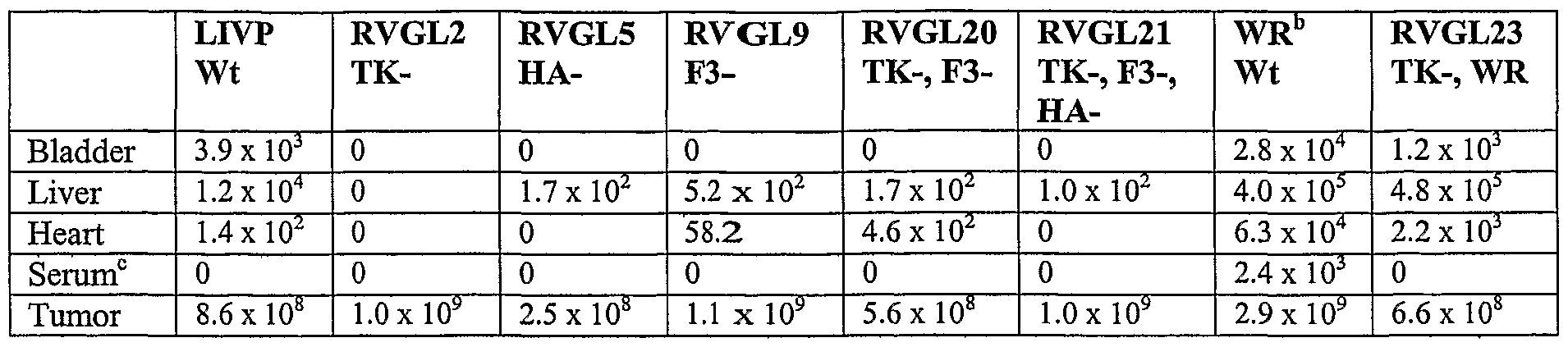


Claims
Priority Applications (8)
| Application Number | Priority Date | Filing Date | Title |
|---|---|---|---|
| AU2004289953A AU2004289953B2 (en) | 2003-06-18 | 2004-06-18 | Modified recombinant vaccina viruses and other microorganisms, uses thereof |
| MXPA05013879A MXPA05013879A (en) | 2003-06-18 | 2004-06-18 | Microorganisms for therapy. |
| JP2006517504A JP3934673B1 (en) | 2003-06-18 | 2004-06-18 | Modified recombinant vaccinia virus and other microorganisms, use thereof |
| EP04816752A EP1644492B1 (en) | 2003-06-18 | 2004-06-18 | Modified recombinant vaccinia viruses, uses thereof |
| BRPI0411526-0A BRPI0411526A (en) | 2003-06-18 | 2004-06-18 | vaccinia virus and other modified recombinant microorganisms and uses thereof |
| CA2527225A CA2527225C (en) | 2003-06-18 | 2004-06-18 | Modified recombinant vaccinia viruses and other microorganisms, uses thereof |
| DE602004018927T DE602004018927D1 (en) | 2003-06-18 | 2004-06-18 | MODIFIED RECOMBINANT VACCINIA VIRUSES, USES THEREOF |
| HK06106250.6A HK1084691A1 (en) | 2003-06-18 | 2006-05-29 | Modified recombinant vaccinia viruses, uses thereof |
Applications Claiming Priority (6)
| Application Number | Priority Date | Filing Date | Title |
|---|---|---|---|
| EP03013826A EP1489164A1 (en) | 2003-06-18 | 2003-06-18 | Recombinant vaccinia viruses with modified F3 genes, uses thereof |
| EP03013826.7 | 2003-06-18 | ||
| EP03018478.2 | 2003-08-14 | ||
| EP03018478A EP1512746B1 (en) | 2003-08-14 | 2003-08-14 | Method for the production of a polypeptide, RNA or other compound in tumor tissue |
| EP03024283A EP1526185B1 (en) | 2003-10-22 | 2003-10-22 | Use of a microorganism or cell to induce autoimmunization of an organism against a tumor |
| EP03024283.8 | 2003-10-22 |
Publications (3)
| Publication Number | Publication Date |
|---|---|
| WO2005047458A2 true WO2005047458A2 (en) | 2005-05-26 |
| WO2005047458A3 WO2005047458A3 (en) | 2005-09-15 |
| WO2005047458A9 WO2005047458A9 (en) | 2006-04-20 |
Family
ID=34119412
Family Applications (1)
| Application Number | Title | Priority Date | Filing Date |
|---|---|---|---|
| PCT/US2004/019866 WO2005047458A2 (en) | 2003-06-18 | 2004-06-18 | Modified recombinant vaccina viruses and other microorganisms, uses thereof |
Country Status (14)
| Country | Link |
|---|---|
| US (11) | US7588767B2 (en) |
| EP (2) | EP2028270B9 (en) |
| JP (4) | JP3934673B1 (en) |
| CN (1) | CN100562570C (en) |
| AT (2) | ATE420160T1 (en) |
| AU (2) | AU2004289953B2 (en) |
| BR (1) | BRPI0411526A (en) |
| CA (1) | CA2527225C (en) |
| DE (1) | DE602004018927D1 (en) |
| HK (2) | HK1084691A1 (en) |
| MX (1) | MXPA05013879A (en) |
| RU (1) | RU2376371C2 (en) |
| SG (1) | SG179291A1 (en) |
| WO (1) | WO2005047458A2 (en) |
Cited By (18)
| Publication number | Priority date | Publication date | Assignee | Title |
|---|---|---|---|---|
| EP1979000A2 (en) * | 2005-12-22 | 2008-10-15 | Memorial Sloan-Kettering Cancer Center | Method for detection of cancer cells using virus |
| WO2008156655A2 (en) * | 2007-06-15 | 2008-12-24 | Genelux Corporation | Vaccinia virus vectors encoding a sodium-dependent transporter protein for imaging and/or treatment of tumors |
| CN101903399A (en) * | 2007-06-08 | 2010-12-01 | 澳大利亚家禽Crc私人有限公司 | Clostridial toxin netb |
| WO2014055960A1 (en) | 2012-10-05 | 2014-04-10 | Genelux Corporation | Energy absorbing-based diagnostic and therapeutic methods employing nucleic acid molecules encoding chromophore-producing enzymes |
| WO2015103438A2 (en) | 2014-01-02 | 2015-07-09 | Genelux Corporation | Oncolytic virus adjunct therapy with agents that increase virus infectivity |
| WO2017209053A1 (en) | 2016-05-30 | 2017-12-07 | アステラス製薬株式会社 | New genetically-modified vaccinia virus |
| US10463730B2 (en) | 2003-06-18 | 2019-11-05 | Genelux Corporation | Microorganisms for therapy |
| CN110747174A (en) * | 2019-10-30 | 2020-02-04 | 青岛宁逸生物科技有限公司 | Recombinant virus for tumor treatment |
| WO2020067085A1 (en) | 2018-09-26 | 2020-04-02 | アステラス製薬株式会社 | Cancer therapy where oncolytic vaccinia virus and immune checkpoint inhibitor are used in combination, and pharmaceutical composition and combination drug used therein |
| WO2020148612A1 (en) | 2019-01-14 | 2020-07-23 | Ignite Immunotherapy, Inc. | Recombinant vaccinia virus and methods of use thereof |
| WO2020165730A1 (en) | 2019-02-14 | 2020-08-20 | Ignite Immunotherapy, Inc. | Recombinant vaccinia virus and methods of use thereof |
| WO2021040056A1 (en) | 2019-08-29 | 2021-03-04 | Astellas Pharma Inc. | Genetically engineered oncolytic vaccinia viruses and methods of uses thereof |
| WO2021116943A1 (en) | 2019-12-12 | 2021-06-17 | Ignite Immunotherapy, Inc. | Variant oncolytic vaccinia virus and methods of use thereof |
| WO2021140435A1 (en) | 2020-01-09 | 2021-07-15 | Pfizer Inc. | Recombinant vaccinia virus |
| US11149254B2 (en) | 2011-04-15 | 2021-10-19 | Genelux Corporation | Clonal strains of attenuated vaccinia viruses and methods of use thereof |
| WO2022013696A2 (en) | 2020-07-14 | 2022-01-20 | Pfizer Inc. | Recombinant vaccinia virus |
| US11773396B2 (en) | 2012-04-11 | 2023-10-03 | The U.S.A., As Represented By The Secretary, Department Of Health And Human Services | Chimeric antigen receptors targeting B-cell maturation antigen |
| WO2024011250A1 (en) | 2022-07-08 | 2024-01-11 | Viromissile, Inc. | Oncolytic vaccinia viruses and recombinant viruses and methods of use thereof |
Families Citing this family (194)
| Publication number | Priority date | Publication date | Assignee | Title |
|---|---|---|---|---|
| GB0109515D0 (en) * | 2001-04-17 | 2001-06-06 | Neg Micon As | A method for transporting a set of large longitudinal items, a package system to be used by the method and use of such a package system |
| EP1281767A3 (en) * | 2001-07-31 | 2003-05-28 | Aladar A. Szalay | Light emitting microorganisms and cells for diagnosis and therapy of tumors |
| US20030228261A1 (en) * | 2002-06-05 | 2003-12-11 | Aladar Szalay | Light emitting microorganisms and cells for diagnosis and therapy of diseases associated with wounded or inflamed tissue |
| EP1369491A1 (en) * | 2002-06-05 | 2003-12-10 | Aladar A. Szalay | Light emitting microorganisms and cells for diagnosis and therapy of diseases associated with wounded or inflamed tissue |
| WO2004014314A2 (en) * | 2002-08-12 | 2004-02-19 | David Kirn | Methods and compositions concerning poxviruses and cancer |
| US20070134202A1 (en) * | 2003-10-15 | 2007-06-14 | The New Industry Reserch Organization | Cancer gene therapeutic drug |
| US8221739B2 (en) | 2004-04-29 | 2012-07-17 | Botanic Oil Innovations, Inc. | Method of cancer treatment |
| JP5489459B2 (en) | 2005-08-01 | 2014-05-14 | ジ・オハイオ・ステイト・ユニバーシティ・リサーチ・ファウンデイション | MicroRNA-based methods and compositions for diagnosis, prognosis and treatment of breast cancer |
| WO2007030668A2 (en) * | 2005-09-07 | 2007-03-15 | Jennerex Biotherapeutics Ulc | Systemic treatment of metastatic and/or systemically-disseminated cancers using gm-csf-expressing poxviruses |
| US8980246B2 (en) | 2005-09-07 | 2015-03-17 | Sillajen Biotherapeutics, Inc. | Oncolytic vaccinia virus cancer therapy |
| EP1937280B1 (en) | 2005-09-12 | 2014-08-27 | The Ohio State University Research Foundation | Compositions for the therapy of bcl2-associated cancers |
| US20070077231A1 (en) * | 2005-09-30 | 2007-04-05 | Contag Christopher H | Immune effector cells pre-infected with oncolytic virus |
| US20070098828A1 (en) * | 2005-11-01 | 2007-05-03 | Gonzalez-Villasenor Lucia I | Methods and compositions for the prevention and regression of neovascularization |
| US8682619B2 (en) * | 2005-12-14 | 2014-03-25 | The Invention Science Fund I, Llc | Device including altered microorganisms, and methods and systems of use |
| US8734823B2 (en) * | 2005-12-14 | 2014-05-27 | The Invention Science Fund I, Llc | Device including altered microorganisms, and methods and systems of use |
| US20110183351A1 (en) | 2010-01-22 | 2011-07-28 | Hyde Roderick A | Compositions and methods for therapeutic delivery with microorganisms |
| US8278094B2 (en) | 2005-12-14 | 2012-10-02 | The Invention Science Fund I, Llc | Bone semi-permeable device |
| ES2536423T3 (en) | 2006-01-05 | 2015-05-25 | The Ohio State University Research Foundation | MicroRNA based methods for the diagnosis of prostate cancer |
| ES2429404T3 (en) | 2006-01-05 | 2013-11-14 | The Ohio State University Research Foundation | MicroRNA-based procedures for the diagnosis and prognosis of lung cancer |
| EP2591794A1 (en) | 2006-01-05 | 2013-05-15 | The Ohio State University Research Foundation | MicroRNA expressions abnormalities in pancreatic endocrine and acinar tumors |
| EP1996731A2 (en) | 2006-03-20 | 2008-12-03 | The Ohio State University Research Foundation | Microrna fingerprints during human megakaryocytopoiesis |
| TW200819540A (en) | 2006-07-11 | 2008-05-01 | Genelux Corp | Methods and compositions for detection of microorganisms and cells and treatment of diseases and disorders |
| AU2007272947B2 (en) | 2006-07-13 | 2013-06-06 | The Government Of The United States Of America As Represented By The Secretary Of The Department Of Health And Human Services | Micro-RNA-based methods and compositions for the diagnosis and treatment of colon cancer-related diseases |
| US8071292B2 (en) | 2006-09-19 | 2011-12-06 | The Ohio State University Research Foundation | Leukemia diagnostic methods |
| EP2415783B1 (en) | 2006-10-16 | 2016-12-14 | Genelux Corporation | Modified vaccinia virus strains for use in a diagnostic and therapeutic method |
| CA2667617A1 (en) | 2006-11-01 | 2008-05-08 | The Ohio State University Research Foundation | Microrna expression signature for predicting survival and metastases in hepatocellular carcinoma |
| US8034560B2 (en) | 2007-01-31 | 2011-10-11 | The Ohio State University Research Foundation | MicroRNA-based methods and compositions for the diagnosis, prognosis and treatment of acute myeloid leukemia (AML) |
| KR100858913B1 (en) * | 2007-03-09 | 2008-09-17 | 한국과학기술원 | L-threonine Overproducing Microorganism Which Constructed by Specific Genetic Modifications and Method for Preparing ??threonine Using the Same |
| KR20080084528A (en) * | 2007-03-15 | 2008-09-19 | 제네렉스 바이오테라퓨틱스 인크. | Oncolytic vaccinia virus cancer therapy |
| EP2150616A4 (en) | 2007-05-10 | 2011-07-27 | Univ Arizona | Regulated synthesis of antigen and/or regulated attentuation to enhance vaccine immunogenics and/or safety |
| WO2008141226A2 (en) * | 2007-05-10 | 2008-11-20 | The Arizona Board Of Regents For And On Behalf Of Arizona State University | Recombinant bacteria comprising vectors for expression of nucleic acid sequences encoding antigens |
| US8003364B2 (en) | 2007-05-14 | 2011-08-23 | Bavarian Nordic A/S | Purification of vaccinia viruses using hydrophobic interaction chromatography |
| EP3988651A1 (en) * | 2007-05-14 | 2022-04-27 | Bavarian Nordic A/S | Purification of vaccinia virus- and recombinant vaccinia virus-based vaccines |
| WO2008144067A1 (en) * | 2007-05-21 | 2008-11-27 | Board Of Regents, University Of Texas System | Methods and compositions for treatment of cancer using oncolytic rsv activity |
| WO2008150496A2 (en) * | 2007-05-31 | 2008-12-11 | Genelux Corporation | Assay for sensitivity to chemotherapeutic agents |
| CN101711287B (en) | 2007-06-08 | 2016-04-27 | 由卫生与公众服务部代表的美利坚合众国政府 | Determine the method for hepatocellular carcinoma hypotype and detection liver-cancer stem cell |
| JP5480132B2 (en) | 2007-06-15 | 2014-04-23 | ジ・オハイオ・ステイト・ユニバーシティ・リサーチ・ファウンデイション | Oncogenic ALL-1 fusion protein for targeting DROSHA-mediated microRNA processing |
| WO2009011924A1 (en) | 2007-07-18 | 2009-01-22 | Genelux Corporation | Use of a chemotherapeutic agent in the preparation of a medicament for treating or ameliorating an adverse side effect associated with oncolytic viral therapy |
| CN101809169B (en) | 2007-07-31 | 2013-07-17 | 俄亥俄州立大学研究基金会 | Methods for reverting methylation by targeting DNMT3A and DNMT3B |
| EP2650383A1 (en) | 2007-08-03 | 2013-10-16 | The Ohio State University Research Foundation | Ultraconserved regions encoding ncRNAs |
| US7807180B2 (en) | 2007-08-21 | 2010-10-05 | University Of Massachusetts | Poxvirus methods and compositions |
| CA2926831A1 (en) | 2007-08-22 | 2009-02-26 | The Ohio State University Research Foundation | Methods and compositions for inducing deregulation of epha7 and erk phosphorylation in human acute leukemias |
| US9040059B2 (en) | 2007-10-05 | 2015-05-26 | The Arizona Board Of Regents For And On Behalf Of Arizona State University | Recombinant bacterium capable of eliciting a protective immune response against C. perfringens |
| US20090136917A1 (en) * | 2007-10-25 | 2009-05-28 | Szalay Aladar A | Systems and methods for viral therapy |
| CN103898069A (en) | 2007-10-26 | 2014-07-02 | 俄亥俄州立大学研究基金会 | Methods for identifying fragile histidine triad (fhit) interaction and uses thereof |
| AU2008328258B2 (en) * | 2007-11-19 | 2013-09-26 | Transgene Sa | Poxviral oncolytic vectors |
| EP2212345B1 (en) * | 2007-11-19 | 2016-01-27 | Transgene SA | Poxviral oncolytic vectors |
| EP2242516A1 (en) * | 2008-01-11 | 2010-10-27 | Genelux Corporation | Methods and compositions for detection of bacteria and treatment of diseases and disorders |
| DK2276774T3 (en) | 2008-03-14 | 2016-11-28 | Retrotope Inc | Drugs that modulate genome methylation. |
| WO2009114809A1 (en) * | 2008-03-14 | 2009-09-17 | Retrotope, Inc. | Therapies for cancer using isotopically substituted lysine |
| US20110172107A1 (en) * | 2008-04-30 | 2011-07-14 | Fox Chase Cancer Center | Assay for identifying agents that modulate epigenetic silencing, and agents identified thereby |
| EP2300977A4 (en) * | 2008-05-15 | 2014-04-30 | Youdocs Llc | Methods and systems for improving human health using targeted probiotics |
| WO2009139921A2 (en) * | 2008-05-16 | 2009-11-19 | Genelux Corporation | Microorganisms for preventing and treating neoplasms accompanying cellular therapy |
| JP5745401B2 (en) | 2008-06-11 | 2015-07-08 | アメリカ合衆国 | Use of the MiR-26 family as a predictive marker for hepatocellular carcinoma and responsiveness to therapy |
| CA2729080C (en) * | 2008-07-03 | 2018-09-11 | Ricardo Rosenbusch | Cattle vaccines |
| WO2010030293A1 (en) * | 2008-09-15 | 2010-03-18 | Gentron, Inc. | Methods, systems, and compositions for cancer diagnosis |
| US8647642B2 (en) | 2008-09-18 | 2014-02-11 | Aviex Technologies, Llc | Live bacterial vaccines resistant to carbon dioxide (CO2), acidic PH and/or osmolarity for viral infection prophylaxis or treatment |
| EP2326712B1 (en) * | 2008-09-19 | 2016-05-04 | ETAT FRANCAIS représenté par le Directeur central du Service de Santé des Armées | Viral strains derived from the vaccinia virus lister vacv-107 and uses thereof |
| WO2010045620A1 (en) | 2008-10-17 | 2010-04-22 | The Arizona Board Of Regents For And On Behalf Of Arizona State University | Recombinant bacterium capable of eliciting an immune response against streptococcus pneumoniae |
| US9163219B2 (en) * | 2009-04-14 | 2015-10-20 | Arizona Board Of Regents On Behalf Of Arizona State University | Single expression vector for generation of a virus with a segmented genome |
| WO2010135563A1 (en) | 2009-05-22 | 2010-11-25 | The Arizona Board Of Regents For And On Behalf Of Arizona State University | Recombinant bacterium and methods of antigen and nucleic acid delivery |
| US9045742B2 (en) * | 2009-05-29 | 2015-06-02 | The Arizona Board Of Regents For And On Behalf Of Arizona State University | Recombinant Edwardsiella bacterium |
| EP3409119B1 (en) | 2009-09-14 | 2020-08-12 | SillaJen Biotherapeutics, Inc. | Oncolytic vaccinia virus combination cancer therapy |
| AU2010321555B2 (en) | 2009-11-23 | 2015-10-15 | The Ohio State University | Materials and methods useful for affecting tumor cell growth, migration and invasion |
| US9062297B2 (en) | 2010-01-13 | 2015-06-23 | The Arizona Board Of Regents For And On Behalf Of Arizona State University | Yersinia pestis vaccine |
| WO2011091291A1 (en) | 2010-01-22 | 2011-07-28 | The Arizona Board Of Regents For And On Behalf Of Arizona State University | BACTERIUM COMPRISING A REGULATED rfaH NUCLEIC ACID |
| WO2011150421A2 (en) * | 2010-05-28 | 2011-12-01 | The Arizona Board Of Regents For And On Behalf Of Arizona State University | Recombinant bacterium to decrease tumor growth |
| EP2637673B1 (en) | 2010-11-12 | 2016-10-26 | The Ohio State University Research Foundation | Methods related to microrna-21 and mismatch repair in colorectal cancer |
| US10758619B2 (en) | 2010-11-15 | 2020-09-01 | The Ohio State University | Controlled release mucoadhesive systems |
| RU2447150C1 (en) * | 2010-11-26 | 2012-04-10 | Учреждение Российской академии наук Институт биоорганической химии им. академиков М.М. Шемякина и Ю.А. Овчинникова РАН | Agent for gene therapy of malignant growths |
| WO2012094386A1 (en) | 2011-01-04 | 2012-07-12 | Jennerex Inc. | Generation of antibodies to tumor antigens and generation of tumor specific complement dependent cytotoxicity by administration of oncolytic vaccinia virus |
| WO2012122239A1 (en) | 2011-03-07 | 2012-09-13 | The Ohio State University | MUTATOR ACTIVITY INDUCED BY MICRORNA-155 (miR-155) LINKS INFLAMMATION AND CANCER |
| CN102685768B (en) | 2011-03-08 | 2016-02-03 | 华为技术有限公司 | The processing method of heartbeat message, the acquisition methods of heart beat cycle and access network equipment |
| EP2720721B1 (en) | 2011-06-17 | 2017-08-30 | President and Fellows of Harvard College | Frizzled 2 as a target for therapeutic antibodies in the treatment of cancer |
| CN102286415B (en) * | 2011-09-07 | 2014-03-05 | 天津工业生物技术研究所 | Bacterial strain for high yield of succinic acid and application thereof |
| US8859256B2 (en) | 2011-10-05 | 2014-10-14 | Genelux Corporation | Method for detecting replication or colonization of a biological therapeutic |
| JP2014530612A (en) | 2011-10-14 | 2014-11-20 | ジ・オハイオ・ステート・ユニバーシティ | Methods and materials for ovarian cancer |
| KR102385013B1 (en) * | 2011-11-18 | 2022-04-12 | 알닐람 파마슈티칼스 인코포레이티드 | RNAi AGENTS, COMPOSITIONS AND METHODS OF USE THEREOF FOR TREATING TRANSTHYRETIN (TTR) ASSOCIATED DISEASES |
| AU2012352265B2 (en) | 2011-12-13 | 2017-02-16 | Ohio State Innovation Foundation | Methods and compositions related to miR-21 and miR-29a, exosome inhibition, and cancer metastasis |
| US9023612B2 (en) | 2012-01-13 | 2015-05-05 | Bell Biosystems, Inc. | Eukaryotic cells with artificial endosymbionts for multimodal detection |
| US10076579B2 (en) | 2012-01-13 | 2018-09-18 | Bell Biosystems, Inc. | Host cells with artificial endosymbionts |
| US8956873B2 (en) * | 2012-01-13 | 2015-02-17 | Bell Biosystems, Inc. | Host cells with artificial endosymbionts |
| US8828681B2 (en) | 2012-01-13 | 2014-09-09 | Bell Biosystems, Inc. | Host cells with artificial endosymbionts |
| EP2804960A4 (en) | 2012-01-20 | 2015-08-19 | Univ Ohio State | Breast cancer biomarker signatures for invasiveness and prognosis |
| AU2012372783B2 (en) * | 2012-03-08 | 2017-11-23 | Trimed Inc. | System and method for treating bone fractures |
| WO2013138522A2 (en) | 2012-03-16 | 2013-09-19 | Genelux Corporation | Methods for assessing effectiveness and monitoring oncolytic virus treatment |
| US20130280170A1 (en) | 2012-04-20 | 2013-10-24 | Aladar A. Szalay | Imaging methods for oncolytic virus therapy |
| US9303264B2 (en) | 2012-05-18 | 2016-04-05 | The Arizona Board Of Regents For And On Behalf Of Arizona State University | Photosynthetic microorganisms expressing thermostable lipase |
| WO2014145785A1 (en) | 2013-03-15 | 2014-09-18 | Bell Biosystems, Llc | Host cells with artificial endosymbionts |
| CN103589656A (en) * | 2013-05-28 | 2014-02-19 | 湖北省农业科学院畜牧兽医研究所 | Porcine contagious pleuropneumonia actinobacillus serum 1-type zinc ion binding protein A deletion strain, construction method therefor and applications thereof |
| US9580718B2 (en) | 2013-06-17 | 2017-02-28 | Arizona Board Of Regents On Behalf Of Arizona State University | Attenuated live bacteria with increased acid resistance and methods of use thereof |
| GB201310917D0 (en) * | 2013-06-19 | 2013-07-31 | Cancer Res Inst Royal | Vaccinia virus for gene-directed enzyme prodrug therapy |
| WO2015034962A1 (en) | 2013-09-03 | 2015-03-12 | Bell Biosystems, Inc. | Host cell modification with artificial endosymbionts |
| WO2015035244A1 (en) * | 2013-09-05 | 2015-03-12 | Braskem S/A | Modified microorganism and methods of using same for producing butadiene and 1-propanol and/or 1,2-propanediol |
| US10028649B2 (en) | 2013-12-02 | 2018-07-24 | Welch Allyn, Inc. | Digital colposcope system |
| CN104694541A (en) * | 2013-12-09 | 2015-06-10 | 江苏命码生物科技有限公司 | Avian influenza virus miRNA as well as identification, detection and application thereof |
| EP3090067B1 (en) * | 2013-12-30 | 2020-07-22 | The Henry M. Jackson Foundation for the Advancement of Military Medicine, Inc. | Genomic rearrangements associated with prostate cancer and methods of using the same |
| CN103743903A (en) * | 2014-01-08 | 2014-04-23 | 江苏省苏北人民医院 | Detection method and kit of predictive marker Capn4 of postoperative recurrence of liver cancer |
| CA2951514A1 (en) | 2014-06-18 | 2015-12-23 | Clear Gene, Inc. | Methods, compositions, and devices for rapid analysis of biological markers |
| JP2017522022A (en) * | 2014-07-15 | 2017-08-10 | ベル・バイオシステムズ,インコーポレーテッド | Eukaryotic cells containing artificial endosymbiosis for multimodal detection |
| US9616114B1 (en) | 2014-09-18 | 2017-04-11 | David Gordon Bermudes | Modified bacteria having improved pharmacokinetics and tumor colonization enhancing antitumor activity |
| CN105586319B (en) * | 2014-10-20 | 2021-01-08 | 中国疾病预防控制中心性病艾滋病预防控制中心 | Replicative vaccinia virus vector AIDS vaccine |
| US11285194B2 (en) | 2014-10-24 | 2022-03-29 | Calidi Biotherapeutics, Inc. | Combination immunotherapy approach for treatment of cancer |
| WO2016102569A1 (en) | 2014-12-22 | 2016-06-30 | Veterinærinstituttet | Salmon gill poxvirus |
| CN106134158B (en) | 2015-01-18 | 2020-03-03 | Lg电子株式会社 | Broadcast signal transmission device, broadcast signal reception device, broadcast signal transmission method, and broadcast signal reception method |
| CN107735103B (en) | 2015-02-25 | 2022-05-27 | 纪念斯隆-凯特琳癌症中心 | Use of inactivated non-replicative modified vaccinia virus ankara (MVA) as a sole immunotherapy of solid tumors or in combination with immune checkpoint blockers |
| KR101835180B1 (en) | 2015-03-18 | 2018-03-06 | 아주대학교산학협력단 | Recombinant BTG2 protein and production method thereof |
| AU2016249521B2 (en) | 2015-04-17 | 2021-08-19 | Memorial Sloan Kettering Cancer Center | Use of MVA or MVADELTAE3L as immunotherapeutic agents against solid tumors |
| US10676723B2 (en) | 2015-05-11 | 2020-06-09 | David Gordon Bermudes | Chimeric protein toxins for expression by therapeutic bacteria |
| KR20160140075A (en) * | 2015-05-29 | 2016-12-07 | 코오롱생명과학 주식회사 | Promoter derived from the poxviridae and vector including the same |
| MX2018001755A (en) | 2015-08-11 | 2018-08-01 | Stemimmune Incorporated | Smallpox vaccine for use in cancer treatment. |
| WO2017074517A1 (en) * | 2015-10-30 | 2017-05-04 | Seracare Life Sciences, Inc. | Adenovirus control virus |
| AU2016369603A1 (en) | 2015-12-18 | 2018-07-05 | Clear Gene, Inc. | Methods, compositions, kits and devices for rapid analysis of biological markers |
| CN106978397A (en) * | 2016-01-18 | 2017-07-25 | 上海宇研生物技术有限公司 | A kind of people DC-CIK immunocompetent cells and preparation method thereof |
| US10512662B2 (en) | 2016-02-25 | 2019-12-24 | Memorial Sloan Kettering Cancer Center | Replication competent attenuated vaccinia viruses with deletion of thymidine kinase with and without the expression of human Flt3L or GM-CSF for cancer immunotherapy |
| SG11201807022XA (en) | 2016-02-25 | 2018-09-27 | Memorial Sloan Kettering Cancer Center | Recombinant mva or mvadele3l expressing human flt3l and use thereof as immuno-therapeutic agents against solid tumors |
| WO2017172713A1 (en) | 2016-03-28 | 2017-10-05 | Minjie Hu | Anti-cancer oncolytic virus combination therapies and elite responder selection platforms |
| SG11201808783XA (en) | 2016-04-15 | 2018-11-29 | Alpine Immune Sciences Inc | Cd80 variant immunomodulatory proteins and uses thereof |
| WO2017181148A2 (en) | 2016-04-15 | 2017-10-19 | Alpine Immune Sciences, Inc. | Icos ligand variant immunomodulatory proteins and uses thereof |
| KR102306553B1 (en) * | 2016-07-21 | 2021-09-30 | 코오롱생명과학 주식회사 | Recombinant vaccinia virus and use thereof |
| US11471488B2 (en) | 2016-07-28 | 2022-10-18 | Alpine Immune Sciences, Inc. | CD155 variant immunomodulatory proteins and uses thereof |
| CN110088127A (en) | 2016-07-28 | 2019-08-02 | 高山免疫科学股份有限公司 | CD155 variant immune modulator and application thereof |
| US11834490B2 (en) | 2016-07-28 | 2023-12-05 | Alpine Immune Sciences, Inc. | CD112 variant immunomodulatory proteins and uses thereof |
| WO2018031694A1 (en) * | 2016-08-09 | 2018-02-15 | City Of Hope | Chimeric poxvirus compositions and uses thereof |
| CN110352245A (en) | 2016-10-20 | 2019-10-18 | 高山免疫科学股份有限公司 | Variant immune modulator and engineering cell therapy can be secreted |
| CN110300801B (en) * | 2016-11-24 | 2023-12-08 | 上海凯赛生物技术股份有限公司 | Control of protein-protein interactions of acid decarboxylases |
| JP7249279B2 (en) | 2016-12-07 | 2023-03-30 | サルスペラ,リミティド ライアビリティ カンパニー | Synergistic treatment methods for cancer |
| US11180535B1 (en) | 2016-12-07 | 2021-11-23 | David Gordon Bermudes | Saccharide binding, tumor penetration, and cytotoxic antitumor chimeric peptides from therapeutic bacteria |
| US11129906B1 (en) | 2016-12-07 | 2021-09-28 | David Gordon Bermudes | Chimeric protein toxins for expression by therapeutic bacteria |
| RU2658606C1 (en) * | 2017-01-26 | 2018-06-21 | Рашит Накипович Кадыров | Streptococcus pyogenes n b-7612, the producer of the complex of biologically active compounds with immunostimulating properties |
| WO2018148462A1 (en) | 2017-02-09 | 2018-08-16 | Indapta Therapeutics, Inc. | Engineered natural killer (nk) cells and compositions and methods thereof |
| US20190134105A1 (en) * | 2017-11-09 | 2019-05-09 | Richard Postrel | Method for Precise Identification, Targeting and Delivery of Directed Therapies with the Use of Bacteria for the Destruction of Cancerous Cells |
| NZ756395A (en) | 2017-03-16 | 2024-01-26 | Alpine Immune Sciences Inc | Cd80 variant immunomodulatory proteins and uses thereof |
| ES2963638T3 (en) | 2017-03-16 | 2024-04-01 | Alpine Immune Sciences Inc | Immunomodulatory proteins of PD-L1 variants and their uses |
| WO2018170023A1 (en) | 2017-03-16 | 2018-09-20 | Alpine Immune Sciences, Inc. | Pd-l2 variant immunomodulatory proteins and uses thereof |
| WO2018209315A1 (en) | 2017-05-12 | 2018-11-15 | Memorial Sloan Kettering Cancer Center | Vaccinia virus mutants useful for cancer immunotherapy |
| US10851350B1 (en) | 2017-06-27 | 2020-12-01 | Genelux Corporation | Bioreactor production of virus from adherent cells |
| CN107164338A (en) * | 2017-06-27 | 2017-09-15 | 镇江市卫克生物科技有限公司 | A kind of recombination oncolytic virus and its application |
| CA3198255A1 (en) | 2017-10-10 | 2019-04-18 | Alpine Immune Sciences, Inc. | Ctla-4 variant immunomodulatory proteins and uses thereof |
| WO2019079520A2 (en) | 2017-10-18 | 2019-04-25 | Alpine Immune Sciences, Inc. | Variant icos ligand immunomodulatory proteins and related compositions and methods |
| JP2021511068A (en) * | 2017-12-20 | 2021-05-06 | ラディミューン セラピュティックス, インク.Radimmune Therapeutics, Inc. | Antibodies to Centrin-1, preparation methods, and their use |
| CN108489941B (en) * | 2018-01-31 | 2021-07-27 | 吉林大学 | Application of bromophenol blue in aspects of serving as probe for detecting bovine insulin amyloid fiber and serving as inhibitor of bovine insulin amyloid fiber |
| WO2019173204A1 (en) * | 2018-03-06 | 2019-09-12 | Danisco Us Inc | Reduction in acetate production by yeast over-expressing pab1 |
| CN110386985B (en) * | 2018-04-19 | 2022-09-20 | 四川大学华西医院 | Tumor-targeting apoptosis-promoting fusion protein and application thereof |
| KR20210028149A (en) * | 2018-05-17 | 2021-03-11 | 투불 메디테라피 피.씨. | Diagnostic blood test |
| CN112533621A (en) | 2018-06-04 | 2021-03-19 | 卡利迪生物治疗有限公司 | Cell-based agents for enhancing viral therapy |
| US11505782B2 (en) | 2018-06-04 | 2022-11-22 | Calidi Biotherapeutics, Inc. | Cell-based vehicles for potentiation of viral therapy |
| US20210363219A1 (en) | 2018-06-15 | 2021-11-25 | Alpine Immune Sciences, Inc. | Pd-1 variant immunomodulatory proteins and uses thereof |
| CN108676890B (en) * | 2018-07-12 | 2022-01-28 | 吉林大学 | Female breast malignant tumor susceptibility prediction kit and system |
| CN109172613B (en) * | 2018-08-14 | 2022-04-22 | 景岳生物科技(中国)有限公司 | Skin external composition containing lactobacillus dead bacteria culture and its use for promoting wound healing and reducing scar generation |
| WO2020047161A2 (en) | 2018-08-28 | 2020-03-05 | Actym Therapeutics, Inc. | Engineered immunostimulatory bacterial strains and uses thereof |
| KR20210089178A (en) * | 2018-10-30 | 2021-07-15 | 소마로직, 인크. | How to evaluate sample quality |
| EP3876951A1 (en) * | 2018-11-06 | 2021-09-15 | Calidi Biotherapeutics, Inc. | Enhanced systems for cell-mediated oncolytic viral therapy |
| CN109546191B (en) * | 2018-11-07 | 2021-06-18 | 大连理工大学 | Mixed matrix type anion membrane and preparation method thereof |
| JP2022511735A (en) | 2018-11-21 | 2022-02-01 | インダプタ セラピューティクス インコーポレイテッド | Methods and Related Compositions and Methods for Enlargement of Natural Killer (NK) Cell Subsets |
| JP2022510276A (en) | 2018-11-30 | 2022-01-26 | アルパイン イミューン サイエンシズ インコーポレイテッド | CD86 variant immunomodulatory protein and its use |
| CN113661236A (en) * | 2019-01-07 | 2021-11-16 | 卡利威尔免疫治疗公司 | Methods of treating cancer |
| WO2020172461A1 (en) * | 2019-02-22 | 2020-08-27 | Arizona Board Of Regents On Behalf Of Arizona State University | Using tumor-navigating salmonella to modulate tumor metabolism |
| CA3131017A1 (en) | 2019-02-27 | 2020-09-03 | Actym Therapeutics, Inc. | Immunostimulatory bacteria engineered to colonize tumors, tumor-resident immune cells, and the tumor microenvironment |
| CN109810991B (en) * | 2019-03-02 | 2021-11-12 | 昆明理工大学 | Application of dihydropteroate synthase gene folP |
| US11471497B1 (en) | 2019-03-13 | 2022-10-18 | David Gordon Bermudes | Copper chelation therapeutics |
| CN113874029A (en) * | 2019-04-01 | 2021-12-31 | 汉阳大学校产学协力团 | CP 2C-targeting peptide-based anticancer agents |
| CN109957551B (en) * | 2019-04-03 | 2021-03-23 | 浙江省医学科学院 | Recombinant vaccinia virus expressing human beta-defensin 2 and application thereof |
| EP3969047A4 (en) * | 2019-05-15 | 2022-11-09 | Codagenix Inc. | Attenuated yellow fever virus and uses thereof for the treatment of cancer |
| US20220218818A1 (en) * | 2019-06-03 | 2022-07-14 | Immunolux International Corp. | Smallpox vaccine and stem cells for treatment of disease |
| CN110585427B (en) * | 2019-09-06 | 2023-06-06 | 刘慧宁 | Composition for improving immunity of organism and application of composition in resisting adult T cell leukemia or nasopharyngeal carcinoma |
| CN110713962B (en) * | 2019-09-06 | 2022-06-21 | 南京农业大学 | Genetic engineering bacterium for high-yield production of malonyl coenzyme A and construction method and application thereof |
| US11271912B2 (en) * | 2019-09-27 | 2022-03-08 | Envistacom, Llc | Anonymous communication over virtual, modular, and distributed satellite communications network |
| CN110551672B (en) * | 2019-09-30 | 2023-05-23 | 中国科学院成都生物研究所 | Coli strain for high-yield trans-4-hydroxy-L-proline and construction method thereof |
| CN110564867B (en) * | 2019-10-10 | 2022-06-24 | 扬州大学 | SNP molecular marker of Qinchuan cattle CFL1 gene and detection method thereof |
| CN111254219B (en) * | 2019-12-27 | 2023-08-04 | 上海华盈生物医药科技有限公司 | Method for detecting virus packaging efficiency |
| CN111019877B (en) * | 2019-12-31 | 2022-04-19 | 浙江工业大学 | Genetically engineered bacterium for producing L-cysteine, construction method and application |
| JP2023523429A (en) | 2020-04-22 | 2023-06-05 | インダプタ セラピューティクス インコーポレイテッド | Natural killer (NK) cell compositions and methods for producing same |
| US10973908B1 (en) | 2020-05-14 | 2021-04-13 | David Gordon Bermudes | Expression of SARS-CoV-2 spike protein receptor binding domain in attenuated salmonella as a vaccine |
| CN111875699B (en) * | 2020-07-03 | 2022-07-05 | 江南大学 | Method for improving bacillus subtilis ovalbumin expression level |
| CN111705144B (en) * | 2020-07-17 | 2022-11-18 | 扬州大学 | SINE transposon polymorphic molecular marker in ZNF2 gene related to pig backfat thickness and detection method thereof |
| CN111913584B (en) * | 2020-08-19 | 2022-04-01 | 福州大学 | Mouse cursor control method and system based on gesture recognition |
| CN112011555A (en) * | 2020-09-09 | 2020-12-01 | 西南大学 | Recombinant gene and recombinant plasmid for regulating and controlling salmonella self-lysis and application thereof |
| WO2022071966A1 (en) | 2020-10-02 | 2022-04-07 | Genelux Corporation | Bioreactor production of virus from adherent cells |
| CN112149041B (en) * | 2020-10-20 | 2022-12-16 | 贵州电网有限责任公司 | Fault analysis method based on PSD-BPA data and graphical interaction technology |
| CN114584999B (en) * | 2020-11-30 | 2023-08-15 | 中国移动通信集团山西有限公司 | Monitoring system, method, equipment and computer storage medium |
| CN113322275A (en) * | 2021-03-24 | 2021-08-31 | 中国人民解放军空军军医大学 | CRISPR/Cas9 appearance editing system and method of macrophage, edited macrophage and application |
| CN113238045B (en) * | 2021-04-27 | 2021-12-28 | 南方医科大学南方医院 | Applications of CRMP2 and anti-CRMP 2 antibody |
| CN113363963B (en) * | 2021-05-20 | 2022-05-20 | 南昌大学 | Optimized three-phase SAPF direct current side control method by improving sparrow search algorithm |
| CN113249286B (en) * | 2021-05-25 | 2023-12-08 | 洛阳华荣生物技术有限公司 | Method for constructing L-sarcosine producing strain |
| CN113322213B (en) * | 2021-07-09 | 2022-05-13 | 吉林大学 | Lactobacillus paracasei Jlus66 microbial inoculum with function of improving dysmnesia and application |
| CN114395574B (en) * | 2022-01-18 | 2023-08-11 | 长沙爱科博生物科技有限公司 | Porcine epidemic diarrhea virus fusion protein, and encoding gene and application thereof |
| US20230233299A1 (en) * | 2022-01-27 | 2023-07-27 | EdgeEndo, LLC | Dental, endodontic, and periodontic treatment methods and systems |
| CN114540521B (en) * | 2022-03-23 | 2022-09-02 | 广东省科学院微生物研究所(广东省微生物分析检测中心) | Lactobacillus plantarum 84-3 specific molecular target with blood sugar and blood fat reducing effects and detection method thereof |
| CN114774468B (en) * | 2022-04-20 | 2022-12-20 | 温氏食品集团股份有限公司 | Allele molecular marker and anti-blue-ear-disease pig group construction method |
| CN115011601B (en) * | 2022-06-27 | 2023-07-21 | 山东大学齐鲁医院 | shRNA (short hairpin ribonucleic acid) interfering with JUND expression, recombinant adeno-associated virus vector and application thereof |
| CN117625547A (en) * | 2022-08-25 | 2024-03-01 | 广州百吉生物制药有限公司 | New generation chimeric antigen receptor for combined expression of GPX4 and application thereof |
| CN116622577B (en) * | 2023-05-26 | 2023-10-27 | 山东奈思健康科技有限责任公司 | Metagen for promoting COL1A2 and COL3A1 expression and improving collagen synthesis and preparation method thereof |
| CN117589991B (en) * | 2024-01-18 | 2024-03-29 | 天津云检医学检验所有限公司 | Biomarker, model, kit and application for identifying HER2 expression state of breast cancer patient |
Citations (5)
| Publication number | Priority date | Publication date | Assignee | Title |
|---|---|---|---|---|
| WO1992022327A1 (en) * | 1991-06-17 | 1992-12-23 | The Regents Of The University Of California | Double recombinant vaccinia virus vaccines |
| WO2000073479A1 (en) * | 1999-05-28 | 2000-12-07 | The Government Of The United States Of America, As Represented By The Secretary, Department Of Health And Human Services | A combined growth factor-deleted and thymidine kinase-deleted vaccinia virus vector |
| US6491905B1 (en) * | 1993-09-14 | 2002-12-10 | The Uab Research Foundation | Recombinant bacterial cells for delivery of PNP to tumor cells |
| WO2003014380A2 (en) * | 2001-07-31 | 2003-02-20 | Genelux Gmbh | Microoroganisms and cells for diagnosis and therapy of tumors |
| EP1369491A1 (en) * | 2002-06-05 | 2003-12-10 | Aladar A. Szalay | Light emitting microorganisms and cells for diagnosis and therapy of diseases associated with wounded or inflamed tissue |
Family Cites Families (198)
| Publication number | Priority date | Publication date | Assignee | Title |
|---|---|---|---|---|
| US83293A (en) * | 1868-10-20 | Improvement in button-hole cutters | ||
| US54865A (en) * | 1866-05-22 | Improvement in cow-milkers | ||
| US8025A (en) * | 1851-04-08 | Apparatus eor boltiitg flouk | ||
| US59400A (en) * | 1866-11-06 | Improvement | ||
| US213741A (en) * | 1879-04-01 | Improvement in portable platforms for fire and other ladders | ||
| US29023A (en) * | 1860-07-03 | Cornelius j | ||
| US228330A (en) * | 1880-06-01 | Advertising checker-board | ||
| US101480A (en) * | 1870-04-05 | Improvement in hand seed-planter | ||
| US44384A (en) * | 1864-09-27 | Improved carpet-fastening | ||
| US143861A (en) * | 1873-10-21 | Improvement in picture-frames | ||
| US161788A (en) * | 1875-04-06 | Improvement in bird-cages | ||
| US213007A (en) * | 1879-03-04 | Improvement in safety-pins | ||
| US31643A (en) * | 1861-03-05 | Bons l | ||
| US165477A (en) * | 1875-07-13 | Improvement in smelting-furnaces | ||
| US69491A (en) * | 1867-10-01 | Newark | ||
| US9015A (en) * | 1852-06-15 | Manufacture of granular fuel from brush-wood and twigs | ||
| US31628A (en) * | 1861-03-05 | Improvement in sewing-machines | ||
| US160970A (en) * | 1875-03-16 | Improvement in key-rings | ||
| US31681A (en) * | 1861-03-12 | Puhchifg-machine | ||
| US76622A (en) * | 1868-04-14 | Bobeet john gaines | ||
| US165465A (en) * | 1875-07-13 | Improvement in paper-weights | ||
| US228261A (en) * | 1880-06-01 | lewis | ||
| US33617A (en) * | 1861-10-29 | Improvement in grain-separators | ||
| US86906A (en) * | 1869-02-16 | Improvement in velocipedes | ||
| US160410A (en) * | 1875-03-02 | Improvement in carbureters or hydrocarbon-diffusers | ||
| JPS5535004Y2 (en) | 1975-07-28 | 1980-08-19 | ||
| JPS542336A (en) * | 1977-06-06 | 1979-01-09 | Meiji Seika Kaisha Ltd | Preventive and remedy for viral diseases |
| JPS5535004A (en) | 1978-09-02 | 1980-03-11 | Wakunaga Yakuhin Kk | Cell-immunity activator and its preparation |
| EP0037441B1 (en) | 1980-03-10 | 1984-05-09 | Seiji Arakawa | Pharmaceutical compositions useful as cellular immunopotentiator and anti-tumor agent, process for production thereof and microorganism used therein |
| US4315914A (en) * | 1980-04-04 | 1982-02-16 | Seiji Arakawa | Pharmaceutical compositions useful as cellular immunopotentiator and antitumor agent and process for production thereof |
| US4442203A (en) | 1981-06-30 | 1984-04-10 | Massachusetts Institute Of Technology | Gene amplification assay for detecting tumor promoters |
| US4603112A (en) | 1981-12-24 | 1986-07-29 | Health Research, Incorporated | Modified vaccinia virus |
| US5364773A (en) | 1991-03-07 | 1994-11-15 | Virogenetics Corporation | Genetically engineered vaccine strain |
| US5155020A (en) | 1989-03-08 | 1992-10-13 | Health Research Inc. | Recombinant poxvirus host range selection system |
| US5378457A (en) | 1981-12-24 | 1995-01-03 | Virogenetics Corporation | Interferon sensitive recombinant poxvirus vaccine |
| US4722848A (en) | 1982-12-08 | 1988-02-02 | Health Research, Incorporated | Method for immunizing animals with synthetically modified vaccinia virus |
| US5833975A (en) | 1989-03-08 | 1998-11-10 | Virogenetics Corporation | Canarypox virus expressing cytokine and/or tumor-associated antigen DNA sequence |
| US5110587A (en) | 1981-12-24 | 1992-05-05 | Health Research, Incorporated | Immunogenic composition comprising synthetically modified vaccinia virus |
| US4769330A (en) | 1981-12-24 | 1988-09-06 | Health Research, Incorporated | Modified vaccinia virus and methods for making and using the same |
| US4778759A (en) | 1982-07-09 | 1988-10-18 | Boyce, Thompson Institute For Plant Research, Inc. | Genetic engineering in cyanobacteria |
| US7045313B1 (en) | 1982-11-30 | 2006-05-16 | The United States Of America As Represented By The Department Of Health And Human Services | Recombinant vaccinia virus containing a chimeric gene having foreign DNA flanked by vaccinia regulatory DNA |
| US5221623A (en) | 1986-07-22 | 1993-06-22 | Boyce Thompson Institute For Plant Research, Inc. | Use of bacterial luciferase structural genes for cloning and monitoring gene expression in microorganisms and for tagging and identification of genetically engineered organisms |
| EP0274527B1 (en) | 1986-07-22 | 1994-05-04 | Boyce Thompson Institute For Plant Research, Inc. | Use of bacterial luciferase structural genes for cloning and monitoring gene expression in microorganisms and for tagging and identification of genetically engineered organisms |
| US5866136A (en) | 1986-08-01 | 1999-02-02 | Commonwealth Scientific And Industrial Organisation | Recombinant vaccine |
| US6007806A (en) | 1986-08-13 | 1999-12-28 | Transgene S.A. | Expression of a tumor-specific antigen by a recombinant vector virus and use thereof in preventive or curative treatment of the corresponding tumor |
| US5283187A (en) | 1987-11-17 | 1994-02-01 | Brown University Research Foundation | Cell culture-containing tubular capsule produced by co-extrusion |
| ATE115187T1 (en) | 1988-02-12 | 1994-12-15 | Commw Scient Ind Res Org | POXVIRUS VECTORS. |
| US5650148A (en) | 1988-12-15 | 1997-07-22 | The Regents Of The University Of California | Method of grafting genetically modified cells to treat defects, disease or damage of the central nervous system |
| DE69033975T2 (en) | 1989-01-23 | 2002-10-02 | Chiron Corp | Recombinant therapies for infections and hyperproliferative disorders |
| WO1990013658A1 (en) | 1989-04-28 | 1990-11-15 | The University Of Alberta | Plant hormone regulated expression of genetically engineered gene products in plant cells |
| WO1991007989A1 (en) | 1989-11-28 | 1991-06-13 | Universite Laval | A method of inactivating human immunodeficiency virus |
| CA2085127C (en) | 1990-06-15 | 2002-12-10 | Barbara Cordell | Transgenic non-human mammal displaying the amyloid-forming pathology of alzheimer's disease |
| US5618531A (en) | 1990-10-19 | 1997-04-08 | New York University | Method for increasing the viability of cells which are administered to the brain or spinal cord |
| US5830702A (en) | 1990-10-31 | 1998-11-03 | The Trustees Of The University Of Pennsylvania | Live, recombinant listeria monocytogenes and production of cytotoxic T-cell response |
| US5863542A (en) | 1991-03-07 | 1999-01-26 | Virogenetics Corporation | Recombinant attenuated ALVAC canaryopox virus containing heterologous HIV or SIV inserts |
| CA2105277C (en) | 1991-03-07 | 2006-12-12 | William I. Cox | Genetically engineered vaccine strain |
| WO1992015672A1 (en) * | 1991-03-07 | 1992-09-17 | Virogenetics Corporation | Genetically engineered vaccine strain |
| US5212082A (en) | 1991-03-13 | 1993-05-18 | New York University | Genetically modified tyrosine hydroxylase and uses thereof |
| EP0585368B1 (en) | 1991-04-25 | 1997-08-06 | Brown University Research Foundation | Implantable biocompatible immunoisolatory vehicle for delivery of selected therapeutic products |
| US5800829A (en) | 1991-04-25 | 1998-09-01 | Brown University Research Foundation | Methods for coextruding immunoisolatory implantable vehicles with a biocompatible jacket and a biocompatible matrix core |
| US5776943A (en) * | 1991-05-14 | 1998-07-07 | American Home Products Corporation | Rapamycin metabolites |
| GB9114468D0 (en) * | 1991-07-04 | 1991-08-21 | Wellcome Found | Antibody production |
| US5411860A (en) | 1992-04-07 | 1995-05-02 | The Johns Hopkins University | Amplification of human MDM2 gene in human tumors |
| WO1993024112A1 (en) | 1992-05-29 | 1993-12-09 | Vivorx, Incorporated | Microencapsulation of cells |
| WO1994010302A1 (en) | 1992-10-29 | 1994-05-11 | Boehringer Mannheim Gmbh | Amplifiable vector against hiv replication |
| DE9302762U1 (en) * | 1993-02-25 | 1993-04-15 | Kelm, Eckehart, 8031 Gilching, De | |
| CA2169292C (en) | 1993-08-12 | 2010-11-23 | E. Edward Baetge | Improved compositions and methods for the delivery of biologically active molecules using genetically altered cells contained in biocompatible immunoisolatory capsules |
| FR2710536B1 (en) | 1993-09-29 | 1995-12-22 | Transgene Sa | Anti-cancer use of a viral vector comprising a gene which modulates the immune and / or inflammatory response. |
| US6984513B2 (en) | 1994-03-03 | 2006-01-10 | The Board Of Trustees Of The Leland Stanford Junior University | Anaerobe targeted enzyme-mediated prodrug therapy |
| US6416754B1 (en) | 1994-03-03 | 2002-07-09 | The Board Of Trustees Of The Leland Stanford Junior University | Anaerobe targeted enzyme-mediated prodrug therapy |
| US5550050A (en) | 1994-04-15 | 1996-08-27 | Cytotherapeutics, Inc. | Method for implanting encapsulated cells in a host |
| WO1995031105A1 (en) | 1994-05-13 | 1995-11-23 | Thomas Jefferson University | A method of inducing an immune response using vaccinia virus recombinants |
| US6093700A (en) | 1995-05-11 | 2000-07-25 | Thomas Jefferson University | Method of inducing an immune response using vaccinia virus recombinants encoding GM-CSF |
| JPH07313187A (en) | 1994-05-24 | 1995-12-05 | Sagami Chem Res Center | Preparation of antibody |
| US6455673B1 (en) | 1994-06-08 | 2002-09-24 | President And Fellows Of Harvard College | Multi-mutant diphtheria toxin vaccines |
| US5650135A (en) | 1994-07-01 | 1997-07-22 | The Board Of Trustees Of The Leland Stanford Junior University | Non-invasive localization of a light-emitting conjugate in a mammal |
| US6649143B1 (en) | 1994-07-01 | 2003-11-18 | The Board Of Trustees Of The Leland Stanford Junior University | Non-invasive localization of a light-emitting conjugate in a mammal |
| US5776747A (en) | 1994-07-20 | 1998-07-07 | Cytotherapeutics, Inc. | Method for controlling the distribution of cells within a bioartificial organ using polycthylene oxide-poly (dimethylsiloxane) copolymer |
| US5853385A (en) | 1994-08-26 | 1998-12-29 | Cytotherapeutics, Inc. | Encapsulated PC12 cell transplants for treatment of Parkinson's disease |
| CA2201592A1 (en) | 1994-10-03 | 1996-04-18 | The Government Of The United States Of America, Represented By The Secre Tary, Department Of Health And Human Services | Enhanced immune response by introduction of cytokine gene and/or costimulatory molecule b7 gene in a recombinant virus expressing system |
| US6045802A (en) | 1994-10-03 | 2000-04-04 | The United States Of America As Represented By The Secretary Of The Department Of Health And Human Services | Enhanced immune response to an antigen by a composition of a recombinant virus expressing the antigen with a recombinant virus expressing an immunostimulatory molecule |
| EP1016418B1 (en) | 1994-10-03 | 2009-12-30 | THE GOVERNMENT OF THE UNITED STATES OF AMERICA, as represented by THE SECRETARY, DEPARTMENT OF HEALTH AND HUMAN SERVICES | Host cell comprising a recombinant virus expressing an antigen and a recombinant virus expressing an immunostimulatory molecule |
| US5693533A (en) | 1994-12-07 | 1997-12-02 | The Goodwin Institue For Cancer Research | Human breast carcinoma cell line capable of production of a spontaneously metastasizing tumor in animals for use in anticancer drug testing |
| US6503703B1 (en) * | 1995-05-19 | 2003-01-07 | Mount Sinai School Of Medicine Of New York University | Identification and use of antiviral compounds that inhibit interaction of host cell proteins and viral proteins required for viral replication |
| US5704910A (en) | 1995-06-05 | 1998-01-06 | Nephros Therapeutics, Inc. | Implantable device and use therefor |
| US5837234A (en) | 1995-06-07 | 1998-11-17 | Cytotherapeutics, Inc. | Bioartificial organ containing cells encapsulated in a permselective polyether suflfone membrane |
| US6190657B1 (en) | 1995-06-07 | 2001-02-20 | Yale University | Vectors for the diagnosis and treatment of solid tumors including melanoma |
| US5646298A (en) | 1995-06-07 | 1997-07-08 | Procoron, Inc. | Cyclopropylindole prodrugs |
| US6077697A (en) | 1996-04-10 | 2000-06-20 | Chromos Molecular Systems, Inc. | Artificial chromosomes, uses thereof and methods for preparing artificial chromosomes |
| US20030033617A1 (en) | 1996-04-10 | 2003-02-13 | Gyula Hadlaczky | Artificial chromosomes, uses thereof and methods for preparing artificial chromosomes |
| US6025155A (en) | 1996-04-10 | 2000-02-15 | Chromos Molecular Systems, Inc. | Artificial chromosomes, uses thereof and methods for preparing artificial chromosomes |
| US20020160970A1 (en) | 1996-04-10 | 2002-10-31 | Gyula Hadlaczky | Artificial chromosomes, uses thereof and methods for preparing artificial chromosomes |
| US5710137A (en) | 1996-08-16 | 1998-01-20 | The Trustees Of Columbia University In The City Of New York | Use of a melanoma differentiation associated gene (mda 7) for reversing a cancerous phenotype |
| US5976796A (en) | 1996-10-04 | 1999-11-02 | Loma Linda University | Construction and expression of renilla luciferase and green fluorescent protein fusion genes |
| WO1999032646A1 (en) | 1997-12-22 | 1999-07-01 | Oxford Biomedica (Uk) Limited | Equine infectious anaemia virus (eiav) based |
| US5842431A (en) | 1997-02-19 | 1998-12-01 | Wu; Chong-Ming | Rotating shuttle and presser plate arrangement |
| US6232523B1 (en) | 1997-04-28 | 2001-05-15 | Anticancer, Inc. | Metastasis models using green fluorescent protein (GFP) as a marker |
| US6251384B1 (en) | 1997-04-28 | 2001-06-26 | Anticancer, Inc. | Metastasis models using green fluorescent protein (GFP) as a marker |
| JP2001517090A (en) | 1997-04-28 | 2001-10-02 | アンチキャンサー インコーポレーテッド | Cancer metastasis model using green fluorescent protein (GFP) as a marker |
| US6884414B1 (en) | 1997-04-30 | 2005-04-26 | Mount Sinai School Of Medicine Of New York University | Recombinant influenza viruses expressing tumor-associated antigens as antitumor agents |
| US6265557B1 (en) | 1997-05-09 | 2001-07-24 | Loma Linda University Medical Center | ABO histo-blood group O alleles of the baboon |
| US6713284B2 (en) | 1997-06-25 | 2004-03-30 | The United States Of America As Represented By The Secretary Of The Army | Bacterial superantigen vaccines |
| US6136307A (en) | 1997-08-13 | 2000-10-24 | Oncolytics Biotech Inc. | Reovirus for the treatment of cellular proliferative disorders |
| US6080849A (en) | 1997-09-10 | 2000-06-27 | Vion Pharmaceuticals, Inc. | Genetically modified tumor-targeted bacteria with reduced virulence |
| WO1999018799A1 (en) | 1997-10-09 | 1999-04-22 | Pro-Virus, Inc. | Treatment of neoplasms with viruses |
| US20030044384A1 (en) | 1997-10-09 | 2003-03-06 | Pro-Virus, Inc. | Treatment of neoplasms with viruses |
| US7780962B2 (en) | 1997-10-09 | 2010-08-24 | Wellstat Biologics Corporation | Treatment of neoplasms with RNA viruses |
| US7470426B1 (en) | 1997-10-09 | 2008-12-30 | Wellstat Biologics Corporation | Treatment of neoplasms with viruses |
| US6099848A (en) | 1997-11-18 | 2000-08-08 | The Trustees Of The University Of Pennsylvania | Immunogenic compositions comprising DAL/DAT double-mutant, auxotrophic, attenuated strains of Listeria and their methods of use |
| US6106826A (en) | 1997-12-17 | 2000-08-22 | Wisconsin Alumni Research Foundation | Replication competent, avirulent Herpes simplex virus as a vector for neural and ocular gene therapy |
| US6065557A (en) * | 1998-04-01 | 2000-05-23 | Von Keyserling; Peter H. | Power assist assembly for wheeled vehicles |
| US6713293B1 (en) | 1999-02-08 | 2004-03-30 | Friedrich Grummt | Encapsulated cells containing an amplified expression vector as a drug delivery device |
| US6428968B1 (en) | 1999-03-15 | 2002-08-06 | The Trustees Of The University Of Pennsylvania | Combined therapy with a chemotherapeutic agent and an oncolytic virus for killing tumor cells in a subject |
| CA2370187C (en) | 1999-04-15 | 2011-08-02 | Pro-Virus, Inc. | Treatment of neoplasms with viruses |
| US6511967B1 (en) | 1999-04-23 | 2003-01-28 | The General Hospital Corporation | Use of an internalizing transferrin receptor to image transgene expression |
| US6627190B2 (en) | 1999-07-12 | 2003-09-30 | Saint Louis University | Recombinant adenovirus vectors that are replication-competent in tert-expressing cells |
| US6544781B1 (en) | 1999-07-15 | 2003-04-08 | The General Hospital Corporation | Non-defective Epstein-Barr viral vector |
| WO2001012234A1 (en) | 1999-08-16 | 2001-02-22 | Uab Research Foundation | Gene transfer imaging and uses thereof |
| WO2001014579A2 (en) | 1999-08-26 | 2001-03-01 | Vion Pharmaceuticals, Inc. | Compositions and methods for delivery of an agent using attenuated salmonella containing phage |
| ATE448309T1 (en) | 1999-09-08 | 2009-11-15 | Xenogen Corp | LUCIFERASE EXPRESSION CASSETTES AND METHODS OF USE THEREOF |
| DE60043547D1 (en) | 1999-09-24 | 2010-01-28 | Mayo Foundation | THERAPEUTIC PROCEDURES AND COMPOSITIONS USING VIRUSES OF THE RECOMBINANT FAMILY OF PARAMYXOVIRIDAE |
| AU7856100A (en) | 1999-10-04 | 2001-05-10 | Vion Pharmaceuticals, Inc. | Methods for treating solid tumors with irradiation and bacteria |
| AU7993600A (en) | 1999-10-04 | 2001-05-10 | Sloan-Kettering Institute For Cancer Research | Non-invasive tumor imaging by tumor-targeted bacteria |
| MXPA02003384A (en) * | 1999-10-04 | 2002-08-20 | Vion Pharmaceuticals Inc | Compositions and methods for tumor-targeted delivery of effector molecules. |
| WO2001035970A1 (en) | 1999-11-12 | 2001-05-25 | Oncolytics Biotech Inc. | Viruses for the treatment of cellular proliferative disorders |
| AU2462101A (en) | 1999-12-29 | 2001-07-09 | Vanderbilt University | Genetic mutation underlying orthostatic intolerance and diagnostic and therapeutic methods relating thereto |
| US6589531B1 (en) * | 2000-01-21 | 2003-07-08 | The Regents Of The University Of California | Recombinant yellow fever virus and method of use thereof |
| ES2238421T3 (en) | 2000-01-26 | 2005-09-01 | Loma Linda University | METHOD FOR THE EVALUATION OF IMPLANTABLE MATERIALS. |
| US6448073B1 (en) * | 2000-01-28 | 2002-09-10 | Ludwig Institute For Cancer Research | Isolated nucleic acid molecules encoding cancer associated antigens, the antigens per se, and uses thereof |
| US6649159B2 (en) | 2000-03-17 | 2003-11-18 | Anticancer, Inc. | Whole-body optical imaging of gene expression and uses thereof |
| DE10013511A1 (en) * | 2000-03-20 | 2001-10-11 | Brand Gmbh & Co Kg | Multiple channel pipetting arrangement used for microtitration plates has pipette shafts each having a sealing receiver on the upper end with a plunger seal arranged in it |
| WO2001075182A1 (en) | 2000-04-04 | 2001-10-11 | Nippon Steel Corporation | Rolled h-shaped steel having uniform microstructure and uniform mechanical properties and method for producing the same |
| CA2341356C (en) | 2000-04-14 | 2011-10-11 | Transgene S.A. | Poxvirus with targeted infection specificity |
| CA2342040C (en) * | 2000-09-21 | 2012-07-10 | Kyowa Hakko Kogyo Co., Ltd. | Anaerobic bacterium as a drug for cancer gene therapy |
| JP3642755B2 (en) | 2000-09-21 | 2005-04-27 | 純 天野 | Drugs for gene therapy using anaerobic bacteria |
| US7118740B1 (en) * | 2000-09-22 | 2006-10-10 | Mayo Foundation For Medical Education And Research | Method for limiting the growth of cancer cells using an attenuated measles virus |
| AR035227A1 (en) | 2001-02-20 | 2004-05-05 | Oncolytics Biotech Inc | USE OF A CHEMOTHERAPEUTIC AGENT FOR THE MANUFACTURE OF A MEDICINAL PRODUCT FOR THE SENSITIZATION OF NEOPLASSIC CELLS RESISTANT TO CHEMOTHERAPEUTIC AGENTS WITH REOVIRUS |
| DE60226160T2 (en) * | 2001-02-22 | 2009-07-02 | Alfmeier Präzision AG Baugruppen und Systemlösungen | MEMBRANE OF MEMORY METAL WITH IMPROVED TEMPERATURE CONTROL |
| US20040091995A1 (en) * | 2001-06-15 | 2004-05-13 | Jeffrey Schlom | Recombinant non-replicating virus expressing gm-csf and uses thereof to enhance immune responses |
| CN1738649A (en) | 2001-07-09 | 2006-02-22 | 抗癌公司 | Imaging infection using fluorescent protein as a marker |
| EP1281772A1 (en) | 2001-07-31 | 2003-02-05 | Aladar A. Szalay | Light-emitting microorganisms and cells for tumour diagnosis/therapy |
| US20030198627A1 (en) | 2001-09-01 | 2003-10-23 | Gert-Jan Arts | siRNA knockout assay method and constructs |
| US6840758B2 (en) * | 2001-10-26 | 2005-01-11 | Mold-Masters Limited | Valve bushing assembly |
| US7344710B2 (en) | 2001-11-21 | 2008-03-18 | The Johns Hopkins University | Combination bacteriolytic therapy for the treatment of tumors |
| JP2005517639A (en) * | 2001-11-30 | 2005-06-16 | イシス イノベーション リミテッド | vaccine |
| IL158297A0 (en) * | 2001-12-05 | 2004-05-12 | Cangene Corp | Pharmaceutical compositions containing immune globulin and methods for the preparation thereof |
| JP4350519B2 (en) | 2001-12-31 | 2009-10-21 | アンチキャンサー インコーポレーテッド | System for monitoring tumor treatment with bacteria |
| WO2003063593A1 (en) | 2002-01-28 | 2003-08-07 | Vion Pharmaceuticals, Inc. | Methods for treating cancer by administering tumor-targetted bacteria and an immunomodulatory agent |
| CA2477411A1 (en) * | 2002-03-02 | 2003-09-12 | Board Of Regents, The University Of Texas | Local production and/or delivery of anti-cancer agents by stromal cell precursors |
| AU2003219057A1 (en) | 2002-03-15 | 2003-09-29 | Baxter Healthcare S.A. | Recombinant drug-sensitive vaccinia virus as smallpox vaccine |
| US20030213007A1 (en) | 2002-03-27 | 2003-11-13 | Slattery Charles Wilbur | Human milk produced by human mammary tissue implanted in non-human host animals and uses thereof |
| US7425449B2 (en) | 2002-04-30 | 2008-09-16 | The Regents Of The University Of California | Site specific Listeria integration vectors and methods for using the same |
| EP1513924A4 (en) | 2002-05-29 | 2008-05-21 | Univ California | Attenuated listeria spp. and methods for using the same |
| US20030228261A1 (en) * | 2002-06-05 | 2003-12-11 | Aladar Szalay | Light emitting microorganisms and cells for diagnosis and therapy of diseases associated with wounded or inflamed tissue |
| AU2003247658A1 (en) | 2002-06-25 | 2004-01-06 | The Ohio State University Research Foundation | Modified sodium iodide symporter proteins and genes for imaging and cancer therapy |
| US7355162B2 (en) * | 2002-07-02 | 2008-04-08 | Optitune Plc | Optical wavelength measuring device using guiding body and diffractive structure |
| WO2004014314A2 (en) | 2002-08-12 | 2004-02-19 | David Kirn | Methods and compositions concerning poxviruses and cancer |
| WO2004019866A2 (en) | 2002-08-28 | 2004-03-11 | Immunex Corporation | Compositions and methods for treating cardiovascular disease |
| US7318917B2 (en) * | 2002-09-06 | 2008-01-15 | Vanderbilt University | Rapid assays for neurotransmitter transporters |
| EP1558282A4 (en) | 2002-10-01 | 2006-04-19 | Chiron Corp | Anti-cancer and anti-infectious disease compositions and methods for using same |
| US20040224370A1 (en) | 2002-11-12 | 2004-11-11 | Ping Jiang | Fluorescence guided cell capture |
| WO2004098534A2 (en) | 2003-02-24 | 2004-11-18 | Therion Biologics Corporation | Methods for delivering angiogenic modulators using pox viruses |
| WO2004078124A2 (en) * | 2003-03-03 | 2004-09-16 | Board Of Regents, The University Of Texas System | Methods and compositions involving mda-7 |
| US8452294B2 (en) | 2003-04-23 | 2013-05-28 | Qualcomm Incorporated | In-band ate indicator methods and apparatus |
| EP1526185B1 (en) | 2003-10-22 | 2012-09-12 | Genelux Corporation | Use of a microorganism or cell to induce autoimmunization of an organism against a tumor |
| EP1512746B1 (en) | 2003-08-14 | 2009-10-07 | Genelux Corporation | Method for the production of a polypeptide, RNA or other compound in tumor tissue |
| ATE420160T1 (en) * | 2003-06-18 | 2009-01-15 | Genelux Corp | MODIFIED RECOMBINANT VACCINIA VIRUSES, USES THEREOF |
| EP1489164A1 (en) | 2003-06-18 | 2004-12-22 | Genelux GmbH | Recombinant vaccinia viruses with modified F3 genes, uses thereof |
| WO2005057488A2 (en) | 2003-12-03 | 2005-06-23 | Anticancer, Inc. | Modular system for multi-color, whole body fluorescence imaging |
| KR20070003879A (en) | 2004-01-26 | 2007-01-05 | 안티캔서, 인코포레이티드 | Whole body imaging using portable observation systems |
| JP4855250B2 (en) | 2004-03-31 | 2012-01-18 | ジェノミディア株式会社 | Composition having antitumor activity |
| US20060063993A1 (en) * | 2004-08-09 | 2006-03-23 | Dejin Yu | Method and apparatus for non-invasive measurement of blood analytes |
| WO2006050274A2 (en) | 2004-11-02 | 2006-05-11 | Uab Research Foundation | Methods and compositions for cytokine expression and treatment of tumors |
| US20060193832A1 (en) * | 2005-02-04 | 2006-08-31 | University Of Iowa Research Foundation | Use of the sodium iodine symporter to effect uptake of iodine |
| JP2006260179A (en) * | 2005-03-17 | 2006-09-28 | Matsushita Electric Ind Co Ltd | Trackball device |
| AU2006279633B2 (en) | 2005-08-17 | 2011-03-03 | The Government Of The United States Of America As Represented By The Secretary Of The Department Of Health And Human Services | Human monoclonal antibodies that specifically bind IGF-II |
| EP1979000B1 (en) | 2005-12-22 | 2011-07-13 | Memorial Sloan-Kettering Cancer Center | Method for detection of cancer cells using virus |
| ES2299362B1 (en) | 2006-07-11 | 2009-04-01 | Bestile, S.L. | TILE FOR COATING AND PAVING. |
| TW200819540A (en) * | 2006-07-11 | 2008-05-01 | Genelux Corp | Methods and compositions for detection of microorganisms and cells and treatment of diseases and disorders |
| EP2043529A4 (en) | 2006-07-12 | 2016-10-05 | Niti Surgical Solutions Ltd | Compression assemblies and applicators for use therewith |
| DE102006034854A1 (en) | 2006-07-25 | 2008-01-31 | Ovd Kinegram Ag | A method for generating a laser mark in a security document and such a security document |
| US7947255B2 (en) | 2006-08-09 | 2011-05-24 | Vanderbilt University | Fluorescent substrates for neurotransmitter transporters |
| US7569396B1 (en) * | 2006-09-08 | 2009-08-04 | Purplecow Llc | Caffeine detection using internally referenced competitive assays |
| WO2008043851A1 (en) | 2006-10-13 | 2008-04-17 | N.V. Organon | A non-homogeneous quencher free assay method for the noradrenaline transporter |
| EP2415783B1 (en) | 2006-10-16 | 2016-12-14 | Genelux Corporation | Modified vaccinia virus strains for use in a diagnostic and therapeutic method |
| WO2008150496A2 (en) * | 2007-05-31 | 2008-12-11 | Genelux Corporation | Assay for sensitivity to chemotherapeutic agents |
| EP2171071B1 (en) | 2007-06-15 | 2015-08-05 | Genelux Corporation | Vaccinia virus vectors encoding a sodium-dependent transporter protein for imaging and/or treatment of tumors |
| WO2009011924A1 (en) * | 2007-07-18 | 2009-01-22 | Genelux Corporation | Use of a chemotherapeutic agent in the preparation of a medicament for treating or ameliorating an adverse side effect associated with oncolytic viral therapy |
| US20090136917A1 (en) | 2007-10-25 | 2009-05-28 | Szalay Aladar A | Systems and methods for viral therapy |
| EP2242516A1 (en) * | 2008-01-11 | 2010-10-27 | Genelux Corporation | Methods and compositions for detection of bacteria and treatment of diseases and disorders |
| WO2009139921A2 (en) * | 2008-05-16 | 2009-11-19 | Genelux Corporation | Microorganisms for preventing and treating neoplasms accompanying cellular therapy |
| WO2012142529A2 (en) | 2011-04-15 | 2012-10-18 | Genelux Corporation | Clonal strains of attenuated vaccinia viruses and methods of use thereof |
| US8859256B2 (en) * | 2011-10-05 | 2014-10-14 | Genelux Corporation | Method for detecting replication or colonization of a biological therapeutic |
| WO2013138522A2 (en) * | 2012-03-16 | 2013-09-19 | Genelux Corporation | Methods for assessing effectiveness and monitoring oncolytic virus treatment |
| US20130280170A1 (en) | 2012-04-20 | 2013-10-24 | Aladar A. Szalay | Imaging methods for oncolytic virus therapy |
| WO2014031693A1 (en) * | 2012-08-20 | 2014-02-27 | Genelux Corporation | Compositions containing protein polymers and vaccinia virus, and methods of use thereof |
| WO2014055960A1 (en) * | 2012-10-05 | 2014-04-10 | Genelux Corporation | Energy absorbing-based diagnostic and therapeutic methods employing nucleic acid molecules encoding chromophore-producing enzymes |
| US20140271549A1 (en) | 2013-03-15 | 2014-09-18 | Aladar A. Szalay | Use of Antibiotics to Enhance Treatment With Therapeutic Viruses |
-
2004
- 2004-06-18 AT AT04816752T patent/ATE420160T1/en not_active IP Right Cessation
- 2004-06-18 JP JP2006517504A patent/JP3934673B1/en active Active
- 2004-06-18 WO PCT/US2004/019866 patent/WO2005047458A2/en active Search and Examination
- 2004-06-18 AT AT08019998T patent/ATE545699T1/en active
- 2004-06-18 CN CNB2004800236108A patent/CN100562570C/en active Active
- 2004-06-18 EP EP08019998A patent/EP2028270B9/en active Active
- 2004-06-18 BR BRPI0411526-0A patent/BRPI0411526A/en not_active IP Right Cessation
- 2004-06-18 MX MXPA05013879A patent/MXPA05013879A/en active IP Right Grant
- 2004-06-18 US US10/872,156 patent/US7588767B2/en active Active
- 2004-06-18 RU RU2006100679/13A patent/RU2376371C2/en not_active IP Right Cessation
- 2004-06-18 CA CA2527225A patent/CA2527225C/en active Active
- 2004-06-18 DE DE602004018927T patent/DE602004018927D1/en active Active
- 2004-06-18 SG SG2007190150A patent/SG179291A1/en unknown
- 2004-06-18 AU AU2004289953A patent/AU2004289953B2/en not_active Ceased
- 2004-06-18 EP EP04816752A patent/EP1644492B1/en active Active
-
2005
- 2005-09-27 US US11/238,025 patent/US7588771B2/en active Active
-
2006
- 2006-05-29 HK HK06106250.6A patent/HK1084691A1/en not_active IP Right Cessation
- 2006-09-27 US US11/529,662 patent/US7662398B2/en active Active
- 2006-10-23 JP JP2006287210A patent/JP4961190B2/en not_active Expired - Fee Related
-
2007
- 2007-04-25 US US11/796,027 patent/US8021662B2/en not_active Expired - Fee Related
- 2007-04-25 US US11/796,028 patent/US20070202572A1/en not_active Abandoned
-
2008
- 2008-01-29 JP JP2008017869A patent/JP4294711B2/en active Active
- 2008-04-17 US US12/148,542 patent/US8323959B2/en active Active
- 2008-12-24 AU AU2008264201A patent/AU2008264201B2/en not_active Ceased
-
2009
- 2009-06-08 JP JP2009136914A patent/JP5342932B2/en not_active Expired - Fee Related
- 2009-07-07 HK HK09106084.5A patent/HK1126818A1/en not_active IP Right Cessation
- 2009-07-28 US US12/462,074 patent/US7754221B2/en active Active
- 2009-10-26 US US12/589,694 patent/US8221769B2/en active Active
-
2012
- 2012-07-10 US US13/507,572 patent/US8784836B2/en active Active
-
2014
- 2014-06-11 US US14/301,813 patent/US9492534B2/en active Active
-
2016
- 2016-10-21 US US15/331,742 patent/US10463730B2/en active Active
Patent Citations (5)
| Publication number | Priority date | Publication date | Assignee | Title |
|---|---|---|---|---|
| WO1992022327A1 (en) * | 1991-06-17 | 1992-12-23 | The Regents Of The University Of California | Double recombinant vaccinia virus vaccines |
| US6491905B1 (en) * | 1993-09-14 | 2002-12-10 | The Uab Research Foundation | Recombinant bacterial cells for delivery of PNP to tumor cells |
| WO2000073479A1 (en) * | 1999-05-28 | 2000-12-07 | The Government Of The United States Of America, As Represented By The Secretary, Department Of Health And Human Services | A combined growth factor-deleted and thymidine kinase-deleted vaccinia virus vector |
| WO2003014380A2 (en) * | 2001-07-31 | 2003-02-20 | Genelux Gmbh | Microoroganisms and cells for diagnosis and therapy of tumors |
| EP1369491A1 (en) * | 2002-06-05 | 2003-12-10 | Aladar A. Szalay | Light emitting microorganisms and cells for diagnosis and therapy of diseases associated with wounded or inflamed tissue |
Non-Patent Citations (16)
| Title |
|---|
| DATABASE BIOSIS [Online] BIOSCIENCES INFORMATION SERVICE, PHILADELPHIA, PA, US; 1980, YAMAMOTO A ET AL: "PRODUCTION OF L FORMS OF STREPTOCOCCUS-PYOGENES AND THEIR ANTI TUMOR EFFECTS" XP002331819 Database accession no. PREV198171075184 & JAPANESE JOURNAL OF EXPERIMENTAL MEDICINE, vol. 50, no. 5, 1980, pages 383-388, ISSN: 0021-5031 * |
| DATABASE BIOSIS [Online] BIOSCIENCES INFORMATION SERVICE, PHILADELPHIA, PA, US; 1989, SHIMIZU Y ET AL: "SIGNIFICANCE OF PRIMING OF HOSTS WITH VIRUS IN THE TUMOR-SPECIFIC IMMUNOTHERAPY MODEL UTILIZING VIRUS-REACTIVE HELPER T CELL ACTIVITY" XP002331821 Database accession no. PREV198988132538 & JOURNAL OF JAPAN SOCIETY FOR CANCER THERAPY, vol. 24, no. 5, 1989, pages 1007-1014, ISSN: 0021-4671 * |
| DATABASE BIOSIS [Online] BIOSCIENCES INFORMATION SERVICE, PHILADELPHIA, PA, US; December 2002 (2002-12), ZEH HERBERT J ET AL: "Development of a replication-selective, oncolytic poxvirus for the treatment of human cancers." XP002262641 Database accession no. PREV200300070352 & CANCER GENE THERAPY, vol. 9, no. 12, December 2002 (2002-12), pages 1001-1012, ISSN: 0929-1903 * |
| DATABASE BIOSIS [Online] BIOSCIENCES INFORMATION SERVICE, PHILADELPHIA, PA, US; January 2000 (2000-01), PUHLMANN MARKUS ET AL: "Vaccinia as a vector for tumor-directed gene therapy: Biodistribution of a thymidine kinase-deleted mutant" XP002257647 Database accession no. PREV200000179026 & CANCER GENE THERAPY, vol. 7, no. 1, January 2000 (2000-01), pages 66-73, ISSN: 0929-1903 * |
| DATABASE BIOSIS [Online] BIOSCIENCES INFORMATION SERVICE, PHILADELPHIA, PA, US; July 1998 (1998-07), SIVANANDHAM MUTHUKUMARAN ET AL: "Colon cancer cell vaccine prepared with replication-deficient vaccinia viruses encoding B7.1 and interleukin-2 induce antitumor response in syngeneic mice" XP002324951 Database accession no. PREV199800394715 & CANCER IMMUNOLOGY IMMUNOTHERAPY, vol. 46, no. 5, July 1998 (1998-07), pages 261-267, ISSN: 0340-7004 * |
| DATABASE BIOSIS [Online] BIOSCIENCES INFORMATION SERVICE, PHILADELPHIA, PA, US; May 2000 (2000-05), MUKHERJEE SUTAPA ET AL: "Replication-restricted vaccinia as a cytokine gene therapy vector in cancer: Persistent transgene expression despite antibody generation" XP002257646 Database accession no. PREV200000285526 & CANCER GENE THERAPY, vol. 7, no. 5, May 2000 (2000-05), pages 663-670, ISSN: 0929-1903 * |
| DATABASE EMBASE [Online] ELSEVIER SCIENCE PUBLISHERS, AMSTERDAM, NL; 1974, COLLINS J L ET AL: "Suppression of SV40 tumors after immunization with Group A Streptococcus pyogenes and Bordetella pertussis" XP002331817 Database accession no. EMB-1975014814 & CANCER RESEARCH 1974, vol. 34, no. 5, 1974, pages 932-937, * |
| DATABASE EMBASE [Online] ELSEVIER SCIENCE PUBLISHERS, AMSTERDAM, NL; 1995, FENG X ET AL: "The antitumor activity of a mixed bacterial vaccine against mouse hepatoma" XP002331818 Database accession no. EMB-1995319198 & CHINESE PHARMACEUTICAL JOURNAL 1995 CHINA, vol. 30, no. 7, 1995, pages 405-407, ISSN: 1001-2494 * |
| DATABASE MEDLINE [Online] US NATIONAL LIBRARY OF MEDICINE (NLM), BETHESDA, MD, US; 1 October 1980 (1980-10-01), TANAKA R ET AL: "Preliminary evaluation of intratumoral injection of a Streptococcus pyrogenes preparation in patients with malignant brain tumors." XP002331820 Database accession no. NLM6998559 & CANCER. 1 OCT 1980, vol. 46, no. 7, 1 October 1980 (1980-10-01), pages 1688-1694, ISSN: 0008-543X * |
| MCCART J ANDREA ET AL: "Systemic cancer therapy with a tumor-selective vaccinia virus mutant lacking thymidine kinase and vaccinia growth factor genes" CANCER RESEARCH, vol. 61, no. 24, 15 December 2001 (2001-12-15), pages 8751-8757, XP002331814 ISSN: 0008-5472 * |
| PUHLMANN MARKUS ET AL: "Thymidine kinase-deleted vaccinia virus expressing purine nucleoside phosphorylase as a vector for tumor-directed gene therapy" HUMAN GENE THERAPY, vol. 10, no. 4, 1 March 1999 (1999-03-01), pages 649-657, XP002331815 ISSN: 1043-0342 * |
| TARTAGLIA J ET AL: "NYVAC: A HIGHLY ATTENUATED STRAIN OF VACCINIA VIRUS" VIROLOGY, ACADEMIC PRESS,ORLANDO, US, vol. 188, no. 1, 1 May 1992 (1992-05-01), pages 217-232, XP002027067 ISSN: 0042-6822 * |
| TIMIRYASOVA ET AL: "VISUALIZATION OF VACCINIA VIRUS INFECTION USING THE RENILLA-LUCIFERASE-GFP FUSION PROTEIN" PROCEEDINGS OF THE 11TH INTERNATIONAL SYMPOSIUM ON BIOLUMINESCENCE AND CHEMILUMINESCENCE, 2000, pages 457-460, XP008023166 cited in the application * |
| TIMIRYASOVA TATYANA M ET AL: "Construction of recombinant vaccinia viruses using PUV-inactivated virus as a helper" BIOTECHNIQUES, vol. 31, no. 3, September 2001 (2001-09), pages 534-540, XP008022947 ISSN: 0736-6205 cited in the application * |
| YU Y A ET AL: "OPTICAL IMAGING: BACTERIA, VIRUSES, AND MAMMALIAN CELLS ENCODING LIGHT-EMITTING PROTEINS REVEAL THE LOCATIONS OF PRIMARY TUMORS AND METASTASES IN ANIMALS" ANALYTICAL AND BIOANALYTICAL CHEMISTRY, DE, vol. 377, no. 6, November 2003 (2003-11), pages 964-972, XP008045750 ISSN: 1618-2642 * |
| YU YONG A ET AL: "Visualization of tumors and metastases in live animals with bacteria and vaccinia virus encoding light-emitting proteins." NATURE BIOTECHNOLOGY, vol. 22, no. 3, March 2004 (2004-03), pages 313-320, XP002331816 ISSN: 1087-0156 cited in the application * |
Cited By (25)
| Publication number | Priority date | Publication date | Assignee | Title |
|---|---|---|---|---|
| US10463730B2 (en) | 2003-06-18 | 2019-11-05 | Genelux Corporation | Microorganisms for therapy |
| EP1979000A2 (en) * | 2005-12-22 | 2008-10-15 | Memorial Sloan-Kettering Cancer Center | Method for detection of cancer cells using virus |
| EP1979000B1 (en) * | 2005-12-22 | 2011-07-13 | Memorial Sloan-Kettering Cancer Center | Method for detection of cancer cells using virus |
| CN101903399A (en) * | 2007-06-08 | 2010-12-01 | 澳大利亚家禽Crc私人有限公司 | Clostridial toxin netb |
| CN101903399B (en) * | 2007-06-08 | 2014-09-10 | 澳大利亚家禽Crc私人有限公司 | Clostridial toxin netb |
| WO2008156655A2 (en) * | 2007-06-15 | 2008-12-24 | Genelux Corporation | Vaccinia virus vectors encoding a sodium-dependent transporter protein for imaging and/or treatment of tumors |
| WO2008156655A3 (en) * | 2007-06-15 | 2009-06-18 | Genelux Corp | Vaccinia virus vectors encoding a sodium-dependent transporter protein for imaging and/or treatment of tumors |
| JP2010530745A (en) * | 2007-06-15 | 2010-09-16 | ジェネラックス・コーポレイション | Microorganisms for tumor imaging and / or treatment |
| US11149254B2 (en) | 2011-04-15 | 2021-10-19 | Genelux Corporation | Clonal strains of attenuated vaccinia viruses and methods of use thereof |
| US11773396B2 (en) | 2012-04-11 | 2023-10-03 | The U.S.A., As Represented By The Secretary, Department Of Health And Human Services | Chimeric antigen receptors targeting B-cell maturation antigen |
| US11827889B2 (en) | 2012-04-11 | 2023-11-28 | The U.S.A., As Represented By The Secretary, Department Of Health And Human Services | Chimeric antigen receptors targeting B-cell maturation antigen |
| WO2014055960A1 (en) | 2012-10-05 | 2014-04-10 | Genelux Corporation | Energy absorbing-based diagnostic and therapeutic methods employing nucleic acid molecules encoding chromophore-producing enzymes |
| US10238700B2 (en) | 2014-01-02 | 2019-03-26 | Genelux Corporation | Oncolytic virus adjunct therapy with agents that increase virus infectivity |
| WO2015103438A2 (en) | 2014-01-02 | 2015-07-09 | Genelux Corporation | Oncolytic virus adjunct therapy with agents that increase virus infectivity |
| WO2017209053A1 (en) | 2016-05-30 | 2017-12-07 | アステラス製薬株式会社 | New genetically-modified vaccinia virus |
| WO2020067085A1 (en) | 2018-09-26 | 2020-04-02 | アステラス製薬株式会社 | Cancer therapy where oncolytic vaccinia virus and immune checkpoint inhibitor are used in combination, and pharmaceutical composition and combination drug used therein |
| WO2020148612A1 (en) | 2019-01-14 | 2020-07-23 | Ignite Immunotherapy, Inc. | Recombinant vaccinia virus and methods of use thereof |
| WO2020165730A1 (en) | 2019-02-14 | 2020-08-20 | Ignite Immunotherapy, Inc. | Recombinant vaccinia virus and methods of use thereof |
| US11685904B2 (en) | 2019-02-14 | 2023-06-27 | Ignite Immunotherapy, Inc. | Recombinant vaccinia virus and methods of use thereof |
| WO2021040056A1 (en) | 2019-08-29 | 2021-03-04 | Astellas Pharma Inc. | Genetically engineered oncolytic vaccinia viruses and methods of uses thereof |
| CN110747174A (en) * | 2019-10-30 | 2020-02-04 | 青岛宁逸生物科技有限公司 | Recombinant virus for tumor treatment |
| WO2021116943A1 (en) | 2019-12-12 | 2021-06-17 | Ignite Immunotherapy, Inc. | Variant oncolytic vaccinia virus and methods of use thereof |
| WO2021140435A1 (en) | 2020-01-09 | 2021-07-15 | Pfizer Inc. | Recombinant vaccinia virus |
| WO2022013696A2 (en) | 2020-07-14 | 2022-01-20 | Pfizer Inc. | Recombinant vaccinia virus |
| WO2024011250A1 (en) | 2022-07-08 | 2024-01-11 | Viromissile, Inc. | Oncolytic vaccinia viruses and recombinant viruses and methods of use thereof |
Also Published As
Similar Documents
| Publication | Publication Date | Title |
|---|---|---|
| CA2527225C (en) | Modified recombinant vaccinia viruses and other microorganisms, uses thereof | |
| CA2375189C (en) | A combined growth factor-deleted and thymidine kinase-deleted vaccinia virus vector | |
| ES2733211T3 (en) | Attenuated clonal strains of vaccinia virus and methods of use thereof | |
| WO2005047458A9 (en) | Modified recombinant vaccina viruses and other microorganisms, uses thereof | |
| ZA200510193B (en) | Modified recobinant vaccinia viruses and other micro-organisms, uses thereof |
Legal Events
| Date | Code | Title | Description |
|---|---|---|---|
| WWE | Wipo information: entry into national phase |
Ref document number: 200480023610.8 Country of ref document: CN |
|
| AK | Designated states |
Kind code of ref document: A2 Designated state(s): AE AG AL AM AT AU AZ BA BB BG BR BW BY BZ CA CH CN CO CR CU CZ DE DK DM DZ EC EE EG ES FI GB GD GE GH GM HR HU ID IL IN IS JP KE KG KP KR KZ LC LK LR LS LT LU LV MA MD MG MK MN MW MX MZ NA NI NO NZ OM PG PH PL PT RO RU SC SD SE SG SK SL SY TJ TM TN TR TT TZ UA UG US UZ VC VN YU ZA ZM ZW |
|
| AL | Designated countries for regional patents |
Kind code of ref document: A2 Designated state(s): GM KE LS MW MZ NA SD SL SZ TZ UG ZM ZW AM AZ BY KG KZ MD RU TJ TM AT BE BG CH CY CZ DE DK EE ES FI FR GB GR HU IE IT LU MC NL PL PT RO SE SI SK TR BF BJ CF CG CI CM GA GN GQ GW ML MR NE SN TD TG |
|
| 121 | Ep: the epo has been informed by wipo that ep was designated in this application | ||
| WWE | Wipo information: entry into national phase |
Ref document number: 171859 Country of ref document: IL |
|
| WWE | Wipo information: entry into national phase |
Ref document number: 2005/10193 Country of ref document: ZA Ref document number: 2527225 Country of ref document: CA Ref document number: 2004289953 Country of ref document: AU Ref document number: 200510193 Country of ref document: ZA |
|
| WWE | Wipo information: entry into national phase |
Ref document number: PA/a/2005/013879 Country of ref document: MX |
|
| WWE | Wipo information: entry into national phase |
Ref document number: 2006517504 Country of ref document: JP |
|
| WWE | Wipo information: entry into national phase |
Ref document number: 2004816752 Country of ref document: EP |
|
| WWE | Wipo information: entry into national phase |
Ref document number: 52/MUMNP/2006 Country of ref document: IN |
|
| WWE | Wipo information: entry into national phase |
Ref document number: 2006100679 Country of ref document: RU |
|
| ENP | Entry into the national phase |
Ref document number: 2004289953 Country of ref document: AU Date of ref document: 20040618 Kind code of ref document: A |
|
| WWP | Wipo information: published in national office |
Ref document number: 2004289953 Country of ref document: AU |
|
| WWP | Wipo information: published in national office |
Ref document number: 2004816752 Country of ref document: EP |
|
| COP | Corrected version of pamphlet |
Free format text: PAGES 1-2, 4-9, 14-16, 21, 26, 29, 32-33, 35-38, 40-43, 45-47, 50-52, 85-89, 93-101, 107, 114, 116, 118, 120, 122, 125, 127, 130, 134, 138, 144, 147, 151-153, 157-158, 162, 165-167, 169-171, 175-177, 182-187, 189-191, DESCRIPTION, REPLACED BY CORRECT PAGES 1-2, 4-9, 14-16, 21, 26, 29, 32-33, 35-38, 40-43, 45-47, 50-52, 85-89, 93-101, 107, 114, 116, 118, 120, 122, 125, 127, 130, 134, 138, 144, 147, 151-153, 157-158, 162, 165-167, 169-171, 175-177, 182-187, 189-191; PAGES 193, 195-201, CLAIMS, REPLACED BY NEW PAGES 193, 195-201 PAGES 1-2, 4-9, 14-16, 21, 26, 29, 32-33, 35-38, 40-43, 45-47, 50-52, 85-89, 93-101, 107, 114, 116, 118, 120, 122, 125, 127, 130, 134, 138, 144, 147, 151-153, 157-158, |
|
| DPEN | Request for preliminary examination filed prior to expiration of 19th month from priority date (pct application filed from 20040101) | ||
| ENP | Entry into the national phase |
Ref document number: PI0411526 Country of ref document: BR |