US4900300A - Surgical instrument - Google Patents
Surgical instrument Download PDFInfo
- Publication number
- US4900300A US4900300A US07/315,190 US31519089A US4900300A US 4900300 A US4900300 A US 4900300A US 31519089 A US31519089 A US 31519089A US 4900300 A US4900300 A US 4900300A
- Authority
- US
- United States
- Prior art keywords
- hollow shaft
- surgical instrument
- tissue
- eye
- instrument
- Prior art date
- Legal status (The legal status is an assumption and is not a legal conclusion. Google has not performed a legal analysis and makes no representation as to the accuracy of the status listed.)
- Expired - Fee Related
Links
Images
Classifications
-
- A—HUMAN NECESSITIES
- A61—MEDICAL OR VETERINARY SCIENCE; HYGIENE
- A61B—DIAGNOSIS; SURGERY; IDENTIFICATION
- A61B10/00—Other methods or instruments for diagnosis, e.g. instruments for taking a cell sample, for biopsy, for vaccination diagnosis; Sex determination; Ovulation-period determination; Throat striking implements
- A61B10/02—Instruments for taking cell samples or for biopsy
- A61B10/04—Endoscopic instruments
-
- A—HUMAN NECESSITIES
- A61—MEDICAL OR VETERINARY SCIENCE; HYGIENE
- A61B—DIAGNOSIS; SURGERY; IDENTIFICATION
- A61B17/00—Surgical instruments, devices or methods, e.g. tourniquets
- A61B17/32—Surgical cutting instruments
- A61B17/320016—Endoscopic cutting instruments, e.g. arthroscopes, resectoscopes
-
- A—HUMAN NECESSITIES
- A61—MEDICAL OR VETERINARY SCIENCE; HYGIENE
- A61F—FILTERS IMPLANTABLE INTO BLOOD VESSELS; PROSTHESES; DEVICES PROVIDING PATENCY TO, OR PREVENTING COLLAPSING OF, TUBULAR STRUCTURES OF THE BODY, e.g. STENTS; ORTHOPAEDIC, NURSING OR CONTRACEPTIVE DEVICES; FOMENTATION; TREATMENT OR PROTECTION OF EYES OR EARS; BANDAGES, DRESSINGS OR ABSORBENT PADS; FIRST-AID KITS
- A61F9/00—Methods or devices for treatment of the eyes; Devices for putting-in contact lenses; Devices to correct squinting; Apparatus to guide the blind; Protective devices for the eyes, carried on the body or in the hand
Definitions
- Glaucoma is a major cause of irreversible blindness in the United States. Approximately one percent of the general population has this disease. Visual loss from glaucoma is usually due to elevated intraocular pressure which damages the optic nerve. The cause for the elevated intraocular pressure is not entirely known, but, at least in many cases, appears to be due to an increase in the resistance to the outflow of aqueous humor from the eye.
- Aqueous humor is a fluid which circulates within the anterior portions of the eye and provides nourishment to and removes waste products from various avascular structures in the eye such as the cornea and the lens.
- Aqueous humor is continuously being formed within the ciliary body epithelium and is normally continuously drained out of the eye through the trabecular meshwork and Schlemm's Canal into the venous system. It is believed that the site of resistance to the outflow of aqueous humor is in the juxtacanalicular portion of the trabecular meshwork.
- Glaucoma Treatment of glaucoma is, therefore, usually directed at lowering the intraocular pressure and thereby preventing progressive optic nerve damage and visual field loss. If medical therapy (topical eyedrops and oral tablets) and laser therapy fail to adequately lower the intraocular pressure, then glaucoma surgery is required. There are several different types of glaucoma surgery (filtration, goniotomy, trabeculotomy, cyclodialysis, etc.), but all of them have a common purpose: that is, to bypass or eliminate any blockage and thereby restore the outflow of aqueous humor from the eye.
- Goniotomy and trabeculotomy surgery involve incising the abnormal tissue which is overlying or inside the trabecular meshwork in order to allow aqueous humor to flow freely into the normal outflow system.
- These two procedures are most commonly used in treating childhood glaucomas, but also have limited success in adult-onset glaucomas.
- the failure is usually due to scarring which blocks the incision in the trabecular meshwork.
- a technique was developed in which an intraocular diathermy probe is used to cauterize the incision edges in an attempt to reduce or eliminate scarring.
- this technique has not been proven to be any more successful than the other surgical procedures.
- U.S. Pat. No. 3,996,935 (Banko II) is perhaps the most representative of the prior art in this field, and of its limitations.
- Banko II discloses a multi-purpose instrument designed for ophthalmic surgery including removal of foreign matter, blood clots, lenses and other eye tissue.
- the instrument includes an internal probe having a distal end for engaging tissue or foreign matter to be removed from the interior of the eye.
- the internal probe comprises two coaxial shafts, each having a jaw or port at its terminal end, such that the inner shaft is slidable and rotatable with respect to the outer.
- the hollow interior of the inner coaxial shaft can be utilized as a fluid irrigation channel or as a suction canal to facilitate tissue removal.
- the Banko II patent requires suction to engage and remove tissue.
- Banko's "internal probe” or “inner jaw” has cutting edges to cut tissue against the sharp opening edges of the "outer jaw” at the end of the outer hollow shaft resulting in a scissor-like cutting action which could damage the tissue.
- This instrument is designed to cut tissue into small pieces for removal, not to extract a relatively large, undamaged and intact tissue segment. The shape of this instrument also makes it unsuitable for goniectomy surgery.
- This instrument is also relatively complex, requiring an external power source for applying suction and for continuously rotating the "internal probe.” Accordingly, this instrument is not suitable for conducting goniectomy surgery.
- U.S. Pat. No. 3,583,390 (Jascalevich) is an example of a typical biopsy instrument.
- This patent discloses a biopsy device which has generally tapered, two-sided arrowhead-shaped cutting tool with a hole near the tip of the cutting edges.
- the shape of this biopsy instrument makes it unworkable for goniectomy surgery because the opening that would be required to introduce the instrument into the eye would be too large. Also, the large exposed surface area of the blades could cause trauma or injury to adjacent intraocular tissue during the surgical procedure.
- the tissue specimen is cut and separated from adjacent tissue by two blades -- the biopsy blade and the scalpel blade -- operating in a scissor-like fashion.
- U.S. Pat. Nos. 3,844,272 (Banko I); 3,929,123 (Jamshidi); and 3,007,471 (McClure) disclose surgical instruments which utilize tapered and pointed distal cutting tips in combination with coaxial members such that a sheath can slidably expose or cover the cutting tip. McClure discloses the use of an internal stylet (reference numeral 40) inside the sliding coaxial members.
- Other patents which show surgical sampling instruments having slidable, coaxial members, internal stylets or both include U.S. Pat. Nos. 4,308,875 (Young); 2,850,007 (Lingley); 3,893,445 (Hofsess); and 4,282,884 (Boebel).
- a principal object of this invention is to provide a surgical instrument for use in glaucoma surgery.
- a further object of this invention is to provide a surgical instrument suitable for both cutting and extracting relatively large, intact segments of internal eye tissue.
- Still a further object of this invention is to provide an instrument specially designed for goniectomy surgery comprising in combination a hollow, tapered shaft having a cutting edge at one end as an integral part thereof; a retractable stylet contained within the hollow interior of the tapered shaft; and an irrigation port running along the outside of the tapered shaft.
- a surgical instrument for use in glaucoma surgery to excise a piece of tissue from the anterior chamber angle (trabecular meshwork and the inner wall of Schlemm's Canal) to therapeutically relieve the obstruction of the outflow of aqueous humor from the eye and to provide specimens of the abnormal tissues excised for histopathological examination.
- FIG. 1 is a schematic side view of the surgical instrument of this invention.
- FIG. 2 is a schematic bottom view of the surgical instrument of this invention.
- FIG. 3 is an enlarged, sectional side view of the forward end of the surgical instrument of FIGS. 1 and 2 along the axis 3--3 in FIG. 2.
- FIG. 4 is a sectional bottom view of the forward end of the surgical instrument along the axis 4--4 in FIG. 3.
- FIG. 5 is a schematic side view of an eyeball with the surgical instrument of this invention in place and ready to begin cutting and removing a tissue segment from the trabecular meshwork.
- FIG. 6 is a schematic side view of an alternative embodiment of the surgical instrument of this invention showing a slidable sleeve in the open position.
- FIG. 7 is a schematic side view of the alternative embodiment of the surgical instrument of this invention with the slidable sleeve in the closed position.
- FIG. 8 is a schematic bottom view of the forward end of the instrument as shown in FIG. 6.
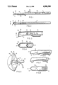
- the surgical instrument of this invention comprises a more or less cylindrical hollow shaft 10 which is tapered from a larger diameter at the handle end 11 to a smaller diameter at the forward cutting edge end.
- the tapered shaft ranges from about 0.5 to 2 mm. in diameter, being widest near the handle 11.
- the length of shaft 10 is about 30 mm. and the overall length of the instrument is about 120 mm.
- the diameter of the handle 11 is about 5 to 7 mm.
- shaft 10 is generally cylindrical, the shaft may have a vertical cross-section shape which ranges from circular to oval to a square or trapezoidal shape with rounded corners.
- the taper of shaft 10 is relatively small of approximately 5 to 15 degrees and is intended to prevent or reduce the leakage of aqueous humor around the paracentesis site.
- the taper is not an essential element of this invention, however; and, the use of shafts which are not tapered is within the scope of this invention.
- the forward end of shaft 10 comprises a parabolic, bowl-like cavity 12 having a sharpened rim which creates a single, more or less U-shaped cutting edge 14 integral with the sides of shaft 10.
- the cutting edge is approximately 2.0 mm. in length and about 0.3 to 0.4 mm. in width.
- the distal end 15 of cutting edge 14 protrudes a distance of about 0.5 to 1.0 mm. for ease of tissue penetration and cutting.
- the cutting edge is softly rounded at its distal end and is generally parabolic in shape in order to avoid damage to the outer wall of Schlemm's Canal.
- the plane of the tip of cutting edge 14 will be at an acute angle of about 5 to 45 degrees with respect to the plane of shaft 10.
- the angle of cutting edge 14, however, may vary from as little as 0 degrees to greater than 45 degrees depending on surgical requirements.
- the bottom of cavity 12 also includes a hole or aperture 16.
- the surgical instrument of this invention also includes a retractable rod or stylet 18 located inside shaft 10.
- the forward end or tip 20 of stylet 18 is preferably made of a relatively soft, non-toxic material such as plastic, silicone or rubber to prevent injury or trauma to a tissue sample.
- the remainder of stylet 18, and all of shaft 10, can be made of any durable and corrosion-resistant metal such as stainless steel.
- both shaft 10 and stylet 18 can be made from plastic, while cutting edge 14 is made from metal and attached to the end of shaft 10 by suitable means.
- lever 24 located on the handle 11 of the instrument which can manually advance and retract the stylet.
- the surgical instrument of this invention further includes an irrigation port 22 which runs along the outside of shaft 10 on the same side of shaft 10 as cutting edge 14.
- the function of this irrigation port is to maintain fluid levels in the anterior chamber of the eye during a surgical procedure and to help protect the cornea and the lens from injury.
- the irrigation port is comprised of flexible irrigation tubing composed of plastic, silastic or similar material; and, such irrigation ports are readily commercially available.
- the end of irrigation port 22 is about 3 to 5 mm. from the beginning of cutting edge 14.
- the diameter of the irrigation port is about 0.25 mm. or 30 gauge.
- the irrigation port is connected to flexible tubing (not shown) near the handle 11 of the instrument, which tubing is connected in turn to an irrigation bottle (not shown) containing sterile balanced salt solution which is suspended in the air by an adjustable intravenous stand.
- the rate of flow of the irrigation fluid can be adjusted by changing the height of the irrigating bottle, i.e. the higher the bottle, the faster the flow.
- the irrigation port has a secondary function of preventing cutting edge 14 from getting caught at the paracentesis site when the instrument is withdrawn from the eye at the end of the procedure.
- the instrument of this invention may be used without the irrigation port or the irrigation port 22 may be incorporated within the shaft 10 of the instrument.
- FIG. 3 is an enlarged sectional side view of the cutting edge end of the surgical instrument shown in FIGS. 1 and 2 along the axis 3--3 in FIG. 2.
- FIG. 3 shows stylet 18 with its soft tip 20 located inside shaft 10.
- FIG. 3 also shows the angle of cutting edge 14 with respect to shaft 10, and the location of irrigation port 22 proximate to the cutting edge.
- FIG. 3 further shows in dotted lines a segment of excised tissue 40 protrucing through aperture 16 and held there in place by tip 20 of stylet 18 when stylet 18 is advanced to its forward position, all as hereinafter described.
- FIG. 4 is a sectional bottom view of the cutting edge end of the surgical instrument along the axis 4--4 in FIG. 3.
- FIG. 4 shows the generally parabolic shape of cutting edge 14, including the softly rounded distal end 15.
- FIG. 4 also clearly shows aperture 16 in the bottom of cavity 12.
- Tip 20 of stylet 18 is preferably of a parabolic shape, as shown, in order to better fit into distal end 15 of cutting edge 14 when stylet 18 is advanced to its forward position (dotted configuration in FIG. 3).
- FIG. 5 is a schematic side view of an eyeball showing the cornea 30, the iris 32, the anterior chamber 34, the trabecular meshwork 36 which rings the iris, and Schwalbe's line 38.
- This instrument is to surgically remove a tissue segment 40 from the anterior chamber angle of the eye and to retrieve this tissue for further examination after the surgical procedure.
- the surgical technique is as follows:
- a surgical goniolens is placed over the cornea and the air bubbles under the lens are removed with saline solution.
- the goniolens is positioned to leave 2 to 3 mm. of limbal cornea exposed for the incision and instrument entry.
- the anterior chamber angle is visualized through the goniolens under magnification with an operating microscope or a binocular head loupe.
- a paracentesis incision is made into the anterior chamber with a sharp knife through clear cornea about 1 mm. anterior to the limbus.
- the length of the incision should be about 2 to 3 mm. and parallel to the limbus.
- the goniectomy instrument of this invention is carefully introduced into the anterior chamber through the paracentesis site under constant irrigation.
- the cutting edge is passed across the center of the anterior chamber to a point in the anterior chamber angle 180 degrees from the entry site.
- the tapered shaft maintains a water tight seal and the irrigation maintains the anterior chamber fluid level.
- the cutting edge 14 is used to excise the angle tissue 40 for approximately one-third of the angle circumference.
- the excised tissue will be guided toward and through aperture 16 in cavity 12 (see FIG. 3) as cutting edge 14 is advanced. This will help to hold the excised tissue in place during removal.
- the paracentesis entry site is used as a pivotal point for the instrument as it swings across the anterior chamber.
- the stylet 18 (not seen in FIG. 5) is advanced so that stylet tip 20 holds tissue 40 firmly against the interior of cavity 12 (see FIG. 3). Then the instrument is carefully withdrawn from the anterior chamber. The anterior chamber may then be deepened with balanced salt solution, air, or a viscoelastic substance. The paracentesis wound may also be closed with 10-0 nylon suture if necessary.
- the instrument may be reinserted into the eye through the existing incision or through a second incision to excise and collect additional tissue samples.
- FIGS. 6, 7, and 8 A modified version of the goniectomy instrument of this invention is illustrated in FIGS. 6, 7, and 8.
- stylet 18 and aperture 16 have been eliminated, and slidable sleeve 50 capable of enclosing the distal end 15 of shaft 10 has been added.
- the forward end of sleeve 50 is also of a generally parabolic shape in order to achieve a good fit over distal end 15.
- the excised tissue specimen does not pass through aperture 16 to the other side of cavity 12. Instead, the tissue specimen is now directed by the interior shape of cavity 12 into hollow shaft 10.
- Metal sleeve 50 around distal end 15 of the shaft helps to hold the specimen in cavity 12.
- the metal sleeve is able to move along the length of the shaft and is controlled by a lever (comparable to lever 24 as shown in FIG. 1) located on the handle of the instrument.
- the sleeve can be retracted to expose the cutting tip so that the tissue specimen can be retrieved.
- the sleeve also serves a secondary purpose by covering the cutting edge to protect it from inadvertently damaging other structures within the eye and to protect the cutting edge from traumatic blunting.
Abstract
This invention relates to the design and application of a goniectomy instrument for the purpose of diagnostically and therapeutically removing tissue from the anterior chamber angle of the eye and for retrieving this tissue for further examination. The surgical instrument of this invention comprises in combination: a hollow, tapered shaft having a cutting edge at one end as an integral part thereof; a retractable stylet contained within the hollow interior of the tapered shaft; and an irrigation port running along the outside of the tapered shaft. This instrument is useful for excising tissue to relieve an obstruction blocking the outflow of aqueous humor from the eye as well as for providing specimens of the excised tissue for histopathological examination.
Description
This is a continuation of co-pending application Ser. No. 07/070,325 filed on July 6, 1987
Glaucoma is a major cause of irreversible blindness in the United States. Approximately one percent of the general population has this disease. Visual loss from glaucoma is usually due to elevated intraocular pressure which damages the optic nerve. The cause for the elevated intraocular pressure is not entirely known, but, at least in many cases, appears to be due to an increase in the resistance to the outflow of aqueous humor from the eye. Aqueous humor is a fluid which circulates within the anterior portions of the eye and provides nourishment to and removes waste products from various avascular structures in the eye such as the cornea and the lens. Aqueous humor is continuously being formed within the ciliary body epithelium and is normally continuously drained out of the eye through the trabecular meshwork and Schlemm's Canal into the venous system. It is believed that the site of resistance to the outflow of aqueous humor is in the juxtacanalicular portion of the trabecular meshwork.
Treatment of glaucoma is, therefore, usually directed at lowering the intraocular pressure and thereby preventing progressive optic nerve damage and visual field loss. If medical therapy (topical eyedrops and oral tablets) and laser therapy fail to adequately lower the intraocular pressure, then glaucoma surgery is required. There are several different types of glaucoma surgery (filtration, goniotomy, trabeculotomy, cyclodialysis, etc.), but all of them have a common purpose: that is, to bypass or eliminate any blockage and thereby restore the outflow of aqueous humor from the eye.
Goniotomy and trabeculotomy surgery involve incising the abnormal tissue which is overlying or inside the trabecular meshwork in order to allow aqueous humor to flow freely into the normal outflow system. These two procedures are most commonly used in treating childhood glaucomas, but also have limited success in adult-onset glaucomas. When these surgical procedures fail, the failure is usually due to scarring which blocks the incision in the trabecular meshwork. In an attempt to avoid this problem, a technique was developed in which an intraocular diathermy probe is used to cauterize the incision edges in an attempt to reduce or eliminate scarring. However, this technique has not been proven to be any more successful than the other surgical procedures.
In identifying the reason for open-angle glaucoma and for failure of these surgical procedures and in determining the extent of the blockage and the preferred treatment, it would be useful to extract relatively large intact samples of undamaged trabecular meshwork and scar tissue and, perhaps, the surrounding tissue for histopathologic examination. Unfortunately, existing instruments for performing this surgery do not simultaneously permit the cutting and removal of relatively large and undamaged tissue specimens. The use of separate instruments as are known in the art for cutting and excision in a delicate operation like glaucoma surgery is not a satisfactory alternative. Such a procedure may entail a plurality of surgical penetrations of the inner eye thereby increasing the dangers of trauma, injury, and infection. In some cases it may also be impossible to locate a severed segment of tissue from the inner eye when using a second instrument to retrieve it. In addition, such a two-step procedure is certain to require more time than would a one-step procedure, a particularly important factor if a plurality of tissue samples are desired.
Those surgical instruments in the prior art which are capable of both cutting and removing tissue specimens in a single operation are, in general, either not adaptable for delicate glaucoma surgery, do not remove large enough tissue samples, or else result in damage to the structure of the excised tissue. For example, the prior art includes a plethora of biopsy instruments which both cut and remove tissue. In testing for carcinomous tissue, however, only relatively small tissue samples are required, and the physical structure of the tissue is usually not important. Therefore, these instruments cannot be adapted for the excision of relatively large, undamaged segments of glaucoma scar tissue. Some of the more pertinent prior art patents in this field are discussed below, and all of these patents are specifically incorporated herein by reference.
U.S. Pat. No. 3,996,935 (Banko II) is perhaps the most representative of the prior art in this field, and of its limitations. Banko II discloses a multi-purpose instrument designed for ophthalmic surgery including removal of foreign matter, blood clots, lenses and other eye tissue. The instrument includes an internal probe having a distal end for engaging tissue or foreign matter to be removed from the interior of the eye. The internal probe comprises two coaxial shafts, each having a jaw or port at its terminal end, such that the inner shaft is slidable and rotatable with respect to the outer. In other embodiments of the Banko II device, the hollow interior of the inner coaxial shaft can be utilized as a fluid irrigation channel or as a suction canal to facilitate tissue removal.
The Banko II patent, however, requires suction to engage and remove tissue. Banko's "internal probe" or "inner jaw" has cutting edges to cut tissue against the sharp opening edges of the "outer jaw" at the end of the outer hollow shaft resulting in a scissor-like cutting action which could damage the tissue. This instrument is designed to cut tissue into small pieces for removal, not to extract a relatively large, undamaged and intact tissue segment. The shape of this instrument also makes it unsuitable for goniectomy surgery. This instrument is also relatively complex, requiring an external power source for applying suction and for continuously rotating the "internal probe." Accordingly, this instrument is not suitable for conducting goniectomy surgery.
U.S. Pat. No. 3,583,390 (Jascalevich) is an example of a typical biopsy instrument. This patent discloses a biopsy device which has generally tapered, two-sided arrowhead-shaped cutting tool with a hole near the tip of the cutting edges. The shape of this biopsy instrument makes it unworkable for goniectomy surgery because the opening that would be required to introduce the instrument into the eye would be too large. Also, the large exposed surface area of the blades could cause trauma or injury to adjacent intraocular tissue during the surgical procedure. With the instrument of this patent, the tissue specimen is cut and separated from adjacent tissue by two blades -- the biopsy blade and the scalpel blade -- operating in a scissor-like fashion. Use of the Jascalevich instrument in the eye would necessitate making three entry wounds, one for the biopsy device and two for the scalpel blade (one for each groove). Because the two blades must be separately manipulated to excise a tissue specimen, unless a high degree of care and skill is exercised by the surgeon severe injury to the surrounding eye tissue could result. Upon withdrawing the biopsy instrument from the eye, the sharp corners of the arrowhead-like blade could catch on and injure eye tissue surrounding the entry incision. Accordingly, this instrument is also not suitable for goniectomy surgery.
U.S. Pat. Nos. 3,844,272 (Banko I); 3,929,123 (Jamshidi); and 3,007,471 (McClure) disclose surgical instruments which utilize tapered and pointed distal cutting tips in combination with coaxial members such that a sheath can slidably expose or cover the cutting tip. McClure discloses the use of an internal stylet (reference numeral 40) inside the sliding coaxial members. Other patents which show surgical sampling instruments having slidable, coaxial members, internal stylets or both include U.S. Pat. Nos. 4,308,875 (Young); 2,850,007 (Lingley); 3,893,445 (Hofsess); and 4,282,884 (Boebel).
In addition, there are a number of recent patents which are specifically directed to instruments for ophthalmic surgery. These patents include U.S. Pat. Nos. 4,590,935 (Ranalli); 4,577,629 (Martinez); 4,538,611 (Kelman); 4,570,632 (Woods); 4,041,947 (Weiss); and 4,320,761 (Haddad). These patents include discussion of such issues as the importance of irrigation during eye surgery and similar matters which have special relevance for ophthalmic surgery. None of these various instruments is suitable, however, both as a surgical tool and as a device for excising relatively large, undamaged tissue segments.
A principal object of this invention is to provide a surgical instrument for use in glaucoma surgery.
A further object of this invention is to provide a surgical instrument suitable for both cutting and extracting relatively large, intact segments of internal eye tissue.
Still a further object of this invention is to provide an instrument specially designed for goniectomy surgery comprising in combination a hollow, tapered shaft having a cutting edge at one end as an integral part thereof; a retractable stylet contained within the hollow interior of the tapered shaft; and an irrigation port running along the outside of the tapered shaft.
Specifically, it is an object of this invention to provide a surgical instrument for use in glaucoma surgery to excise a piece of tissue from the anterior chamber angle (trabecular meshwork and the inner wall of Schlemm's Canal) to therapeutically relieve the obstruction of the outflow of aqueous humor from the eye and to provide specimens of the abnormal tissues excised for histopathological examination.
These and other objects and advantages of this invention will become apparent in the following description.
FIG. 1 is a schematic side view of the surgical instrument of this invention.
FIG. 2 is a schematic bottom view of the surgical instrument of this invention.
FIG. 3 is an enlarged, sectional side view of the forward end of the surgical instrument of FIGS. 1 and 2 along the axis 3--3 in FIG. 2.
FIG. 4 is a sectional bottom view of the forward end of the surgical instrument along the axis 4--4 in FIG. 3.
FIG. 5 is a schematic side view of an eyeball with the surgical instrument of this invention in place and ready to begin cutting and removing a tissue segment from the trabecular meshwork.
FIG. 6 is a schematic side view of an alternative embodiment of the surgical instrument of this invention showing a slidable sleeve in the open position.
FIG. 7 is a schematic side view of the alternative embodiment of the surgical instrument of this invention with the slidable sleeve in the closed position.
FIG. 8 is a schematic bottom view of the forward end of the instrument as shown in FIG. 6.
Referring to FIGS. 1 and 2, in the preferred embodiment the surgical instrument of this invention comprises a more or less cylindrical hollow shaft 10 which is tapered from a larger diameter at the handle end 11 to a smaller diameter at the forward cutting edge end. The tapered shaft ranges from about 0.5 to 2 mm. in diameter, being widest near the handle 11. The length of shaft 10 is about 30 mm. and the overall length of the instrument is about 120 mm. The diameter of the handle 11 is about 5 to 7 mm.
Although in the preferred embodiment shaft 10 is generally cylindrical, the shaft may have a vertical cross-section shape which ranges from circular to oval to a square or trapezoidal shape with rounded corners. The taper of shaft 10 is relatively small of approximately 5 to 15 degrees and is intended to prevent or reduce the leakage of aqueous humor around the paracentesis site. The taper is not an essential element of this invention, however; and, the use of shafts which are not tapered is within the scope of this invention.
The forward end of shaft 10 comprises a parabolic, bowl-like cavity 12 having a sharpened rim which creates a single, more or less U-shaped cutting edge 14 integral with the sides of shaft 10. The cutting edge is approximately 2.0 mm. in length and about 0.3 to 0.4 mm. in width. The distal end 15 of cutting edge 14 protrudes a distance of about 0.5 to 1.0 mm. for ease of tissue penetration and cutting. The cutting edge is softly rounded at its distal end and is generally parabolic in shape in order to avoid damage to the outer wall of Schlemm's Canal.
In the preferred embodiment, as better shown in FIG. 3, the plane of the tip of cutting edge 14 will be at an acute angle of about 5 to 45 degrees with respect to the plane of shaft 10. The angle of cutting edge 14, however, may vary from as little as 0 degrees to greater than 45 degrees depending on surgical requirements. As shown in FIGS. 2 and 4, in the preferred embodiment the bottom of cavity 12 also includes a hole or aperture 16.
The surgical instrument of this invention also includes a retractable rod or stylet 18 located inside shaft 10. The forward end or tip 20 of stylet 18 is preferably made of a relatively soft, non-toxic material such as plastic, silicone or rubber to prevent injury or trauma to a tissue sample. The remainder of stylet 18, and all of shaft 10, can be made of any durable and corrosion-resistant metal such as stainless steel. In another embodiment, both shaft 10 and stylet 18 can be made from plastic, while cutting edge 14 is made from metal and attached to the end of shaft 10 by suitable means. There is also a lever 24 located on the handle 11 of the instrument which can manually advance and retract the stylet. There may also be a lubricated spring operated system 26 to facilitate sliding the stylet back and forth smoothly with minimal friction and force.
The surgical instrument of this invention further includes an irrigation port 22 which runs along the outside of shaft 10 on the same side of shaft 10 as cutting edge 14. The function of this irrigation port is to maintain fluid levels in the anterior chamber of the eye during a surgical procedure and to help protect the cornea and the lens from injury. The irrigation port is comprised of flexible irrigation tubing composed of plastic, silastic or similar material; and, such irrigation ports are readily commercially available.
The end of irrigation port 22 is about 3 to 5 mm. from the beginning of cutting edge 14. The diameter of the irrigation port is about 0.25 mm. or 30 gauge. The irrigation port is connected to flexible tubing (not shown) near the handle 11 of the instrument, which tubing is connected in turn to an irrigation bottle (not shown) containing sterile balanced salt solution which is suspended in the air by an adjustable intravenous stand. The rate of flow of the irrigation fluid can be adjusted by changing the height of the irrigating bottle, i.e. the higher the bottle, the faster the flow. The irrigation port has a secondary function of preventing cutting edge 14 from getting caught at the paracentesis site when the instrument is withdrawn from the eye at the end of the procedure. In some applications the instrument of this invention may be used without the irrigation port or the irrigation port 22 may be incorporated within the shaft 10 of the instrument.
FIG. 3 is an enlarged sectional side view of the cutting edge end of the surgical instrument shown in FIGS. 1 and 2 along the axis 3--3 in FIG. 2. FIG. 3 shows stylet 18 with its soft tip 20 located inside shaft 10. FIG. 3 also shows the angle of cutting edge 14 with respect to shaft 10, and the location of irrigation port 22 proximate to the cutting edge. FIG. 3 further shows in dotted lines a segment of excised tissue 40 protrucing through aperture 16 and held there in place by tip 20 of stylet 18 when stylet 18 is advanced to its forward position, all as hereinafter described.
FIG. 4 is a sectional bottom view of the cutting edge end of the surgical instrument along the axis 4--4 in FIG. 3. FIG. 4 shows the generally parabolic shape of cutting edge 14, including the softly rounded distal end 15. FIG. 4 also clearly shows aperture 16 in the bottom of cavity 12. Tip 20 of stylet 18 is preferably of a parabolic shape, as shown, in order to better fit into distal end 15 of cutting edge 14 when stylet 18 is advanced to its forward position (dotted configuration in FIG. 3).
The application of the instrument of this invention to goniectomy surgery will be described by reference to FIG. 5. FIG. 5 is a schematic side view of an eyeball showing the cornea 30, the iris 32, the anterior chamber 34, the trabecular meshwork 36 which rings the iris, and Schwalbe's line 38.
The purpose of this instrument is to surgically remove a tissue segment 40 from the anterior chamber angle of the eye and to retrieve this tissue for further examination after the surgical procedure. The surgical technique is as follows:
(1) The patient is placed in the supine position on the operating table. General or local anesthesia is given.
(2) The patient' head is rotated slightly away from the side of the surgery and the eye being operated on is slightly abducted. The eye's position is fixed by locking forceps which are being held by the surgical assistant.
(3) A surgical goniolens is placed over the cornea and the air bubbles under the lens are removed with saline solution. The goniolens is positioned to leave 2 to 3 mm. of limbal cornea exposed for the incision and instrument entry. The anterior chamber angle is visualized through the goniolens under magnification with an operating microscope or a binocular head loupe.
(4) A paracentesis incision is made into the anterior chamber with a sharp knife through clear cornea about 1 mm. anterior to the limbus. The length of the incision should be about 2 to 3 mm. and parallel to the limbus.
(5) The goniectomy instrument of this invention is carefully introduced into the anterior chamber through the paracentesis site under constant irrigation. The cutting edge is passed across the center of the anterior chamber to a point in the anterior chamber angle 180 degrees from the entry site. The tapered shaft maintains a water tight seal and the irrigation maintains the anterior chamber fluid level.
(6) The cutting edge 14 is used to excise the angle tissue 40 for approximately one-third of the angle circumference. The excised tissue will be guided toward and through aperture 16 in cavity 12 (see FIG. 3) as cutting edge 14 is advanced. This will help to hold the excised tissue in place during removal. The paracentesis entry site is used as a pivotal point for the instrument as it swings across the anterior chamber.
(7) After the desired strip of angle tissue 40 is excised, the stylet 18 (not seen in FIG. 5) is advanced so that stylet tip 20 holds tissue 40 firmly against the interior of cavity 12 (see FIG. 3). Then the instrument is carefully withdrawn from the anterior chamber. The anterior chamber may then be deepened with balanced salt solution, air, or a viscoelastic substance. The paracentesis wound may also be closed with 10-0 nylon suture if necessary.
Alternatively, if desired, the instrument may be reinserted into the eye through the existing incision or through a second incision to excise and collect additional tissue samples.
A modified version of the goniectomy instrument of this invention is illustrated in FIGS. 6, 7, and 8. In the modified version of the instrument, stylet 18 and aperture 16 have been eliminated, and slidable sleeve 50 capable of enclosing the distal end 15 of shaft 10 has been added. As shown in FIG. 8, the forward end of sleeve 50 is also of a generally parabolic shape in order to achieve a good fit over distal end 15.
With the modified instrument as shown in FIGS. 6, 7 and 8, the excised tissue specimen does not pass through aperture 16 to the other side of cavity 12. Instead, the tissue specimen is now directed by the interior shape of cavity 12 into hollow shaft 10. Metal sleeve 50 around distal end 15 of the shaft helps to hold the specimen in cavity 12. The metal sleeve is able to move along the length of the shaft and is controlled by a lever (comparable to lever 24 as shown in FIG. 1) located on the handle of the instrument. After the tissue specimen 40 is cut and directed up the hollow shaft 10 of the instrument, sleeve 50 is advanced to the distal end 15 of the instrument to cover cutting edge 14 and to thereby contain the tissue specimen in cavity 12 and in the hollow shaft. After the instrument is removed from the eye, the sleeve can be retracted to expose the cutting tip so that the tissue specimen can be retrieved. The sleeve also serves a secondary purpose by covering the cutting edge to protect it from inadvertently damaging other structures within the eye and to protect the cutting edge from traumatic blunting.
Many other variations and modifications of my basic design will be readily apparent to those skilled in the art. All such variations and modifications are within the spirit and the scope of this invention and, therefore, are intended to be encompassed by the following claims:
Claims (24)
1. A surgical instrument designed for cutting and removing intact an elongated strip of internal tissue greater in length than the width of the instrument cutting edge, said instrument comprising:
(a) a hollow shaft with rounded sides having a central, longitudinal axis, a handle at the rear end thereof and, at the forward end, a generally parabolic-shaped cavity which forms a bowl;
(b) said bowl having closed sides, a closed forward wall, a closed bottom, and an open mouth such that the sides, forward wall and bottom form a continuous inner wall integral with the adjacent sides of said hollow shaft except for an opening in said bowl which communicates with the interior of said hollow shaft; and,
(c) further wherein said open mouth is defined by a generally U-shaped rim integral with the adjacent sides of said hollow shaft and said rim is sharpened so as to form a cutting blade having side cutting edges and a distal tip.
2. The surgical instrument of claim 1 wherein said hollow shaft is tapered from a larger diameter at said handle end to a smaller diameter at said cutting edge end.
3. The surgical instrument of claim 1 wherein the distal tip of said cutting blade is rounded.
4. The surgical instrument of claim 1 further including an irrigation port along the outside of said hollow shaft.
5. The surgical instrument of claim 1 further including tissue securing and removal for securing and removing intact and elongated strip of internal tissue.
6. The surgical instrument of claim 5 wherein said tissue securing and removal means comprises a stylet slidably positioned inside said hollow shaft.
7. The surgical instrument of claim 6 wherein said stylet has a soft blunt tip at the forward end thereof designed to engage without cutting a strip of tissue between said soft tip and the inner wall of said bowl.
8. The surgical instrument of claim 6 further including lever and spring means for moving said stylet inside said shaft.
9. The surgical instrument of claim 6 further including an aperture located in the bottom of said bowl.
10. The surgical instrument of claim 5 wherein said tissue securing and removal means comprises a sleeve slidably positioned along the outside of said hollow shaft.
11. The surgical instrument of claim 10 further including lever means for moving said sleeve.
12. The surgical instrument of claim 1 wherein the mouth of said bowl is outwardly sloping with respect to the adjacent sides of said hollow shaft at an acute angle to the central longitudinal axis of said shaft.
13. A surgical instrument of comprising: a tubular member having a central, longitudinal axis and a forwardly extending leading end having an open mouth, said open mouth being outwardly sloping at an acute angle with respect to the central, longitudinal axis of said tubular member and having sharpened cutting edges; and, holding means slidably positioned within said tubular member, said holding means having a blunt front end for contacting and fixing without cutting an excised tissue segment within said tubular member.
14. The surgical instrument of claim 13 wherein said open mouth comprises a continuous cutting edge.
15. The surgical instrument of claim 13 wherein said leading end has an aperture.
16. The surgical instrument of claim 13 wherein said tubular member is forwardly tapered toward said leading end.
17. The surgical instrument of claim 13 further including an irrigation port attached to said tubular member.
18. In a surgical instrument designed for goniectomy surgery, the improvements which comprise: a hollow shaft having a handle at one end thereof and a parabolic cavity at the other end, said cavity having a sharpened rim which forms a U-shaped cutting edge integral with the sides of said shaft; a soft-tipped stylet slidably positioned inside said hollow shaft; and an irrigation port along the outside of said hollow shaft proximate to said cutting edge.
19. A method for performing goniectomy surgery on an eye and for retrieving an elongated excised tissue segment comprising the following steps: p1 (a) introducing into the anterior chamber of the eye through an incision a surgical instrument comprising a hollow shaft with rounded sides and a parabolic cavity at the forward end thereof, said cavity having sides and a bottom integral with the sides of said hollow shaft, and an open mouth having a sharpened rim which defines a generally U-shaped cutting edge, and an aperture in the bottom of said cavity, and a blunt-tipped stylet slidably positioned inside said hollow said shaft;
(b) excising an elongated tissue segment by drawing said cutting edge along the trabecular meshwork such that the end of the tissue segment is directed through said aperture in said cavity;
(c) advancing said stylet such that the blunt tip engages said tissue segment and holds it securely against the inside of including tissue securing and removal means for securing and removing intact an elongated strip of internal tissue.
20. The method of claim 19 further comprising he step of irrigating the anterior chamber of the eye during the surgical procedure with a sterile balanced salt solution which is introduced into the eye through an irrigation port along the outside of said hollow shaft.
21. The method of claim 19 further comprising the step of irrigating the anterior chamber of the eye during the surgical procedure with a sterile balanced salt solution which is introduced into the eye through an irrigation port located inside said hollow shaft.
22. A method for performing goniectomy surgery on an eye and for retrieving an elongated excised tissue segment comprising the following steps:
(a) introducing into the anterior chamber of the eye through an incision a surgical instrument comprising: a hollow shaft with rounded sides having a central, longitudinal axis, a handle at the rear end thereof and, at the forward end, a generally parabolic-shaped cavity which forms a bowl; said bowl having closed sides, a closed forward wall, a closed bottom, and an open mouth such that the sides, forward wall and bottom form a continuous inner wall integral with the adjacent sides of said hollow shaft except for an opening in said bowl which communicates with the interior of said hollow shaft; further wherein said open mouth is defined by a generally U-shaped rim integral with the adjacent sides of said hollow shaft and said rim is sharpened so as to form a cutting blade having side cutting edges and a distal tip; and further including a sleeve slidably positioned along the outside of said hollow shaft;
(b) excising an elongated tissue segment by drawing said cutting edge along a trabecular meshwork such that the end of the tissue segment is directed into the parabolic cavity and up the hollow shaft;
(c) advancing said sleeve along the hollow shaft so as to enclose the parabolic cavity and the tissue segment contained therein; and,
(d) removing said surgical instrument from the eye together with said elongated tissue segment intact.
23. The method of claim 22 further comprising the step of irrigating the anterior chamber of the eye during the surgical procedure with a sterile balanced salt solution which is introduced into the eye through an irrigation port along the outside of said hollow shaft.
24. The method of claim 22 further comprising the step of irrigating the anterior chamber of the eye during the surgical procedure with a sterile balanced salt solution which is introduced into the eye through an irrigation port located inside said hollow shaft.
Priority Applications (1)
| Application Number | Priority Date | Filing Date | Title |
|---|---|---|---|
| US07/315,190 US4900300A (en) | 1987-07-06 | 1989-02-24 | Surgical instrument |
Applications Claiming Priority (2)
| Application Number | Priority Date | Filing Date | Title |
|---|---|---|---|
| US7032587A | 1987-07-06 | 1987-07-06 | |
| US07/315,190 US4900300A (en) | 1987-07-06 | 1989-02-24 | Surgical instrument |
Related Parent Applications (1)
| Application Number | Title | Priority Date | Filing Date |
|---|---|---|---|
| US7032587A Continuation | 1987-07-06 | 1987-07-06 |
Publications (1)
| Publication Number | Publication Date |
|---|---|
| US4900300A true US4900300A (en) | 1990-02-13 |
Family
ID=26751021
Family Applications (1)
| Application Number | Title | Priority Date | Filing Date |
|---|---|---|---|
| US07/315,190 Expired - Fee Related US4900300A (en) | 1987-07-06 | 1989-02-24 | Surgical instrument |
Country Status (1)
| Country | Link |
|---|---|
| US (1) | US4900300A (en) |
Cited By (112)
| Publication number | Priority date | Publication date | Assignee | Title |
|---|---|---|---|---|
| WO1991002499A1 (en) * | 1989-08-14 | 1991-03-07 | Central Institute For The Deaf | Surgical instrument and cell isolation and transplantation |
| US5248297A (en) * | 1992-02-26 | 1993-09-28 | Haruo Takase | Suction tube for use in surgical operation |
| US5335671A (en) * | 1989-11-06 | 1994-08-09 | Mectra Labs, Inc. | Tissue removal assembly with provision for an electro-cautery device |
| US5338292A (en) * | 1989-11-06 | 1994-08-16 | Mectra Labs, Inc. | Disposable lavage with instrument shield |
| US5360399A (en) * | 1992-01-10 | 1994-11-01 | Robert Stegmann | Method and apparatus for maintaining the normal intraocular pressure |
| US5409013A (en) * | 1989-11-06 | 1995-04-25 | Mectra Labs, Inc. | Tissue removal assembly |
| US5527332A (en) | 1994-11-02 | 1996-06-18 | Mectra Labs, Inc. | Tissue cutter for surgery |
| US5662603A (en) * | 1996-05-29 | 1997-09-02 | Gelbfish; Gary A. | Medical material removal method and associated instrumentation |
| US5797907A (en) | 1989-11-06 | 1998-08-25 | Mectra Labs, Inc. | Electrocautery cutter |
| US5817075A (en) * | 1989-08-14 | 1998-10-06 | Photogenesis, Inc. | Method for preparation and transplantation of planar implants and surgical instrument therefor |
| US5868728A (en) * | 1995-02-28 | 1999-02-09 | Photogenesis, Inc. | Medical linear actuator for surgical delivery, manipulation, and extraction |
| US5893862A (en) * | 1997-04-10 | 1999-04-13 | Pratt; Arthur William | Surgical apparatus |
| US5962027A (en) * | 1989-08-14 | 1999-10-05 | Photogenesis, Inc. | Retinal cell transplant |
| US5984889A (en) * | 1996-02-23 | 1999-11-16 | Allergan Sales, Inc. | Apparatus and method for delivering viscoelastic material to an eye |
| US6045791A (en) * | 1992-03-06 | 2000-04-04 | Photogenesis, Inc. | Retinal pigment epithelium transplantation |
| US6193672B1 (en) | 1993-05-11 | 2001-02-27 | Mectra Labs, Inc. | Lavage |
| US20020121281A1 (en) * | 2000-10-18 | 2002-09-05 | Mark Humayun | Soft tip cannula and methods for use thereof |
| WO2002074052A2 (en) * | 2001-03-16 | 2002-09-26 | Glaukos Corporation | Applicator and methods for placing a trabecular shunt for glaucoma treatment |
| US20020143284A1 (en) * | 2001-04-03 | 2002-10-03 | Hosheng Tu | Drug-releasing trabecular implant for glaucoma treatment |
| US20020169130A1 (en) * | 2001-05-03 | 2002-11-14 | Hosheng Tu | Medical device and methods of use for glaucoma treatment |
| US20020188308A1 (en) * | 2001-04-07 | 2002-12-12 | Hosheng Tu | Glaucoma stent and methods thereof for glaucoma treatment |
| US20030055372A1 (en) * | 1999-04-26 | 2003-03-20 | Lynch Mary G. | Shunt device and method for treating glaucoma |
| US20030054023A1 (en) * | 1989-08-14 | 2003-03-20 | Photogenesis, Inc. | Volute subretinal implants |
| US20030088260A1 (en) * | 2001-11-08 | 2003-05-08 | Smedley Gregory T. | Combined treatment for cataract and glaucoma treatment |
| US20030097151A1 (en) * | 2001-10-25 | 2003-05-22 | Smedley Gregory T. | Apparatus and mitochondrial treatment for glaucoma |
| US20030144678A1 (en) * | 1996-02-07 | 2003-07-31 | Hellenkamp Johann F. | Automatic surgical device and control assembly for cutting a cornea |
| US6626858B2 (en) | 1999-04-26 | 2003-09-30 | Gmp Vision Solutions, Inc. | Shunt device and method for treating glaucoma |
| US20030187385A1 (en) * | 2000-04-14 | 2003-10-02 | Bergheim Olav B. | Implant with anchor |
| US20030216747A1 (en) * | 2002-02-14 | 2003-11-20 | Kaplan Henry J. | Subretinal implantation device and surgical cannulas for use therewith |
| US6666841B2 (en) | 2001-05-02 | 2003-12-23 | Glaukos Corporation | Bifurcatable trabecular shunt for glaucoma treatment |
| US20040024345A1 (en) * | 2002-04-19 | 2004-02-05 | Morteza Gharib | Glaucoma implant with valveless flow bias |
| ES2200623A1 (en) * | 2001-02-16 | 2004-03-01 | Univ Miguel Hernandez De Elche | Apparatus for the obtaining of eye tissue samples in vertebrate animals. (Machine-translation by Google Translate, not legally binding) |
| US20040050392A1 (en) * | 2001-08-28 | 2004-03-18 | Hosheng Tu | Glaucoma stent for treating glaucoma and methods of use |
| US20040102729A1 (en) * | 2002-04-08 | 2004-05-27 | David Haffner | Devices and methods for glaucoma treatment |
| US20040111050A1 (en) * | 2000-04-14 | 2004-06-10 | Gregory Smedley | Implantable ocular pump to reduce intraocular pressure |
| US20040127843A1 (en) * | 2000-04-14 | 2004-07-01 | Hosheng Tu | Glaucoma implant with therapeutic agents |
| US20040147870A1 (en) * | 2002-04-08 | 2004-07-29 | Burns Thomas W. | Glaucoma treatment kit |
| US20040236358A1 (en) * | 2003-04-07 | 2004-11-25 | Barrile-Josephson Craig A. | Bar-link drive system for a microkeratome |
| US20040254598A1 (en) * | 2003-06-16 | 2004-12-16 | Schumacher Brian S. | Suture cutter |
| WO2004110501A2 (en) | 2003-06-10 | 2004-12-23 | Neomedix Corporation | Tubular cutting device for cutting and removing tissue |
| US6852092B2 (en) | 2002-10-02 | 2005-02-08 | Advanced Medical Optics, Inc. | Handpiece system for multiple phacoemulsification techniques |
| US20050049578A1 (en) * | 2000-04-14 | 2005-03-03 | Hosheng Tu | Implantable ocular pump to reduce intraocular pressure |
| US20050119636A1 (en) * | 2001-05-02 | 2005-06-02 | David Haffner | Implant with intraocular pressure sensor for glaucoma treatment |
| US20050192527A1 (en) * | 2001-05-02 | 2005-09-01 | Morteza Gharib | Glaucoma implant with extending members |
| US20050250788A1 (en) * | 2004-01-30 | 2005-11-10 | Hosheng Tu | Aqueous outflow enhancement with vasodilated aqueous cavity |
| US20050266047A1 (en) * | 2002-04-08 | 2005-12-01 | Hosheng Tu | Injectable glaucoma implants with multiple openings |
| US20050271704A1 (en) * | 2002-04-08 | 2005-12-08 | Hosheng Tu | Injectable glaucoma implants with multiple openings |
| US20050277864A1 (en) * | 2000-04-14 | 2005-12-15 | David Haffner | Injectable gel implant for glaucoma treatment |
| US6979328B2 (en) | 2001-01-18 | 2005-12-27 | The Regents Of The University Of California | Minimally invasive glaucoma surgical instrument and method |
| US20060058703A1 (en) * | 2002-12-10 | 2006-03-16 | Universitaetsklinikum Charite Medizinische Fakultaet Der Humboldt-Universitaet Zu Berlin | Optical biopsy instrument |
| US7186232B1 (en) | 2002-03-07 | 2007-03-06 | Glaukoa Corporation | Fluid infusion methods for glaucoma treatment |
| US20070088242A1 (en) * | 2003-11-14 | 2007-04-19 | Coroneo Minas T | Ocular pressure regulation |
| US20070106310A1 (en) * | 2005-11-10 | 2007-05-10 | Goldin Mark A | Suture cutter |
| US20070149915A1 (en) * | 2003-05-05 | 2007-06-28 | Judith Yablonski | Internal shunt and method for treating glaucoma |
| US20070191863A1 (en) * | 2006-01-17 | 2007-08-16 | De Juan Eugene Jr | Glaucoma Treatment Device |
| US20070213755A1 (en) * | 2006-03-07 | 2007-09-13 | Beckman Andrew T | Device for minimally invasive internal tissue removal |
| US20070213630A1 (en) * | 2006-03-07 | 2007-09-13 | Beckman Andrew T | Device for minimally invasive internal tissue removal |
| US20070233037A1 (en) * | 2006-01-17 | 2007-10-04 | Gifford Hanson S Iii | Drug Delivery Treatment Device |
| US20070239064A1 (en) * | 2006-03-29 | 2007-10-11 | Cicenas Chris W | Device for minimally invasive internal tissue removal |
| US20070244496A1 (en) * | 1996-02-07 | 2007-10-18 | Hellenkamp Johann F | Automatic surgical device and control assembly for cutting a cornea |
| US20070293807A1 (en) * | 2006-05-01 | 2007-12-20 | Lynch Mary G | Dual drainage pathway shunt device and method for treating glaucoma |
| US20080172204A1 (en) * | 2007-01-15 | 2008-07-17 | Fujitsu Limited | Step counter and method of counting steps |
| US7488303B1 (en) | 2002-09-21 | 2009-02-10 | Glaukos Corporation | Ocular implant with anchor and multiple openings |
| US20090182421A1 (en) * | 2007-07-17 | 2009-07-16 | Tom Silvestrini | Ocular implant with hydrogel expansion capabilities |
| US20090216244A1 (en) * | 2004-11-30 | 2009-08-27 | Joel Pynson | Two Stage Plunger for Intraocular Lens Injector |
| US20090287143A1 (en) * | 2008-05-15 | 2009-11-19 | Casey Line | Small Gauge Mechanical Tissue Cutter/Aspirator Probe For Glaucoma Surgery |
| US20090287233A1 (en) * | 2008-05-15 | 2009-11-19 | Huculak John C | Small Gauge Mechanical Tissue Cutter/Aspirator Probe For Glaucoma Surgery |
| US20100125278A1 (en) * | 2008-11-19 | 2010-05-20 | Wagner Christopher E | Hard and Soft Tip Intraocular Lens Injector System and Method |
| US20100137981A1 (en) * | 2008-06-25 | 2010-06-03 | Silvestrini Thomas A | Ocular implant with shape change capabilities |
| US20100274258A1 (en) * | 2009-01-28 | 2010-10-28 | Silvestrini Thomas A | Ocular implant with stiffness qualities, methods of implantation and system |
| US20100312252A1 (en) * | 2009-06-03 | 2010-12-09 | Guangyao Jia | Capsularhexis device with flexible heating element having an angled transitional neck |
| US20100312232A1 (en) * | 2009-06-03 | 2010-12-09 | Guangyao Jia | Capsulotomy Repair Device and Method for Capsulotomy Repair |
| US20110105990A1 (en) * | 2009-11-04 | 2011-05-05 | Silvestrini Thomas A | Zonal drug delivery device and method |
| US7951155B2 (en) | 2002-03-15 | 2011-05-31 | Glaukos Corporation | Combined treatment for cataract and glaucoma treatment |
| US20110202049A1 (en) * | 2010-02-18 | 2011-08-18 | Alcon Research, Ltd. | Small Gauge Ablation Probe For Glaucoma Surgery |
| US20110238075A1 (en) * | 2009-12-23 | 2011-09-29 | Luke Clauson | Drug delivery devices and methods |
| US8137344B2 (en) | 2008-12-10 | 2012-03-20 | Alcon Research, Ltd. | Flexible, automated capsulorhexis device |
| US8157797B2 (en) | 2009-01-12 | 2012-04-17 | Alcon Research, Ltd. | Capsularhexis device with retractable bipolar electrodes |
| US8506515B2 (en) | 2006-11-10 | 2013-08-13 | Glaukos Corporation | Uveoscleral shunt and methods for implanting same |
| USD707818S1 (en) | 2013-03-05 | 2014-06-24 | Alcon Research Ltd. | Capsulorhexis handpiece |
| USD737438S1 (en) | 2014-03-04 | 2015-08-25 | Novartis Ag | Capsulorhexis handpiece |
| US9125720B2 (en) | 2008-10-13 | 2015-09-08 | Alcon Research, Ltd. | Capsularhexis device with flexible heating element |
| US9149388B2 (en) | 2010-09-29 | 2015-10-06 | Alcon Research, Ltd. | Attenuated RF power for automated capsulorhexis |
| US9155656B2 (en) | 2012-04-24 | 2015-10-13 | Transcend Medical, Inc. | Delivery system for ocular implant |
| US9173775B2 (en) | 2012-03-26 | 2015-11-03 | Glaukos Corporation | System for delivering multiple ocular implants |
| US9241755B2 (en) | 2010-05-11 | 2016-01-26 | Alcon Research, Ltd. | Capsule polishing device and method for capsule polishing |
| US9282948B2 (en) | 2011-02-22 | 2016-03-15 | Cook Medical Technologies Llc | Total core biopsy device and method of use |
| US9480598B2 (en) | 2012-09-17 | 2016-11-01 | Novartis Ag | Expanding ocular implant devices and methods |
| US9592151B2 (en) | 2013-03-15 | 2017-03-14 | Glaukos Corporation | Systems and methods for delivering an ocular implant to the suprachoroidal space within an eye |
| US9730638B2 (en) | 2013-03-13 | 2017-08-15 | Glaukos Corporation | Intraocular physiological sensor |
| US9757279B2 (en) | 2012-04-24 | 2017-09-12 | The Regents Of The University Of Colorado, A Body Corporate | Intraocular device for dual incisions |
| US9757536B2 (en) | 2012-07-17 | 2017-09-12 | Novartis Ag | Soft tip cannula |
| US9763829B2 (en) | 2012-11-14 | 2017-09-19 | Novartis Ag | Flow promoting ocular implant |
| US9861385B2 (en) | 2012-03-16 | 2018-01-09 | Nfinium Vascular Technologies, Llc | Surgical needle with enhanced ultrasound reflectivity |
| US9872666B2 (en) | 2013-03-14 | 2018-01-23 | Muffin Incorporated | Echogenic surface using reuleaux triangle |
| WO2018068125A1 (en) * | 2016-10-14 | 2018-04-19 | Patrick Gooi | Reusable ab-interno trabeculotomy system |
| US9980699B2 (en) | 2014-09-12 | 2018-05-29 | Cook Medical Technologies Llc | Shaped echogenic needle groove |
| US9980707B2 (en) | 2011-04-04 | 2018-05-29 | Cook Medical Technologies Llc | Endoscopic ultrasound-guided biopsy needle |
| US9987163B2 (en) | 2013-04-16 | 2018-06-05 | Novartis Ag | Device for dispensing intraocular substances |
| US10085633B2 (en) | 2012-04-19 | 2018-10-02 | Novartis Ag | Direct visualization system for glaucoma treatment |
| US10123905B2 (en) | 2003-06-10 | 2018-11-13 | Neomedix | Devices useable for treatment of glaucoma and other surgical procedures |
| US10213342B2 (en) | 2015-12-23 | 2019-02-26 | The Regents Of The University Of Colorado, A Body Corporate | Ophthalmic knife and methods of use |
| USD846738S1 (en) | 2017-10-27 | 2019-04-23 | Glaukos Corporation | Implant delivery apparatus |
| US10327947B2 (en) | 2012-04-24 | 2019-06-25 | The Regents Of The University Of Colorado, A Body Corporate | Modified dual-blade cutting system |
| US10448930B2 (en) | 2011-05-04 | 2019-10-22 | Cook Medical Technologies Llc | Methods and devices for maximizing tissue collection in partial-core biopsy needles |
| US10682254B2 (en) | 2012-04-24 | 2020-06-16 | The Regents Of The University Of Colorado, A Body Corporate | Intraocular device for dual incisions |
| US10779991B2 (en) | 2015-12-23 | 2020-09-22 | The Regents of the University of Colorado, a body corporated | Ophthalmic knife and methods of use |
| US11116625B2 (en) | 2017-09-28 | 2021-09-14 | Glaukos Corporation | Apparatus and method for controlling placement of intraocular implants |
| US11266527B2 (en) | 2017-02-16 | 2022-03-08 | Microsurgical Technology, Inc. | Devices, system and methods for minimally invasive glaucoma surgery |
| US11363951B2 (en) | 2011-09-13 | 2022-06-21 | Glaukos Corporation | Intraocular physiological sensor |
| US11376040B2 (en) | 2017-10-06 | 2022-07-05 | Glaukos Corporation | Systems and methods for delivering multiple ocular implants |
| US11877954B2 (en) | 2022-03-16 | 2024-01-23 | Sight Sciences, Inc. | Devices and methods for intraocular tissue manipulation |
Citations (9)
| Publication number | Priority date | Publication date | Assignee | Title |
|---|---|---|---|---|
| US3542031A (en) * | 1968-06-24 | 1970-11-24 | Marshall B Taylor | Vacuum curette |
| US4011869A (en) * | 1975-08-01 | 1977-03-15 | David Kopf Instruments | Tubular cutting instrument |
| US4043322A (en) * | 1976-05-13 | 1977-08-23 | Robinson Ralph R | Surgical scraping instrument |
| US4210146A (en) * | 1978-06-01 | 1980-07-01 | Anton Banko | Surgical instrument with flexible blade |
| US4220155A (en) * | 1978-05-11 | 1980-09-02 | Colorado State University Research Foundation | Apparatus for spaying large animals |
| FR2450597A1 (en) * | 1979-03-08 | 1980-10-03 | Codman & Shurtleff | Curette for sampling lining of uterus - is hollow tube with curved closed distal end and slit behind it to form cutting edges |
| US4577629A (en) * | 1983-10-28 | 1986-03-25 | Coopervision, Inc. | Surgical cutting instrument for ophthalmic surgery |
| US4655743A (en) * | 1985-05-06 | 1987-04-07 | Hyde Lawrence L | Frog aspirator |
| US4689040A (en) * | 1985-04-29 | 1987-08-25 | Thompson Robert J | Tip for a phacoemulsification needle |
-
1989
- 1989-02-24 US US07/315,190 patent/US4900300A/en not_active Expired - Fee Related
Patent Citations (9)
| Publication number | Priority date | Publication date | Assignee | Title |
|---|---|---|---|---|
| US3542031A (en) * | 1968-06-24 | 1970-11-24 | Marshall B Taylor | Vacuum curette |
| US4011869A (en) * | 1975-08-01 | 1977-03-15 | David Kopf Instruments | Tubular cutting instrument |
| US4043322A (en) * | 1976-05-13 | 1977-08-23 | Robinson Ralph R | Surgical scraping instrument |
| US4220155A (en) * | 1978-05-11 | 1980-09-02 | Colorado State University Research Foundation | Apparatus for spaying large animals |
| US4210146A (en) * | 1978-06-01 | 1980-07-01 | Anton Banko | Surgical instrument with flexible blade |
| FR2450597A1 (en) * | 1979-03-08 | 1980-10-03 | Codman & Shurtleff | Curette for sampling lining of uterus - is hollow tube with curved closed distal end and slit behind it to form cutting edges |
| US4577629A (en) * | 1983-10-28 | 1986-03-25 | Coopervision, Inc. | Surgical cutting instrument for ophthalmic surgery |
| US4689040A (en) * | 1985-04-29 | 1987-08-25 | Thompson Robert J | Tip for a phacoemulsification needle |
| US4655743A (en) * | 1985-05-06 | 1987-04-07 | Hyde Lawrence L | Frog aspirator |
Cited By (319)
| Publication number | Priority date | Publication date | Assignee | Title |
|---|---|---|---|---|
| US6579256B2 (en) | 1989-08-14 | 2003-06-17 | Photogenesis, Inc. | Instrument for subretinal implantation |
| US20060039993A1 (en) * | 1989-08-14 | 2006-02-23 | Hughes Stephen E | Retinal cell grafts and instrument for implanting |
| US20030054023A1 (en) * | 1989-08-14 | 2003-03-20 | Photogenesis, Inc. | Volute subretinal implants |
| US5962027A (en) * | 1989-08-14 | 1999-10-05 | Photogenesis, Inc. | Retinal cell transplant |
| US6955809B2 (en) | 1989-08-14 | 2005-10-18 | Photogenesis, Inc. | Retinal cell grafts |
| US5817075A (en) * | 1989-08-14 | 1998-10-06 | Photogenesis, Inc. | Method for preparation and transplantation of planar implants and surgical instrument therefor |
| WO1991002499A1 (en) * | 1989-08-14 | 1991-03-07 | Central Institute For The Deaf | Surgical instrument and cell isolation and transplantation |
| US5797907A (en) | 1989-11-06 | 1998-08-25 | Mectra Labs, Inc. | Electrocautery cutter |
| US5409013A (en) * | 1989-11-06 | 1995-04-25 | Mectra Labs, Inc. | Tissue removal assembly |
| US5338292A (en) * | 1989-11-06 | 1994-08-16 | Mectra Labs, Inc. | Disposable lavage with instrument shield |
| US5335671A (en) * | 1989-11-06 | 1994-08-09 | Mectra Labs, Inc. | Tissue removal assembly with provision for an electro-cautery device |
| US5360399A (en) * | 1992-01-10 | 1994-11-01 | Robert Stegmann | Method and apparatus for maintaining the normal intraocular pressure |
| US5248297A (en) * | 1992-02-26 | 1993-09-28 | Haruo Takase | Suction tube for use in surgical operation |
| US6045791A (en) * | 1992-03-06 | 2000-04-04 | Photogenesis, Inc. | Retinal pigment epithelium transplantation |
| US6193672B1 (en) | 1993-05-11 | 2001-02-27 | Mectra Labs, Inc. | Lavage |
| US5527332A (en) | 1994-11-02 | 1996-06-18 | Mectra Labs, Inc. | Tissue cutter for surgery |
| US5868728A (en) * | 1995-02-28 | 1999-02-09 | Photogenesis, Inc. | Medical linear actuator for surgical delivery, manipulation, and extraction |
| US6036678A (en) * | 1995-02-28 | 2000-03-14 | Photogenesis, Inc. | Method for preparation and transplantation of planar implants and surgical instrument therefor |
| US20070244496A1 (en) * | 1996-02-07 | 2007-10-18 | Hellenkamp Johann F | Automatic surgical device and control assembly for cutting a cornea |
| US20030144678A1 (en) * | 1996-02-07 | 2003-07-31 | Hellenkamp Johann F. | Automatic surgical device and control assembly for cutting a cornea |
| US5984889A (en) * | 1996-02-23 | 1999-11-16 | Allergan Sales, Inc. | Apparatus and method for delivering viscoelastic material to an eye |
| US5662603A (en) * | 1996-05-29 | 1997-09-02 | Gelbfish; Gary A. | Medical material removal method and associated instrumentation |
| US5893862A (en) * | 1997-04-10 | 1999-04-13 | Pratt; Arthur William | Surgical apparatus |
| US6626858B2 (en) | 1999-04-26 | 2003-09-30 | Gmp Vision Solutions, Inc. | Shunt device and method for treating glaucoma |
| US8771217B2 (en) | 1999-04-26 | 2014-07-08 | Glaukos Corporation | Shunt device and method for treating ocular disorders |
| US20030055372A1 (en) * | 1999-04-26 | 2003-03-20 | Lynch Mary G. | Shunt device and method for treating glaucoma |
| US10568762B2 (en) | 1999-04-26 | 2020-02-25 | Glaukos Corporation | Stent for treating ocular disorders |
| US20100004580A1 (en) * | 1999-04-26 | 2010-01-07 | Glaukos Corporation | Shunt device and method for treating ocular disorders |
| US6783544B2 (en) | 1999-04-26 | 2004-08-31 | Gmp Vision Solutions, Inc. | Stent device and method for treating glaucoma |
| US9492320B2 (en) | 1999-04-26 | 2016-11-15 | Glaukos Corporation | Shunt device and method for treating ocular disorders |
| US20050119601A9 (en) * | 1999-04-26 | 2005-06-02 | Lynch Mary G. | Shunt device and method for treating glaucoma |
| US20050090807A1 (en) * | 1999-04-26 | 2005-04-28 | Gmp Vision Solutions, Inc. | Shunt device and method for treating glaucoma |
| US7850637B2 (en) | 1999-04-26 | 2010-12-14 | Glaukos Corporation | Shunt device and method for treating glaucoma |
| US9827143B2 (en) | 1999-04-26 | 2017-11-28 | Glaukos Corporation | Shunt device and method for treating ocular disorders |
| US20030236484A1 (en) * | 1999-04-26 | 2003-12-25 | Gmp Vision Solutions, Inc. | Inflatable device and method for treating glaucoma |
| US20050090806A1 (en) * | 1999-04-26 | 2005-04-28 | Gmp Vision Solutions Inc. | Shunt device and method for treating glaucoma |
| US8388568B2 (en) | 1999-04-26 | 2013-03-05 | Glaukos Corporation | Shunt device and method for treating ocular disorders |
| US20110196281A1 (en) * | 1999-04-26 | 2011-08-11 | Glaukos Corporation | Shunt device and method for treating ocular disorders |
| US10492950B2 (en) | 1999-04-26 | 2019-12-03 | Glaukos Corporation | Shunt device and method for treating ocular disorders |
| US6827699B2 (en) | 1999-04-26 | 2004-12-07 | Gmp Vision Solutions, Inc. | Shunt device and method for treating glaucoma |
| US6827700B2 (en) | 1999-04-26 | 2004-12-07 | Gmp Vision Solutions, Inc. | Shunt device and method for treating glaucoma |
| US8152752B2 (en) | 1999-04-26 | 2012-04-10 | Glaukos Corporation | Shunt device and method for treating glaucoma |
| US9789001B2 (en) | 2000-04-14 | 2017-10-17 | Dose Medical Corporation | Ocular implant with therapeutic agents and methods thereof |
| US7867205B2 (en) | 2000-04-14 | 2011-01-11 | Glaukos Corporation | Method of delivering an implant for treating an ocular disorder |
| US7297130B2 (en) | 2000-04-14 | 2007-11-20 | Glaukos Corporation | Implant with anchor |
| US20040210185A1 (en) * | 2000-04-14 | 2004-10-21 | Hosheng Tu | Glaucoma implant kit |
| US8273050B2 (en) | 2000-04-14 | 2012-09-25 | Glaukos Corporation | Ocular implant with anchor and therapeutic agent |
| US20040127843A1 (en) * | 2000-04-14 | 2004-07-01 | Hosheng Tu | Glaucoma implant with therapeutic agents |
| US20040111050A1 (en) * | 2000-04-14 | 2004-06-10 | Gregory Smedley | Implantable ocular pump to reduce intraocular pressure |
| US20040249333A1 (en) * | 2000-04-14 | 2004-12-09 | Bergheim Olav B. | Glaucoma implant with bi-directional flow |
| US10485702B2 (en) | 2000-04-14 | 2019-11-26 | Glaukos Corporation | System and method for treating an ocular disorder |
| US8333742B2 (en) | 2000-04-14 | 2012-12-18 | Glaukos Corporation | Method of delivering an implant for treating an ocular disorder |
| US20040254519A1 (en) * | 2000-04-14 | 2004-12-16 | Hosheng Tu | Glaucoma treatment device |
| US8348877B2 (en) | 2000-04-14 | 2013-01-08 | Dose Medical Corporation | Ocular implant with therapeutic agents and methods thereof |
| US20110105987A1 (en) * | 2000-04-14 | 2011-05-05 | Glaukos Corporation | System and method for treating an ocular disorder |
| US20070282244A1 (en) * | 2000-04-14 | 2007-12-06 | Glaukos Corporation | Glaucoma implant with anchor |
| US20050049578A1 (en) * | 2000-04-14 | 2005-03-03 | Hosheng Tu | Implantable ocular pump to reduce intraocular pressure |
| US9993368B2 (en) | 2000-04-14 | 2018-06-12 | Glaukos Corporation | System and method for treating an ocular disorder |
| US6638239B1 (en) | 2000-04-14 | 2003-10-28 | Glaukos Corporation | Apparatus and method for treating glaucoma |
| US20030187385A1 (en) * | 2000-04-14 | 2003-10-02 | Bergheim Olav B. | Implant with anchor |
| US8801648B2 (en) | 2000-04-14 | 2014-08-12 | Glaukos Corporation | Ocular implant with anchor and methods thereof |
| US6780164B2 (en) | 2000-04-14 | 2004-08-24 | Glaukos Corporation | L-shaped implant with bi-directional flow |
| US20050209549A1 (en) * | 2000-04-14 | 2005-09-22 | Bergheim Olav B | Glaucoma implant with multiple openings |
| US20050209550A1 (en) * | 2000-04-14 | 2005-09-22 | Bergheim Olav B | Method of treating glaucoma using an implant having a uniform diameter between the anterior chamber and Schlemm's canal |
| US8808219B2 (en) | 2000-04-14 | 2014-08-19 | Glaukos Corporation | Implant delivery device and methods thereof for treatment of ocular disorders |
| US6955656B2 (en) | 2000-04-14 | 2005-10-18 | Glaukos Corporation | Apparatus and method for treating glaucoma |
| US8814820B2 (en) | 2000-04-14 | 2014-08-26 | Glaukos Corporation | Ocular implant with therapeutic agent and methods thereof |
| US20100234790A1 (en) * | 2000-04-14 | 2010-09-16 | Glaukos Corporation | Ocular implant with therapeutic agents and methods thereof |
| US7708711B2 (en) | 2000-04-14 | 2010-05-04 | Glaukos Corporation | Ocular implant with therapeutic agents and methods thereof |
| US20050277864A1 (en) * | 2000-04-14 | 2005-12-15 | David Haffner | Injectable gel implant for glaucoma treatment |
| US20100056979A1 (en) * | 2000-04-14 | 2010-03-04 | Glaukos Corporation | Implantable ocular pump to reduce intraocular pressure |
| US20100010414A1 (en) * | 2000-04-14 | 2010-01-14 | Glaukos Corporation | Method of delivering an implant for treating an ocular disorder |
| US9066782B2 (en) | 2000-04-14 | 2015-06-30 | Dose Medical Corporation | Ocular implant with therapeutic agents and methods thereof |
| US20070282245A1 (en) * | 2000-04-14 | 2007-12-06 | Glaukos Corporation | Glaucoma implant with valve |
| US20090137983A1 (en) * | 2000-04-14 | 2009-05-28 | Glaukos Corporation | Implant delivery device and methods thereof for treatment of ocular disorders |
| US20080234624A2 (en) * | 2000-04-14 | 2008-09-25 | Glaukos Corporation | Ocular implant with anchor and therapeutic agent |
| US7537593B2 (en) | 2000-10-18 | 2009-05-26 | Retinalabs.Com, Inc. | Soft tip cannula and methods for use thereof |
| US20020121281A1 (en) * | 2000-10-18 | 2002-09-05 | Mark Humayun | Soft tip cannula and methods for use thereof |
| US20050033272A1 (en) * | 2000-10-18 | 2005-02-10 | Retinalabs, Inc. | Soft tip cannula and methods for use thereof |
| US6800076B2 (en) * | 2000-10-18 | 2004-10-05 | Retinalabs, Inc. | Soft tip cannula and methods for use thereof |
| US10085885B2 (en) * | 2001-01-18 | 2018-10-02 | The Regents Of The University Of California | Minimally invasive glaucoma surgical instrument and method |
| US7785321B2 (en) | 2001-01-18 | 2010-08-31 | The Regents Of The University Of California | Minimally invasive glaucoma surgical instrument and method |
| US6979328B2 (en) | 2001-01-18 | 2005-12-27 | The Regents Of The University Of California | Minimally invasive glaucoma surgical instrument and method |
| US8512321B2 (en) | 2001-01-18 | 2013-08-20 | The Regents Of The University Of California | Minimally invasive glaucoma surgical instrument and method |
| US9999544B2 (en) * | 2001-01-18 | 2018-06-19 | The Regents Of The University Of California | Minimally invasive glaucoma surgical instrument and method |
| US20160051408A1 (en) * | 2001-01-18 | 2016-02-25 | The Regents Of The University Of California | Minimally invasive glaucoma surgical instrument and method |
| US20170258636A1 (en) * | 2001-01-18 | 2017-09-14 | The Regents Of The University Of California | Minimally Invasive Glaucoma Surgical Instrument and Method |
| US20110077626A1 (en) * | 2001-01-18 | 2011-03-31 | The Regents Of The University Of California | Minimally invasive glaucoma surgical instrument and method |
| US10744033B2 (en) | 2001-01-18 | 2020-08-18 | The Regents Of The University Of California | Minimally invasive glaucoma surgical instrument and method |
| US9226850B2 (en) | 2001-01-18 | 2016-01-05 | The Regents Of The University Of California | Minimally invasive glaucoma surgical instrument and method |
| ES2200623A1 (en) * | 2001-02-16 | 2004-03-01 | Univ Miguel Hernandez De Elche | Apparatus for the obtaining of eye tissue samples in vertebrate animals. (Machine-translation by Google Translate, not legally binding) |
| WO2002074052A2 (en) * | 2001-03-16 | 2002-09-26 | Glaukos Corporation | Applicator and methods for placing a trabecular shunt for glaucoma treatment |
| WO2002074052A3 (en) * | 2001-03-16 | 2003-05-22 | Glaukos Corp | Applicator and methods for placing a trabecular shunt for glaucoma treatment |
| US20020143284A1 (en) * | 2001-04-03 | 2002-10-03 | Hosheng Tu | Drug-releasing trabecular implant for glaucoma treatment |
| US8075511B2 (en) | 2001-04-07 | 2011-12-13 | Glaukos Corporation | System for treating ocular disorders and methods thereof |
| US20040254520A1 (en) * | 2001-04-07 | 2004-12-16 | Eric Porteous | Coil implant for glaucoma treatment |
| US7857782B2 (en) | 2001-04-07 | 2010-12-28 | Glaukos Corporation | Ocular implant delivery system and method thereof |
| US8118768B2 (en) | 2001-04-07 | 2012-02-21 | Dose Medical Corporation | Drug eluting ocular implant with anchor and methods thereof |
| US9155654B2 (en) | 2001-04-07 | 2015-10-13 | Glaukos Corporation | Ocular system with anchoring implant and therapeutic agent |
| US7135009B2 (en) | 2001-04-07 | 2006-11-14 | Glaukos Corporation | Glaucoma stent and methods thereof for glaucoma treatment |
| US10828473B2 (en) | 2001-04-07 | 2020-11-10 | Glaukos Corporation | Ocular implant delivery system and methods thereof |
| US9572963B2 (en) | 2001-04-07 | 2017-02-21 | Glaukos Corporation | Ocular disorder treatment methods and systems |
| US20090138022A1 (en) * | 2001-04-07 | 2009-05-28 | Glaukos Corporation | Ocular implant delivery system and method thereof |
| US20020188308A1 (en) * | 2001-04-07 | 2002-12-12 | Hosheng Tu | Glaucoma stent and methods thereof for glaucoma treatment |
| US9987472B2 (en) | 2001-04-07 | 2018-06-05 | Glaukos Corporation | Ocular implant delivery systems |
| US8579846B2 (en) | 2001-04-07 | 2013-11-12 | Glaukos Corporation | Ocular implant systems |
| US8062244B2 (en) | 2001-04-07 | 2011-11-22 | Glaukos Corporation | Self-trephining implant and methods thereof for treatment of ocular disorders |
| US20070112292A1 (en) * | 2001-04-07 | 2007-05-17 | Hosheng Tu | Glaucoma stent and methods thereof for glaucoma treatment |
| US20090036819A1 (en) * | 2001-04-07 | 2009-02-05 | Glaukos Corporation | Drug eluting ocular implant with anchor and methods thereof |
| US7563241B2 (en) | 2001-04-07 | 2009-07-21 | Glaukos Corporation | Implant and methods thereof for treatment of ocular disorders |
| US20050192527A1 (en) * | 2001-05-02 | 2005-09-01 | Morteza Gharib | Glaucoma implant with extending members |
| US20090076436A2 (en) * | 2001-05-02 | 2009-03-19 | Glaukos Corporation | Ocular implants with deployable structure |
| US20100106073A1 (en) * | 2001-05-02 | 2010-04-29 | Glaukos Corporation | Method of monitoring intraocular pressure and treating an ocular disorder |
| US7678065B2 (en) | 2001-05-02 | 2010-03-16 | Glaukos Corporation | Implant with intraocular pressure sensor for glaucoma treatment |
| US6981958B1 (en) | 2001-05-02 | 2006-01-03 | Glaukos Corporation | Implant with pressure sensor for glaucoma treatment |
| US6666841B2 (en) | 2001-05-02 | 2003-12-23 | Glaukos Corporation | Bifurcatable trabecular shunt for glaucoma treatment |
| US20050119636A1 (en) * | 2001-05-02 | 2005-06-02 | David Haffner | Implant with intraocular pressure sensor for glaucoma treatment |
| US8142364B2 (en) | 2001-05-02 | 2012-03-27 | Dose Medical Corporation | Method of monitoring intraocular pressure and treating an ocular disorder |
| US8337445B2 (en) | 2001-05-03 | 2012-12-25 | Glaukos Corporation | Ocular implant with double anchor mechanism |
| US20020169130A1 (en) * | 2001-05-03 | 2002-11-14 | Hosheng Tu | Medical device and methods of use for glaucoma treatment |
| US7094225B2 (en) | 2001-05-03 | 2006-08-22 | Glaukos Corporation | Medical device and methods of use of glaucoma treatment |
| US20080015488A1 (en) * | 2001-05-03 | 2008-01-17 | Glaukos Corporation | Glaucoma implant with double anchor mechanism |
| US7273475B2 (en) | 2001-05-03 | 2007-09-25 | Glaukos Corporation | Medical device and methods of use for glaucoma treatment |
| US7331984B2 (en) | 2001-08-28 | 2008-02-19 | Glaukos Corporation | Glaucoma stent for treating glaucoma and methods of use |
| US7879079B2 (en) | 2001-08-28 | 2011-02-01 | Glaukos Corporation | Implant delivery system and methods thereof for treating ocular disorders |
| US20060241749A1 (en) * | 2001-08-28 | 2006-10-26 | Hosheng Tu | Glaucoma stent system |
| US9561131B2 (en) | 2001-08-28 | 2017-02-07 | Glaukos Corporation | Implant delivery system and methods thereof for treating ocular disorders |
| US20040050392A1 (en) * | 2001-08-28 | 2004-03-18 | Hosheng Tu | Glaucoma stent for treating glaucoma and methods of use |
| US20070010827A1 (en) * | 2001-08-28 | 2007-01-11 | Hosheng Tu | Glaucoma stent system |
| US10285856B2 (en) | 2001-08-28 | 2019-05-14 | Glaukos Corporation | Implant delivery system and methods thereof for treating ocular disorders |
| US20030097151A1 (en) * | 2001-10-25 | 2003-05-22 | Smedley Gregory T. | Apparatus and mitochondrial treatment for glaucoma |
| US20030088260A1 (en) * | 2001-11-08 | 2003-05-08 | Smedley Gregory T. | Combined treatment for cataract and glaucoma treatment |
| US7163543B2 (en) | 2001-11-08 | 2007-01-16 | Glaukos Corporation | Combined treatment for cataract and glaucoma treatment |
| US7189245B2 (en) | 2002-02-14 | 2007-03-13 | Photogenesis, Inc. | Subretinal implantation device and surgical cannulas for use therewith |
| US20030216747A1 (en) * | 2002-02-14 | 2003-11-20 | Kaplan Henry J. | Subretinal implantation device and surgical cannulas for use therewith |
| US8617094B2 (en) | 2002-03-07 | 2013-12-31 | Glaukos Corporation | Fluid infusion methods for glaucoma treatment |
| US9220632B2 (en) | 2002-03-07 | 2015-12-29 | Glaukos Corporation | Fluid infusion methods for ocular disorder treatment |
| US7186232B1 (en) | 2002-03-07 | 2007-03-06 | Glaukoa Corporation | Fluid infusion methods for glaucoma treatment |
| US7951155B2 (en) | 2002-03-15 | 2011-05-31 | Glaukos Corporation | Combined treatment for cataract and glaucoma treatment |
| US8882781B2 (en) | 2002-03-15 | 2014-11-11 | Glaukos Corporation | Combined treatment for cataract and glaucoma treatment |
| US7431710B2 (en) | 2002-04-08 | 2008-10-07 | Glaukos Corporation | Ocular implants with anchors and methods thereof |
| US9301875B2 (en) | 2002-04-08 | 2016-04-05 | Glaukos Corporation | Ocular disorder treatment implants with multiple opening |
| US9597230B2 (en) | 2002-04-08 | 2017-03-21 | Glaukos Corporation | Devices and methods for glaucoma treatment |
| US7879001B2 (en) | 2002-04-08 | 2011-02-01 | Glaukos Corporation | Devices and methods for treatment of ocular disorders |
| US20050271704A1 (en) * | 2002-04-08 | 2005-12-08 | Hosheng Tu | Injectable glaucoma implants with multiple openings |
| US20050266047A1 (en) * | 2002-04-08 | 2005-12-01 | Hosheng Tu | Injectable glaucoma implants with multiple openings |
| US20040102729A1 (en) * | 2002-04-08 | 2004-05-27 | David Haffner | Devices and methods for glaucoma treatment |
| US7867186B2 (en) | 2002-04-08 | 2011-01-11 | Glaukos Corporation | Devices and methods for treatment of ocular disorders |
| US10485701B2 (en) | 2002-04-08 | 2019-11-26 | Glaukos Corporation | Devices and methods for glaucoma treatment |
| US20040147870A1 (en) * | 2002-04-08 | 2004-07-29 | Burns Thomas W. | Glaucoma treatment kit |
| US20040024345A1 (en) * | 2002-04-19 | 2004-02-05 | Morteza Gharib | Glaucoma implant with valveless flow bias |
| US7488303B1 (en) | 2002-09-21 | 2009-02-10 | Glaukos Corporation | Ocular implant with anchor and multiple openings |
| US8007459B2 (en) | 2002-09-21 | 2011-08-30 | Glaukos Corporation | Ocular implant with anchoring mechanism and multiple outlets |
| US6852092B2 (en) | 2002-10-02 | 2005-02-08 | Advanced Medical Optics, Inc. | Handpiece system for multiple phacoemulsification techniques |
| US20060058703A1 (en) * | 2002-12-10 | 2006-03-16 | Universitaetsklinikum Charite Medizinische Fakultaet Der Humboldt-Universitaet Zu Berlin | Optical biopsy instrument |
| US20040236358A1 (en) * | 2003-04-07 | 2004-11-25 | Barrile-Josephson Craig A. | Bar-link drive system for a microkeratome |
| US7780689B2 (en) | 2003-04-07 | 2010-08-24 | Technolas Perfect Vision Gmbh | Bar-link drive system for a microkeratome |
| US8444588B2 (en) | 2003-05-05 | 2013-05-21 | Transcend Medical, Inc. | Internal shunt and method for treating glaucoma |
| US8945038B2 (en) | 2003-05-05 | 2015-02-03 | Transcend Medical, Inc. | Internal shunt and method for treating glaucoma |
| US20070149915A1 (en) * | 2003-05-05 | 2007-06-28 | Judith Yablonski | Internal shunt and method for treating glaucoma |
| US9844462B2 (en) | 2003-05-05 | 2017-12-19 | Novartis Ag | Internal shunt and method for treating glaucoma |
| US11559431B2 (en) | 2003-06-10 | 2023-01-24 | Microsurgical Technology, Inc. | Devices and methods useable for treatment of glaucoma and other surgical procedures |
| US10987248B2 (en) | 2003-06-10 | 2021-04-27 | Microsurgical Technology, Inc. | Devices and methods useable for treatment of glaucoma and other surgical procedures |
| US9358155B2 (en) * | 2003-06-10 | 2016-06-07 | Neomedix Corporation | Dual blade ophthalmologic surgery device |
| WO2004110501A2 (en) | 2003-06-10 | 2004-12-23 | Neomedix Corporation | Tubular cutting device for cutting and removing tissue |
| US7959641B2 (en) | 2003-06-10 | 2011-06-14 | Neomedix Corporation | Tubular cutter device and methods for cutting and removing strips of tissue from the body of a patient |
| US20150297400A1 (en) * | 2003-06-10 | 2015-10-22 | Neomedix Corporation | Dual blade ophthalmologic surgery device |
| JP2007501687A (en) * | 2003-06-10 | 2007-02-01 | ネオメディックス コーポレイション | Tubular resection device and method for cutting tissue pieces from a patient's body |
| US20070276420A1 (en) * | 2003-06-10 | 2007-11-29 | Sorensen John T | Tubular Cutter Device And Methods For Cutting And Removing Strips Of Tissue From The Body Of A Patient |
| US10123905B2 (en) | 2003-06-10 | 2018-11-13 | Neomedix | Devices useable for treatment of glaucoma and other surgical procedures |
| US9107729B2 (en) * | 2003-06-10 | 2015-08-18 | Neomedix Corporation | Methods for forming an opening in the trabecular meshwork of the eye of a patient |
| US20160220418A1 (en) * | 2003-06-10 | 2016-08-04 | Neomedix Corporation | Dual blade ophthalmologic surgery device |
| US20140379015A1 (en) * | 2003-06-10 | 2014-12-25 | Neomedix Corporation | Tubular cutter device and methods for cutting and removing strips of tissue from the body of a patient |
| US9820885B2 (en) * | 2003-06-10 | 2017-11-21 | Neomedix Corporation | Dual blade ophthalmologic surgery device |
| EP1631204A4 (en) * | 2003-06-10 | 2009-02-18 | Neomedix Corp | Tubular cutter device and methods for cutting and removing strips of tissue from the body of a patient |
| US11464676B2 (en) | 2003-06-10 | 2022-10-11 | Microsurgical Technology, Inc. | Dual blade ophthalmologic surgery device |
| EP1631204A2 (en) * | 2003-06-10 | 2006-03-08 | Neomedix Corporation | Tubular cutter device and methods for cutting and removing strips of tissue from the body of a patient |
| US10888460B2 (en) | 2003-06-10 | 2021-01-12 | Microsurgical Technology, Inc. | Dual blade ophthalmologic surgery device |
| US11291584B2 (en) | 2003-06-10 | 2022-04-05 | Microsurgical Technology, Inc. | Dual blade ophthalmologic surgery device |
| US20040254598A1 (en) * | 2003-06-16 | 2004-12-16 | Schumacher Brian S. | Suture cutter |
| US8486000B2 (en) | 2003-11-14 | 2013-07-16 | Transcend Medical, Inc. | Ocular pressure regulation |
| US20070106235A1 (en) * | 2003-11-14 | 2007-05-10 | Coroneo Minas T | Ocular Pressure Regulation |
| US20070106236A1 (en) * | 2003-11-14 | 2007-05-10 | Coroneo Minas T | Ocular Pressure Regulation |
| US20110028884A1 (en) * | 2003-11-14 | 2011-02-03 | Minas Theodore Coroneo | Ocular pressure regulation |
| US20110087149A1 (en) * | 2003-11-14 | 2011-04-14 | Minas Theodore Coroneo | Ocular pressure regulation |
| US7815592B2 (en) * | 2003-11-14 | 2010-10-19 | Transcend Medical, Inc. | Ocular pressure regulation |
| US20110087151A1 (en) * | 2003-11-14 | 2011-04-14 | Minas Theodore Coroneo | Ocular pressure regulation |
| US8128588B2 (en) | 2003-11-14 | 2012-03-06 | Transcend Medical, Inc. | Ocular pressure regulation |
| US10226380B2 (en) | 2003-11-14 | 2019-03-12 | Novartis Ag | Ocular pressure regulation |
| US20080195027A1 (en) * | 2003-11-14 | 2008-08-14 | Minas Theodore Coroneo | Ocular pressure regulation |
| US8728021B2 (en) | 2003-11-14 | 2014-05-20 | Transcend Medical, Inc. | Ocular pressure regulation |
| US9351873B2 (en) | 2003-11-14 | 2016-05-31 | Transcend Medical, Inc. | Ocular pressure regulation |
| US8808220B2 (en) | 2003-11-14 | 2014-08-19 | Transcend Medical, Inc. | Ocular pressure regulation |
| US8758289B2 (en) | 2003-11-14 | 2014-06-24 | Transcend Medical, Inc. | Ocular pressure regulation |
| US7850638B2 (en) * | 2003-11-14 | 2010-12-14 | Transcend Medical, Inc. | Ocular pressure regulation |
| US8771218B2 (en) | 2003-11-14 | 2014-07-08 | Transcend Medical, Inc. | Ocular pressure regulation |
| US20070088242A1 (en) * | 2003-11-14 | 2007-04-19 | Coroneo Minas T | Ocular pressure regulation |
| US20050250788A1 (en) * | 2004-01-30 | 2005-11-10 | Hosheng Tu | Aqueous outflow enhancement with vasodilated aqueous cavity |
| US20090216244A1 (en) * | 2004-11-30 | 2009-08-27 | Joel Pynson | Two Stage Plunger for Intraocular Lens Injector |
| US8246631B2 (en) | 2004-11-30 | 2012-08-21 | Bausch & Lomb Incorporated | Two stage plunger for intraocular lens injector |
| US8535332B2 (en) | 2004-11-30 | 2013-09-17 | Bausch & Lomb Incorporated | Two stage plunger for intraocular lens injector |
| US20070106310A1 (en) * | 2005-11-10 | 2007-05-10 | Goldin Mark A | Suture cutter |
| US8734378B2 (en) | 2006-01-17 | 2014-05-27 | Transcend Medical, Inc. | Glaucoma treatment device |
| US20100010416A1 (en) * | 2006-01-17 | 2010-01-14 | Juan Jr Eugene De | Glaucoma treatment device |
| US8814819B2 (en) | 2006-01-17 | 2014-08-26 | Transcend Medical, Inc. | Glaucoma treatment device |
| US11786402B2 (en) | 2006-01-17 | 2023-10-17 | Alcon Inc. | Glaucoma treatment device |
| US8801649B2 (en) | 2006-01-17 | 2014-08-12 | Transcend Medical, Inc. | Glaucoma treatment device |
| US9084662B2 (en) | 2006-01-17 | 2015-07-21 | Transcend Medical, Inc. | Drug delivery treatment device |
| US9421130B2 (en) | 2006-01-17 | 2016-08-23 | Novartis Ag. | Glaucoma treatment device |
| US8721656B2 (en) | 2006-01-17 | 2014-05-13 | Transcend Medical, Inc. | Glaucoma treatment device |
| US9789000B2 (en) | 2006-01-17 | 2017-10-17 | Novartis Ag | Glaucoma treatment device |
| US20110028883A1 (en) * | 2006-01-17 | 2011-02-03 | Juan Jr Eugene De | Glaucoma treatment device |
| US9668917B2 (en) | 2006-01-17 | 2017-06-06 | Novartis Ag | Drug delivery treatment device |
| US20070191863A1 (en) * | 2006-01-17 | 2007-08-16 | De Juan Eugene Jr | Glaucoma Treatment Device |
| US10905590B2 (en) | 2006-01-17 | 2021-02-02 | Alcon Inc. | Glaucoma treatment device |
| US9398977B2 (en) | 2006-01-17 | 2016-07-26 | Transcend Medical, Inc. | Glaucoma treatment device |
| US20070233037A1 (en) * | 2006-01-17 | 2007-10-04 | Gifford Hanson S Iii | Drug Delivery Treatment Device |
| US20070213630A1 (en) * | 2006-03-07 | 2007-09-13 | Beckman Andrew T | Device for minimally invasive internal tissue removal |
| US20070213755A1 (en) * | 2006-03-07 | 2007-09-13 | Beckman Andrew T | Device for minimally invasive internal tissue removal |
| US7670299B2 (en) | 2006-03-07 | 2010-03-02 | Ethincon Endo-Surgery, Inc. | Device for minimally invasive internal tissue removal |
| US7806834B2 (en) | 2006-03-07 | 2010-10-05 | Devicor Medical Products, Inc. | Device for minimally invasive internal tissue removal |
| US20070239064A1 (en) * | 2006-03-29 | 2007-10-11 | Cicenas Chris W | Device for minimally invasive internal tissue removal |
| US7465278B2 (en) | 2006-03-29 | 2008-12-16 | Ethicon Endo-Surgery, Inc. | Device for minimally invasive internal tissue removal |
| US20070293807A1 (en) * | 2006-05-01 | 2007-12-20 | Lynch Mary G | Dual drainage pathway shunt device and method for treating glaucoma |
| US10828195B2 (en) | 2006-11-10 | 2020-11-10 | Glaukos Corporation | Uveoscleral shunt and methods for implanting same |
| US9962290B2 (en) | 2006-11-10 | 2018-05-08 | Glaukos Corporation | Uveoscleral shunt and methods for implanting same |
| US8506515B2 (en) | 2006-11-10 | 2013-08-13 | Glaukos Corporation | Uveoscleral shunt and methods for implanting same |
| US20080172204A1 (en) * | 2007-01-15 | 2008-07-17 | Fujitsu Limited | Step counter and method of counting steps |
| US9585789B2 (en) | 2007-07-17 | 2017-03-07 | Novartis Ag | Ocular implant with hydrogel expansion capabilities |
| US8672870B2 (en) | 2007-07-17 | 2014-03-18 | Transcend Medical, Inc. | Ocular implant with hydrogel expansion capabilities |
| US20090182421A1 (en) * | 2007-07-17 | 2009-07-16 | Tom Silvestrini | Ocular implant with hydrogel expansion capabilities |
| US20090287143A1 (en) * | 2008-05-15 | 2009-11-19 | Casey Line | Small Gauge Mechanical Tissue Cutter/Aspirator Probe For Glaucoma Surgery |
| US20090287233A1 (en) * | 2008-05-15 | 2009-11-19 | Huculak John C | Small Gauge Mechanical Tissue Cutter/Aspirator Probe For Glaucoma Surgery |
| US8617139B2 (en) | 2008-06-25 | 2013-12-31 | Transcend Medical, Inc. | Ocular implant with shape change capabilities |
| US20100137981A1 (en) * | 2008-06-25 | 2010-06-03 | Silvestrini Thomas A | Ocular implant with shape change capabilities |
| US10016301B2 (en) | 2008-06-25 | 2018-07-10 | Novartis Ag | Ocular implant with shape change capabilities |
| US9125720B2 (en) | 2008-10-13 | 2015-09-08 | Alcon Research, Ltd. | Capsularhexis device with flexible heating element |
| US20100125278A1 (en) * | 2008-11-19 | 2010-05-20 | Wagner Christopher E | Hard and Soft Tip Intraocular Lens Injector System and Method |
| US8137344B2 (en) | 2008-12-10 | 2012-03-20 | Alcon Research, Ltd. | Flexible, automated capsulorhexis device |
| US8157797B2 (en) | 2009-01-12 | 2012-04-17 | Alcon Research, Ltd. | Capsularhexis device with retractable bipolar electrodes |
| US9763828B2 (en) | 2009-01-28 | 2017-09-19 | Novartis Ag | Ocular implant with stiffness qualities, methods of implantation and system |
| US11839571B2 (en) | 2009-01-28 | 2023-12-12 | Alcon Inc. | Ocular implant with stiffness qualities, methods of implantation and system |
| US20100274258A1 (en) * | 2009-01-28 | 2010-10-28 | Silvestrini Thomas A | Ocular implant with stiffness qualities, methods of implantation and system |
| US8262726B2 (en) | 2009-01-28 | 2012-09-11 | Transcend Medical, Inc. | Ocular implant with stiffness qualities, methods of implantation and system |
| US8574294B2 (en) | 2009-01-28 | 2013-11-05 | Transcend Medical, Inc. | Ocular implant with stiffness qualities, methods of implantation and system |
| US8167939B2 (en) | 2009-01-28 | 2012-05-01 | Transcend Medical, Inc. | Ocular implant with stiffness qualities, methods of implantation and system |
| US11344448B2 (en) | 2009-01-28 | 2022-05-31 | Alcon Inc. | Ocular implant with stiffness qualities, methods of implantation and system |
| US10531983B2 (en) | 2009-01-28 | 2020-01-14 | Novartis Ag | Ocular implant with stiffness qualities, methods of implantation and system |
| US20110087148A1 (en) * | 2009-01-28 | 2011-04-14 | Silvestrini Thomas A | Ocular implant with stiffness qualities, methods of implantation and system |
| US20110028983A1 (en) * | 2009-01-28 | 2011-02-03 | Silvestrini Thomas A | Ocular implant with stiffness qualities, methods of implantation and system |
| US8172899B2 (en) | 2009-01-28 | 2012-05-08 | Transcend Medical, Inc. | Ocular implant with stiffness qualities, methods of implantation and system |
| US8377122B2 (en) | 2009-01-28 | 2013-02-19 | Transcend Medical, Inc. | Ocular implant with stiffness qualities, methods of implantation and system |
| US8814854B2 (en) | 2009-06-03 | 2014-08-26 | Alcon Research, Ltd. | Capsulotomy repair device and method for capsulotomy repair |
| US20100312232A1 (en) * | 2009-06-03 | 2010-12-09 | Guangyao Jia | Capsulotomy Repair Device and Method for Capsulotomy Repair |
| US20100312252A1 (en) * | 2009-06-03 | 2010-12-09 | Guangyao Jia | Capsularhexis device with flexible heating element having an angled transitional neck |
| US20110105990A1 (en) * | 2009-11-04 | 2011-05-05 | Silvestrini Thomas A | Zonal drug delivery device and method |
| US9089392B2 (en) | 2009-12-23 | 2015-07-28 | Transcend Medical, Inc. | Drug delivery devices and methods |
| US20110238075A1 (en) * | 2009-12-23 | 2011-09-29 | Luke Clauson | Drug delivery devices and methods |
| US8529492B2 (en) | 2009-12-23 | 2013-09-10 | Trascend Medical, Inc. | Drug delivery devices and methods |
| US9549846B2 (en) | 2009-12-23 | 2017-01-24 | Novartis Ag | Drug delivery devices and methods |
| US20110202049A1 (en) * | 2010-02-18 | 2011-08-18 | Alcon Research, Ltd. | Small Gauge Ablation Probe For Glaucoma Surgery |
| US9986981B2 (en) | 2010-04-06 | 2018-06-05 | Cook Medical Technologies Llc | Endoscopic ultrasound-guided notched biopsy needle |
| US9241755B2 (en) | 2010-05-11 | 2016-01-26 | Alcon Research, Ltd. | Capsule polishing device and method for capsule polishing |
| US9149388B2 (en) | 2010-09-29 | 2015-10-06 | Alcon Research, Ltd. | Attenuated RF power for automated capsulorhexis |
| US9351872B2 (en) | 2010-09-29 | 2016-05-31 | Alcon Research, Ltd. | Attenuated RF power for automated capsulorhexis |
| US9282948B2 (en) | 2011-02-22 | 2016-03-15 | Cook Medical Technologies Llc | Total core biopsy device and method of use |
| US9980707B2 (en) | 2011-04-04 | 2018-05-29 | Cook Medical Technologies Llc | Endoscopic ultrasound-guided biopsy needle |
| US10448930B2 (en) | 2011-05-04 | 2019-10-22 | Cook Medical Technologies Llc | Methods and devices for maximizing tissue collection in partial-core biopsy needles |
| US11363951B2 (en) | 2011-09-13 | 2022-06-21 | Glaukos Corporation | Intraocular physiological sensor |
| US9861385B2 (en) | 2012-03-16 | 2018-01-09 | Nfinium Vascular Technologies, Llc | Surgical needle with enhanced ultrasound reflectivity |
| US10271989B2 (en) | 2012-03-26 | 2019-04-30 | Glaukos Corporation | System and method for delivering multiple ocular implants |
| US11197780B2 (en) | 2012-03-26 | 2021-12-14 | Glaukos Corporation | System and method for delivering multiple ocular implants |
| US11944573B2 (en) | 2012-03-26 | 2024-04-02 | Glaukos Corporation | System and method for delivering multiple ocular implants |
| US9554940B2 (en) | 2012-03-26 | 2017-01-31 | Glaukos Corporation | System and method for delivering multiple ocular implants |
| US9173775B2 (en) | 2012-03-26 | 2015-11-03 | Glaukos Corporation | System for delivering multiple ocular implants |
| US10085633B2 (en) | 2012-04-19 | 2018-10-02 | Novartis Ag | Direct visualization system for glaucoma treatment |
| US10945885B2 (en) | 2012-04-24 | 2021-03-16 | The Regents Of The University Of Colorado, A Body Corporate | Intraocular device for dual incisions |
| US10786391B2 (en) | 2012-04-24 | 2020-09-29 | The Regents Of The University Of Colorado, A Body Corporate | Intraocular device for dual incisions |
| US9757279B2 (en) | 2012-04-24 | 2017-09-12 | The Regents Of The University Of Colorado, A Body Corporate | Intraocular device for dual incisions |
| US11110008B2 (en) | 2012-04-24 | 2021-09-07 | The Regents Of The University Of Colorado | Intraocular device for dual incisions |
| US11896530B2 (en) | 2012-04-24 | 2024-02-13 | The Regents Of The University Of Colorado, A Body Corporate | Intraocular device for dual incisions |
| US9907697B2 (en) | 2012-04-24 | 2018-03-06 | Novartis Ag | Delivery system for ocular implant |
| US9872799B2 (en) | 2012-04-24 | 2018-01-23 | The Regents Of The University Of Colorado, A Body Corporate | Intraocular device for dual incisions |
| US10327947B2 (en) | 2012-04-24 | 2019-06-25 | The Regents Of The University Of Colorado, A Body Corporate | Modified dual-blade cutting system |
| US10682254B2 (en) | 2012-04-24 | 2020-06-16 | The Regents Of The University Of Colorado, A Body Corporate | Intraocular device for dual incisions |
| US11896529B2 (en) | 2012-04-24 | 2024-02-13 | The Regents Of The University Of Colorado, A Body Corporate | Intraocular device for dual incisions |
| US9241832B2 (en) | 2012-04-24 | 2016-01-26 | Transcend Medical, Inc. | Delivery system for ocular implant |
| US10912676B2 (en) | 2012-04-24 | 2021-02-09 | Alcon Inc. | Delivery system for ocular implant |
| US9155656B2 (en) | 2012-04-24 | 2015-10-13 | Transcend Medical, Inc. | Delivery system for ocular implant |
| US11547603B2 (en) | 2012-04-24 | 2023-01-10 | The Regents Of The University Of Colorado | Intraocular device for dual incisions |
| US9757536B2 (en) | 2012-07-17 | 2017-09-12 | Novartis Ag | Soft tip cannula |
| US9480598B2 (en) | 2012-09-17 | 2016-11-01 | Novartis Ag | Expanding ocular implant devices and methods |
| US9763829B2 (en) | 2012-11-14 | 2017-09-19 | Novartis Ag | Flow promoting ocular implant |
| USD707818S1 (en) | 2013-03-05 | 2014-06-24 | Alcon Research Ltd. | Capsulorhexis handpiece |
| US10849558B2 (en) | 2013-03-13 | 2020-12-01 | Glaukos Corporation | Intraocular physiological sensor |
| US9730638B2 (en) | 2013-03-13 | 2017-08-15 | Glaukos Corporation | Intraocular physiological sensor |
| US10004475B2 (en) | 2013-03-14 | 2018-06-26 | Muffin Incorporated | Echogenic surfaces with pressed-dimple formations |
| US9872666B2 (en) | 2013-03-14 | 2018-01-23 | Muffin Incorporated | Echogenic surface using reuleaux triangle |
| US10285853B2 (en) | 2013-03-15 | 2019-05-14 | Glaukos Corporation | Systems and methods for delivering an ocular implant to the suprachoroidal space within an eye |
| US10188551B2 (en) | 2013-03-15 | 2019-01-29 | Glaukos Corporation | Systems and methods for delivering an ocular implant to the suprachoroidal space within an eye |
| US11523938B2 (en) | 2013-03-15 | 2022-12-13 | Glaukos Corporation | Systems and methods for delivering an ocular implant to the suprachoroidal space within an eye |
| US9592151B2 (en) | 2013-03-15 | 2017-03-14 | Glaukos Corporation | Systems and methods for delivering an ocular implant to the suprachoroidal space within an eye |
| US9987163B2 (en) | 2013-04-16 | 2018-06-05 | Novartis Ag | Device for dispensing intraocular substances |
| USD737438S1 (en) | 2014-03-04 | 2015-08-25 | Novartis Ag | Capsulorhexis handpiece |
| US9980699B2 (en) | 2014-09-12 | 2018-05-29 | Cook Medical Technologies Llc | Shaped echogenic needle groove |
| US11844727B2 (en) | 2015-12-23 | 2023-12-19 | The Regents Of The University Of Colorado, A Body Corporate | Ophthalmic knife and methods of use |
| US11364148B2 (en) | 2015-12-23 | 2022-06-21 | The Regents Of The University Of Colorado, A Body Corporate | Ophthalmic knife and methods of use |
| US10779991B2 (en) | 2015-12-23 | 2020-09-22 | The Regents of the University of Colorado, a body corporated | Ophthalmic knife and methods of use |
| US10213342B2 (en) | 2015-12-23 | 2019-02-26 | The Regents Of The University Of Colorado, A Body Corporate | Ophthalmic knife and methods of use |
| US10653558B2 (en) | 2015-12-23 | 2020-05-19 | New World Medical, Inc. | Ophthalmic knife and methods of use |
| WO2018068125A1 (en) * | 2016-10-14 | 2018-04-19 | Patrick Gooi | Reusable ab-interno trabeculotomy system |
| US11744735B2 (en) | 2017-02-16 | 2023-09-05 | Microsurgical Technology, Inc. | Devices, systems and methods for minimally invasive glaucoma surgery |
| US11266527B2 (en) | 2017-02-16 | 2022-03-08 | Microsurgical Technology, Inc. | Devices, system and methods for minimally invasive glaucoma surgery |
| US11116625B2 (en) | 2017-09-28 | 2021-09-14 | Glaukos Corporation | Apparatus and method for controlling placement of intraocular implants |
| US11376040B2 (en) | 2017-10-06 | 2022-07-05 | Glaukos Corporation | Systems and methods for delivering multiple ocular implants |
| USD901683S1 (en) | 2017-10-27 | 2020-11-10 | Glaukos Corporation | Implant delivery apparatus |
| USD938585S1 (en) | 2017-10-27 | 2021-12-14 | Glaukos Corporation | Implant delivery apparatus |
| USD846738S1 (en) | 2017-10-27 | 2019-04-23 | Glaukos Corporation | Implant delivery apparatus |
| US11877954B2 (en) | 2022-03-16 | 2024-01-23 | Sight Sciences, Inc. | Devices and methods for intraocular tissue manipulation |
Similar Documents
| Publication | Publication Date | Title |
|---|---|---|
| US4900300A (en) | Surgical instrument | |
| JP7162044B2 (en) | Ophthalmic knife and method of use | |
| US7077848B1 (en) | Sutureless occular surgical methods and instruments for use in such methods | |
| AU2009246553B2 (en) | Small gauge mechanical tissue cutter/aspirator probe for glaucoma surgery | |
| US11844727B2 (en) | Ophthalmic knife and methods of use | |
| JP6054514B2 (en) | Modified double blade cutting system | |
| JP2005521435A (en) | Ophthalmic microsurgery device | |
| WO1994002075A1 (en) | Surgical cutting heads | |
| US5135481A (en) | Ophthamalic cannula | |
| JP2023184744A (en) | Intraocular device for dual incisions | |
| JPH0512939B2 (en) | ||
| WO2021113399A9 (en) | An ophthalmic knife | |
| Verma et al. | 39 Vitreoretinal Surgery: Instrumentation | |
| SU812282A1 (en) | Method of forming pupil | |
| Peyman et al. | A simplified approach to the management of tangential retinal traction bands. | |
| Worst et al. | The irrigating knife: an ultrasharp surgical blade with irrigating system | |
| ARORA | LALIT VERMA |
Legal Events
| Date | Code | Title | Description |
|---|---|---|---|
| CC | Certificate of correction | ||
| FEPP | Fee payment procedure |
Free format text: PAYOR NUMBER ASSIGNED (ORIGINAL EVENT CODE: ASPN); ENTITY STATUS OF PATENT OWNER: SMALL ENTITY |
|
| FPAY | Fee payment |
Year of fee payment: 4 |
|
| FPAY | Fee payment |
Year of fee payment: 8 |
|
| REMI | Maintenance fee reminder mailed | ||
| LAPS | Lapse for failure to pay maintenance fees | ||
| STCH | Information on status: patent discontinuation |
Free format text: PATENT EXPIRED DUE TO NONPAYMENT OF MAINTENANCE FEES UNDER 37 CFR 1.362 |
|
| FP | Lapsed due to failure to pay maintenance fee |
Effective date: 20020213 |