EP1944312A1 - Peptides that specifically bind HGF receptor (CMET) and uses thereof - Google Patents
Peptides that specifically bind HGF receptor (CMET) and uses thereof Download PDFInfo
- Publication number
- EP1944312A1 EP1944312A1 EP08008144A EP08008144A EP1944312A1 EP 1944312 A1 EP1944312 A1 EP 1944312A1 EP 08008144 A EP08008144 A EP 08008144A EP 08008144 A EP08008144 A EP 08008144A EP 1944312 A1 EP1944312 A1 EP 1944312A1
- Authority
- EP
- European Patent Office
- Prior art keywords
- seq
- cmet
- binding
- polypeptide
- amino acid
- Prior art date
- Legal status (The legal status is an assumption and is not a legal conclusion. Google has not performed a legal analysis and makes no representation as to the accuracy of the status listed.)
- Granted
Links
- 108090000765 processed proteins & peptides Proteins 0.000 title claims abstract description 603
- 102000004196 processed proteins & peptides Human genes 0.000 title claims abstract description 450
- 102000027430 HGF receptors Human genes 0.000 title 1
- 108091008603 HGF receptors Proteins 0.000 title 1
- 229920001184 polypeptide Polymers 0.000 claims abstract description 346
- 238000000034 method Methods 0.000 claims abstract description 115
- 150000001413 amino acids Chemical class 0.000 claims description 200
- 239000000203 mixture Substances 0.000 claims description 54
- 206010028980 Neoplasm Diseases 0.000 claims description 51
- 125000003275 alpha amino acid group Chemical group 0.000 claims description 51
- 230000002285 radioactive effect Effects 0.000 claims description 26
- 230000005298 paramagnetic effect Effects 0.000 claims description 18
- 230000004913 activation Effects 0.000 claims description 11
- 241001465754 Metazoa Species 0.000 claims description 10
- 210000000481 breast Anatomy 0.000 claims description 4
- 201000008968 osteosarcoma Diseases 0.000 claims description 4
- 210000002307 prostate Anatomy 0.000 claims description 4
- 208000005017 glioblastoma Diseases 0.000 claims description 3
- 208000006178 malignant mesothelioma Diseases 0.000 claims description 3
- 239000008194 pharmaceutical composition Substances 0.000 claims description 3
- 210000001685 thyroid gland Anatomy 0.000 claims description 3
- 208000008839 Kidney Neoplasms Diseases 0.000 claims 1
- 208000024719 uterine cervix neoplasm Diseases 0.000 claims 1
- 230000027455 binding Effects 0.000 description 392
- 229940024606 amino acid Drugs 0.000 description 211
- 235000001014 amino acid Nutrition 0.000 description 208
- 102000003745 Hepatocyte Growth Factor Human genes 0.000 description 113
- 108090000100 Hepatocyte Growth Factor Proteins 0.000 description 113
- 210000004027 cell Anatomy 0.000 description 92
- -1 e.g. Substances 0.000 description 82
- 108090000623 proteins and genes Proteins 0.000 description 64
- 102000004169 proteins and genes Human genes 0.000 description 55
- 239000007789 gas Substances 0.000 description 53
- 125000005647 linker group Chemical group 0.000 description 51
- 239000003814 drug Substances 0.000 description 49
- MTCFGRXMJLQNBG-REOHCLBHSA-N (2S)-2-Amino-3-hydroxypropansäure Chemical compound OC[C@H](N)C(O)=O MTCFGRXMJLQNBG-REOHCLBHSA-N 0.000 description 46
- WHUUTDBJXJRKMK-UHFFFAOYSA-N Glutamic acid Natural products OC(=O)C(N)CCC(O)=O WHUUTDBJXJRKMK-UHFFFAOYSA-N 0.000 description 39
- 150000001875 compounds Chemical class 0.000 description 39
- 238000003384 imaging method Methods 0.000 description 39
- 239000002738 chelating agent Substances 0.000 description 38
- 239000003795 chemical substances by application Substances 0.000 description 38
- COLNVLDHVKWLRT-QMMMGPOBSA-N L-phenylalanine Chemical compound OC(=O)[C@@H](N)CC1=CC=CC=C1 COLNVLDHVKWLRT-QMMMGPOBSA-N 0.000 description 35
- 229910052751 metal Inorganic materials 0.000 description 35
- 239000002184 metal Substances 0.000 description 35
- 235000018102 proteins Nutrition 0.000 description 35
- 229940124597 therapeutic agent Drugs 0.000 description 35
- XUJNEKJLAYXESH-REOHCLBHSA-N L-Cysteine Chemical compound SC[C@H](N)C(O)=O XUJNEKJLAYXESH-REOHCLBHSA-N 0.000 description 33
- WHUUTDBJXJRKMK-VKHMYHEASA-N L-glutamic acid Chemical compound OC(=O)[C@@H](N)CCC(O)=O WHUUTDBJXJRKMK-VKHMYHEASA-N 0.000 description 32
- 239000000243 solution Substances 0.000 description 32
- AYFVYJQAPQTCCC-GBXIJSLDSA-N L-threonine Chemical compound C[C@@H](O)[C@H](N)C(O)=O AYFVYJQAPQTCCC-GBXIJSLDSA-N 0.000 description 30
- 150000003904 phospholipids Chemical class 0.000 description 30
- 230000033115 angiogenesis Effects 0.000 description 29
- 102000037865 fusion proteins Human genes 0.000 description 29
- 108020001507 fusion proteins Proteins 0.000 description 29
- OUYCCCASQSFEME-QMMMGPOBSA-N L-tyrosine Chemical compound OC(=O)[C@@H](N)CC1=CC=C(O)C=C1 OUYCCCASQSFEME-QMMMGPOBSA-N 0.000 description 28
- KDXKERNSBIXSRK-UHFFFAOYSA-N Lysine Natural products NCCCCC(N)C(O)=O KDXKERNSBIXSRK-UHFFFAOYSA-N 0.000 description 28
- 229910052757 nitrogen Inorganic materials 0.000 description 28
- 125000006850 spacer group Chemical group 0.000 description 28
- 238000002965 ELISA Methods 0.000 description 27
- DHMQDGOQFOQNFH-UHFFFAOYSA-N Glycine Chemical compound NCC(O)=O DHMQDGOQFOQNFH-UHFFFAOYSA-N 0.000 description 27
- 230000015572 biosynthetic process Effects 0.000 description 26
- 208000037265 diseases, disorders, signs and symptoms Diseases 0.000 description 25
- 229910052731 fluorine Inorganic materials 0.000 description 25
- 239000012216 imaging agent Substances 0.000 description 25
- KDXKERNSBIXSRK-YFKPBYRVSA-N L-lysine Chemical compound NCCCC[C@H](N)C(O)=O KDXKERNSBIXSRK-YFKPBYRVSA-N 0.000 description 24
- 235000018417 cysteine Nutrition 0.000 description 24
- 229910052721 tungsten Inorganic materials 0.000 description 24
- 230000000694 effects Effects 0.000 description 23
- 229920001223 polyethylene glycol Polymers 0.000 description 23
- 210000002966 serum Anatomy 0.000 description 23
- YBJHBAHKTGYVGT-ZKWXMUAHSA-N (+)-Biotin Chemical compound N1C(=O)N[C@@H]2[C@H](CCCCC(=O)O)SC[C@@H]21 YBJHBAHKTGYVGT-ZKWXMUAHSA-N 0.000 description 22
- CKLJMWTZIZZHCS-REOHCLBHSA-N L-aspartic acid Chemical compound OC(=O)[C@@H](N)CC(O)=O CKLJMWTZIZZHCS-REOHCLBHSA-N 0.000 description 22
- 239000003446 ligand Substances 0.000 description 22
- 239000000463 material Substances 0.000 description 22
- 238000002360 preparation method Methods 0.000 description 22
- 238000006467 substitution reaction Methods 0.000 description 22
- RTZKZFJDLAIYFH-UHFFFAOYSA-N Diethyl ether Chemical compound CCOCC RTZKZFJDLAIYFH-UHFFFAOYSA-N 0.000 description 21
- 239000004472 Lysine Substances 0.000 description 21
- XUJNEKJLAYXESH-UHFFFAOYSA-N cysteine Natural products SCC(N)C(O)=O XUJNEKJLAYXESH-UHFFFAOYSA-N 0.000 description 21
- 229910052717 sulfur Inorganic materials 0.000 description 21
- 239000003153 chemical reaction reagent Substances 0.000 description 20
- 102000005962 receptors Human genes 0.000 description 20
- 108020003175 receptors Proteins 0.000 description 20
- 230000001225 therapeutic effect Effects 0.000 description 20
- DCXYFEDJOCDNAF-REOHCLBHSA-N L-asparagine Chemical compound OC(=O)[C@@H](N)CC(N)=O DCXYFEDJOCDNAF-REOHCLBHSA-N 0.000 description 19
- 239000002961 echo contrast media Substances 0.000 description 19
- 229920005989 resin Polymers 0.000 description 19
- 239000011347 resin Substances 0.000 description 19
- 125000000510 L-tryptophano group Chemical group [H]C1=C([H])C([H])=C2N([H])C([H])=C(C([H])([H])[C@@]([H])(C(O[H])=O)N([H])[*])C2=C1[H] 0.000 description 18
- 239000002202 Polyethylene glycol Substances 0.000 description 18
- 150000002148 esters Chemical class 0.000 description 18
- 235000018977 lysine Nutrition 0.000 description 18
- 230000004048 modification Effects 0.000 description 18
- 238000012986 modification Methods 0.000 description 18
- 230000008685 targeting Effects 0.000 description 18
- IJGRMHOSHXDMSA-UHFFFAOYSA-N Atomic nitrogen Chemical compound N#N IJGRMHOSHXDMSA-UHFFFAOYSA-N 0.000 description 17
- ZDXPYRJPNDTMRX-VKHMYHEASA-N L-glutamine Chemical group OC(=O)[C@@H](N)CCC(N)=O ZDXPYRJPNDTMRX-VKHMYHEASA-N 0.000 description 17
- JGFZNNIVVJXRND-UHFFFAOYSA-N N,N-Diisopropylethylamine (DIPEA) Chemical compound CCN(C(C)C)C(C)C JGFZNNIVVJXRND-UHFFFAOYSA-N 0.000 description 17
- KZSNJWFQEVHDMF-UHFFFAOYSA-N Valine Chemical compound CC(C)C(N)C(O)=O KZSNJWFQEVHDMF-UHFFFAOYSA-N 0.000 description 17
- 230000004663 cell proliferation Effects 0.000 description 17
- 239000002872 contrast media Substances 0.000 description 17
- 238000002823 phage display Methods 0.000 description 17
- 239000000126 substance Substances 0.000 description 17
- XLYOFNOQVPJJNP-UHFFFAOYSA-N water Substances O XLYOFNOQVPJJNP-UHFFFAOYSA-N 0.000 description 17
- 229910052727 yttrium Inorganic materials 0.000 description 17
- ROHFNLRQFUQHCH-YFKPBYRVSA-N L-leucine Chemical group CC(C)C[C@H](N)C(O)=O ROHFNLRQFUQHCH-YFKPBYRVSA-N 0.000 description 16
- 108010071390 Serum Albumin Proteins 0.000 description 16
- 102000007562 Serum Albumin Human genes 0.000 description 16
- DTQVDTLACAAQTR-UHFFFAOYSA-N Trifluoroacetic acid Chemical compound OC(=O)C(F)(F)F DTQVDTLACAAQTR-UHFFFAOYSA-N 0.000 description 16
- 238000002372 labelling Methods 0.000 description 16
- 125000003277 amino group Chemical group 0.000 description 15
- 238000006243 chemical reaction Methods 0.000 description 15
- 235000014113 dietary fatty acids Nutrition 0.000 description 15
- 229930195729 fatty acid Natural products 0.000 description 15
- 239000000194 fatty acid Substances 0.000 description 15
- 150000004665 fatty acids Chemical class 0.000 description 15
- 238000010647 peptide synthesis reaction Methods 0.000 description 15
- 239000007787 solid Substances 0.000 description 15
- 238000001514 detection method Methods 0.000 description 14
- 208000035475 disorder Diseases 0.000 description 14
- 230000006870 function Effects 0.000 description 14
- 229910052698 phosphorus Inorganic materials 0.000 description 14
- 238000012216 screening Methods 0.000 description 14
- 239000000725 suspension Substances 0.000 description 14
- 210000001519 tissue Anatomy 0.000 description 14
- 238000011282 treatment Methods 0.000 description 14
- AGPKZVBTJJNPAG-WHFBIAKZSA-N L-isoleucine Chemical compound CC[C@H](C)[C@H](N)C(O)=O AGPKZVBTJJNPAG-WHFBIAKZSA-N 0.000 description 13
- 125000000217 alkyl group Chemical group 0.000 description 13
- 239000013522 chelant Substances 0.000 description 13
- OAKJQQAXSVQMHS-UHFFFAOYSA-N hydrazine group Chemical group NN OAKJQQAXSVQMHS-UHFFFAOYSA-N 0.000 description 13
- 230000003463 hyperproliferative effect Effects 0.000 description 13
- 229920000642 polymer Polymers 0.000 description 13
- 125000003295 alanine group Chemical group N[C@@H](C)C(=O)* 0.000 description 12
- 238000003556 assay Methods 0.000 description 12
- 229910052739 hydrogen Inorganic materials 0.000 description 12
- 230000001976 improved effect Effects 0.000 description 12
- 239000007924 injection Substances 0.000 description 12
- 238000002347 injection Methods 0.000 description 12
- 238000002595 magnetic resonance imaging Methods 0.000 description 12
- 230000008569 process Effects 0.000 description 12
- 239000000047 product Substances 0.000 description 12
- 125000006239 protecting group Chemical group 0.000 description 12
- 230000003439 radiotherapeutic effect Effects 0.000 description 12
- 238000002604 ultrasonography Methods 0.000 description 12
- PEDCQBHIVMGVHV-UHFFFAOYSA-N Glycerine Chemical compound OCC(O)CO PEDCQBHIVMGVHV-UHFFFAOYSA-N 0.000 description 11
- 125000000174 L-prolyl group Chemical group [H]N1C([H])([H])C([H])([H])C([H])([H])[C@@]1([H])C(*)=O 0.000 description 11
- 238000013459 approach Methods 0.000 description 11
- 239000011230 binding agent Substances 0.000 description 11
- 229960002685 biotin Drugs 0.000 description 11
- 235000020958 biotin Nutrition 0.000 description 11
- 239000011616 biotin Substances 0.000 description 11
- 201000010099 disease Diseases 0.000 description 11
- 238000001727 in vivo Methods 0.000 description 11
- 108090001008 Avidin Proteins 0.000 description 10
- 108020004414 DNA Proteins 0.000 description 10
- BWGNESOTFCXPMA-UHFFFAOYSA-N Dihydrogen disulfide Chemical compound SS BWGNESOTFCXPMA-UHFFFAOYSA-N 0.000 description 10
- 108010001336 Horseradish Peroxidase Proteins 0.000 description 10
- 239000002253 acid Substances 0.000 description 10
- 238000007792 addition Methods 0.000 description 10
- 229910052783 alkali metal Inorganic materials 0.000 description 10
- 210000004899 c-terminal region Anatomy 0.000 description 10
- 201000011510 cancer Diseases 0.000 description 10
- 238000009472 formulation Methods 0.000 description 10
- 230000001965 increasing effect Effects 0.000 description 10
- 108010087904 neutravidin Proteins 0.000 description 10
- 230000003287 optical effect Effects 0.000 description 10
- 229960003330 pentetic acid Drugs 0.000 description 10
- 230000005855 radiation Effects 0.000 description 10
- 238000003786 synthesis reaction Methods 0.000 description 10
- 238000012384 transportation and delivery Methods 0.000 description 10
- PEEHTFAAVSWFBL-UHFFFAOYSA-N Maleimide Chemical compound O=C1NC(=O)C=C1 PEEHTFAAVSWFBL-UHFFFAOYSA-N 0.000 description 9
- QPCDCPDFJACHGM-UHFFFAOYSA-N N,N-bis{2-[bis(carboxymethyl)amino]ethyl}glycine Chemical compound OC(=O)CN(CC(O)=O)CCN(CC(=O)O)CCN(CC(O)=O)CC(O)=O QPCDCPDFJACHGM-UHFFFAOYSA-N 0.000 description 9
- 210000004369 blood Anatomy 0.000 description 9
- 239000008280 blood Substances 0.000 description 9
- 239000003638 chemical reducing agent Substances 0.000 description 9
- 125000004122 cyclic group Chemical group 0.000 description 9
- 125000000151 cysteine group Chemical group N[C@@H](CS)C(=O)* 0.000 description 9
- 239000000539 dimer Substances 0.000 description 9
- 238000004128 high performance liquid chromatography Methods 0.000 description 9
- 150000002632 lipids Chemical class 0.000 description 9
- 239000002502 liposome Substances 0.000 description 9
- 239000007788 liquid Substances 0.000 description 9
- 125000003588 lysine group Chemical group [H]N([H])C([H])([H])C([H])([H])C([H])([H])C([H])([H])C([H])(N([H])[H])C(*)=O 0.000 description 9
- 238000012634 optical imaging Methods 0.000 description 9
- 239000003381 stabilizer Substances 0.000 description 9
- 125000003088 (fluoren-9-ylmethoxy)carbonyl group Chemical group 0.000 description 8
- PECYZEOJVXMISF-UHFFFAOYSA-N 3-aminoalanine Chemical compound [NH3+]CC(N)C([O-])=O PECYZEOJVXMISF-UHFFFAOYSA-N 0.000 description 8
- WEVYAHXRMPXWCK-UHFFFAOYSA-N Acetonitrile Chemical compound CC#N WEVYAHXRMPXWCK-UHFFFAOYSA-N 0.000 description 8
- KCXVZYZYPLLWCC-UHFFFAOYSA-N EDTA Chemical compound OC(=O)CN(CC(O)=O)CCN(CC(O)=O)CC(O)=O KCXVZYZYPLLWCC-UHFFFAOYSA-N 0.000 description 8
- 125000000393 L-methionino group Chemical group [H]OC(=O)[C@@]([H])(N([H])[*])C([H])([H])C(SC([H])([H])[H])([H])[H] 0.000 description 8
- ZMANZCXQSJIPKH-UHFFFAOYSA-N Triethylamine Chemical compound CCN(CC)CC ZMANZCXQSJIPKH-UHFFFAOYSA-N 0.000 description 8
- 229910002092 carbon dioxide Inorganic materials 0.000 description 8
- 239000010949 copper Substances 0.000 description 8
- 230000008878 coupling Effects 0.000 description 8
- 238000010168 coupling process Methods 0.000 description 8
- 238000005859 coupling reaction Methods 0.000 description 8
- GNBHRKFJIUUOQI-UHFFFAOYSA-N fluorescein Chemical compound O1C(=O)C2=CC=CC=C2C21C1=CC=C(O)C=C1OC1=CC(O)=CC=C21 GNBHRKFJIUUOQI-UHFFFAOYSA-N 0.000 description 8
- 230000002401 inhibitory effect Effects 0.000 description 8
- 229910052740 iodine Inorganic materials 0.000 description 8
- 238000004519 manufacturing process Methods 0.000 description 8
- 108020004707 nucleic acids Proteins 0.000 description 8
- 102000039446 nucleic acids Human genes 0.000 description 8
- 150000007523 nucleic acids Chemical class 0.000 description 8
- 239000003960 organic solvent Substances 0.000 description 8
- 230000035755 proliferation Effects 0.000 description 8
- 102000004506 Blood Proteins Human genes 0.000 description 7
- 108010017384 Blood Proteins Proteins 0.000 description 7
- 108010022366 Carcinoembryonic Antigen Proteins 0.000 description 7
- 102100025475 Carcinoembryonic antigen-related cell adhesion molecule 5 Human genes 0.000 description 7
- 108010069514 Cyclic Peptides Proteins 0.000 description 7
- 102000001189 Cyclic Peptides Human genes 0.000 description 7
- 108010067902 Peptide Library Proteins 0.000 description 7
- FAPWRFPIFSIZLT-UHFFFAOYSA-M Sodium chloride Chemical compound [Na+].[Cl-] FAPWRFPIFSIZLT-UHFFFAOYSA-M 0.000 description 7
- XSQUKJJJFZCRTK-UHFFFAOYSA-N Urea Natural products NC(N)=O XSQUKJJJFZCRTK-UHFFFAOYSA-N 0.000 description 7
- 125000004429 atom Chemical group 0.000 description 7
- 239000000872 buffer Substances 0.000 description 7
- 238000003776 cleavage reaction Methods 0.000 description 7
- 239000000975 dye Substances 0.000 description 7
- 125000000291 glutamic acid group Chemical group N[C@@H](CCC(O)=O)C(=O)* 0.000 description 7
- 150000003951 lactams Chemical group 0.000 description 7
- 230000001613 neoplastic effect Effects 0.000 description 7
- 238000001959 radiotherapy Methods 0.000 description 7
- 229910052702 rhenium Inorganic materials 0.000 description 7
- WUAPFZMCVAUBPE-UHFFFAOYSA-N rhenium atom Chemical compound [Re] WUAPFZMCVAUBPE-UHFFFAOYSA-N 0.000 description 7
- 230000007017 scission Effects 0.000 description 7
- TXEYQDLBPFQVAA-UHFFFAOYSA-N tetrafluoromethane Chemical compound FC(F)(F)F TXEYQDLBPFQVAA-UHFFFAOYSA-N 0.000 description 7
- 150000003573 thiols Chemical class 0.000 description 7
- 229910052720 vanadium Inorganic materials 0.000 description 7
- 239000003981 vehicle Substances 0.000 description 7
- MSXVEPNJUHWQHW-UHFFFAOYSA-N 2-methylbutan-2-ol Chemical compound CCC(C)(C)O MSXVEPNJUHWQHW-UHFFFAOYSA-N 0.000 description 6
- 150000008574 D-amino acids Chemical group 0.000 description 6
- YMWUJEATGCHHMB-UHFFFAOYSA-N Dichloromethane Chemical compound ClCCl YMWUJEATGCHHMB-UHFFFAOYSA-N 0.000 description 6
- AOJJSUZBOXZQNB-TZSSRYMLSA-N Doxorubicin Chemical compound O([C@H]1C[C@@](O)(CC=2C(O)=C3C(=O)C=4C=CC=C(C=4C(=O)C3=C(O)C=21)OC)C(=O)CO)[C@H]1C[C@H](N)[C@H](O)[C@H](C)O1 AOJJSUZBOXZQNB-TZSSRYMLSA-N 0.000 description 6
- 108010043958 Peptoids Proteins 0.000 description 6
- 108010090804 Streptavidin Proteins 0.000 description 6
- 150000001299 aldehydes Chemical group 0.000 description 6
- 150000001408 amides Chemical class 0.000 description 6
- 125000003118 aryl group Chemical group 0.000 description 6
- 230000021615 conjugation Effects 0.000 description 6
- 238000002059 diagnostic imaging Methods 0.000 description 6
- 238000010494 dissociation reaction Methods 0.000 description 6
- 230000005593 dissociations Effects 0.000 description 6
- 229940079593 drug Drugs 0.000 description 6
- 238000010828 elution Methods 0.000 description 6
- 238000002875 fluorescence polarization Methods 0.000 description 6
- IPCSVZSSVZVIGE-UHFFFAOYSA-N hexadecanoic acid Chemical compound CCCCCCCCCCCCCCCC(O)=O IPCSVZSSVZVIGE-UHFFFAOYSA-N 0.000 description 6
- 238000003402 intramolecular cyclocondensation reaction Methods 0.000 description 6
- VLKZOEOYAKHREP-UHFFFAOYSA-N n-Hexane Chemical compound CCCCCC VLKZOEOYAKHREP-UHFFFAOYSA-N 0.000 description 6
- 238000000746 purification Methods 0.000 description 6
- 239000012217 radiopharmaceutical Substances 0.000 description 6
- 229940121896 radiopharmaceutical Drugs 0.000 description 6
- 230000002799 radiopharmaceutical effect Effects 0.000 description 6
- 239000011541 reaction mixture Substances 0.000 description 6
- 238000007363 ring formation reaction Methods 0.000 description 6
- 150000003839 salts Chemical class 0.000 description 6
- 229920006395 saturated elastomer Polymers 0.000 description 6
- 150000003384 small molecules Chemical class 0.000 description 6
- 208000024891 symptom Diseases 0.000 description 6
- 230000004614 tumor growth Effects 0.000 description 6
- 230000002476 tumorcidal effect Effects 0.000 description 6
- 238000012285 ultrasound imaging Methods 0.000 description 6
- AHLPHDHHMVZTML-BYPYZUCNSA-N L-Ornithine Chemical compound NCCC[C@H](N)C(O)=O AHLPHDHHMVZTML-BYPYZUCNSA-N 0.000 description 5
- ODKSFYDXXFIFQN-BYPYZUCNSA-N L-arginine Chemical compound OC(=O)[C@@H](N)CCCN=C(N)N ODKSFYDXXFIFQN-BYPYZUCNSA-N 0.000 description 5
- 125000002059 L-arginyl group Chemical group O=C([*])[C@](N([H])[H])([H])C([H])([H])C([H])([H])C([H])([H])N([H])C(=N[H])N([H])[H] 0.000 description 5
- AHLPHDHHMVZTML-UHFFFAOYSA-N Orn-delta-NH2 Natural products NCCCC(N)C(O)=O AHLPHDHHMVZTML-UHFFFAOYSA-N 0.000 description 5
- UTJLXEIPEHZYQJ-UHFFFAOYSA-N Ornithine Natural products OC(=O)C(C)CCCN UTJLXEIPEHZYQJ-UHFFFAOYSA-N 0.000 description 5
- MTCFGRXMJLQNBG-UHFFFAOYSA-N Serine Natural products OCC(N)C(O)=O MTCFGRXMJLQNBG-UHFFFAOYSA-N 0.000 description 5
- JVHROZDXPAUZFK-UHFFFAOYSA-N TETA Chemical compound OC(=O)CN1CCCN(CC(O)=O)CCN(CC(O)=O)CCCN(CC(O)=O)CC1 JVHROZDXPAUZFK-UHFFFAOYSA-N 0.000 description 5
- 238000002835 absorbance Methods 0.000 description 5
- 239000013543 active substance Substances 0.000 description 5
- 150000001336 alkenes Chemical class 0.000 description 5
- 238000004458 analytical method Methods 0.000 description 5
- 239000002585 base Substances 0.000 description 5
- 230000004071 biological effect Effects 0.000 description 5
- BFGKITSFLPAWGI-UHFFFAOYSA-N chromium(3+) Chemical compound [Cr+3] BFGKITSFLPAWGI-UHFFFAOYSA-N 0.000 description 5
- 230000001268 conjugating effect Effects 0.000 description 5
- 230000004927 fusion Effects 0.000 description 5
- 230000014509 gene expression Effects 0.000 description 5
- 238000001415 gene therapy Methods 0.000 description 5
- 238000010348 incorporation Methods 0.000 description 5
- 230000003993 interaction Effects 0.000 description 5
- JRZJOMJEPLMPRA-UHFFFAOYSA-N olefin Natural products CCCCCCCC=C JRZJOMJEPLMPRA-UHFFFAOYSA-N 0.000 description 5
- 229960003104 ornithine Drugs 0.000 description 5
- 239000012071 phase Substances 0.000 description 5
- 229910052700 potassium Inorganic materials 0.000 description 5
- 239000002243 precursor Substances 0.000 description 5
- 230000002829 reductive effect Effects 0.000 description 5
- 229960001153 serine Drugs 0.000 description 5
- 239000007790 solid phase Substances 0.000 description 5
- 229910052713 technetium Inorganic materials 0.000 description 5
- GKLVYJBZJHMRIY-UHFFFAOYSA-N technetium atom Chemical compound [Tc] GKLVYJBZJHMRIY-UHFFFAOYSA-N 0.000 description 5
- 235000002374 tyrosine Nutrition 0.000 description 5
- 230000029663 wound healing Effects 0.000 description 5
- PORPENFLTBBHSG-MGBGTMOVSA-N 1,2-dihexadecanoyl-sn-glycerol-3-phosphate Chemical compound CCCCCCCCCCCCCCCC(=O)OC[C@H](COP(O)(O)=O)OC(=O)CCCCCCCCCCCCCCC PORPENFLTBBHSG-MGBGTMOVSA-N 0.000 description 4
- RTBFRGCFXZNCOE-UHFFFAOYSA-N 1-methylsulfonylpiperidin-4-one Chemical compound CS(=O)(=O)N1CCC(=O)CC1 RTBFRGCFXZNCOE-UHFFFAOYSA-N 0.000 description 4
- JHALWMSZGCVVEM-UHFFFAOYSA-N 2-[4,7-bis(carboxymethyl)-1,4,7-triazonan-1-yl]acetic acid Chemical compound OC(=O)CN1CCN(CC(O)=O)CCN(CC(O)=O)CC1 JHALWMSZGCVVEM-UHFFFAOYSA-N 0.000 description 4
- YFMDCHTYRWCFRZ-UHFFFAOYSA-N 2-[n-[2-[n-(carboxymethyl)-2-hydroxyanilino]ethyl]-2-hydroxyanilino]acetic acid Chemical compound C=1C=CC=C(O)C=1N(CC(=O)O)CCN(CC(O)=O)C1=CC=CC=C1O YFMDCHTYRWCFRZ-UHFFFAOYSA-N 0.000 description 4
- QTBSBXVTEAMEQO-UHFFFAOYSA-M Acetate Chemical compound CC([O-])=O QTBSBXVTEAMEQO-UHFFFAOYSA-M 0.000 description 4
- 108091035707 Consensus sequence Proteins 0.000 description 4
- CWYNVVGOOAEACU-UHFFFAOYSA-N Fe2+ Chemical compound [Fe+2] CWYNVVGOOAEACU-UHFFFAOYSA-N 0.000 description 4
- XEEYBQQBJWHFJM-UHFFFAOYSA-N Iron Chemical compound [Fe] XEEYBQQBJWHFJM-UHFFFAOYSA-N 0.000 description 4
- ISWSIDIOOBJBQZ-UHFFFAOYSA-N Phenol Chemical compound OC1=CC=CC=C1 ISWSIDIOOBJBQZ-UHFFFAOYSA-N 0.000 description 4
- 108020004511 Recombinant DNA Proteins 0.000 description 4
- RJURFGZVJUQBHK-UHFFFAOYSA-N actinomycin D Natural products CC1OC(=O)C(C(C)C)N(C)C(=O)CN(C)C(=O)C2CCCN2C(=O)C(C(C)C)NC(=O)C1NC(=O)C1=C(N)C(=O)C(C)=C2OC(C(C)=CC=C3C(=O)NC4C(=O)NC(C(N5CCCC5C(=O)N(C)CC(=O)N(C)C(C(C)C)C(=O)OC4C)=O)C(C)C)=C3N=C21 RJURFGZVJUQBHK-UHFFFAOYSA-N 0.000 description 4
- 239000000654 additive Substances 0.000 description 4
- 238000013019 agitation Methods 0.000 description 4
- 239000003570 air Substances 0.000 description 4
- 150000001412 amines Chemical class 0.000 description 4
- 125000000539 amino acid group Chemical group 0.000 description 4
- JFCQEDHGNNZCLN-UHFFFAOYSA-N anhydrous glutaric acid Natural products OC(=O)CCCC(O)=O JFCQEDHGNNZCLN-UHFFFAOYSA-N 0.000 description 4
- YZXBAPSDXZZRGB-DOFZRALJSA-N arachidonic acid Chemical compound CCCCC\C=C/C\C=C/C\C=C/C\C=C/CCCC(O)=O YZXBAPSDXZZRGB-DOFZRALJSA-N 0.000 description 4
- 230000008901 benefit Effects 0.000 description 4
- 125000004432 carbon atom Chemical group C* 0.000 description 4
- 125000003178 carboxy group Chemical group [H]OC(*)=O 0.000 description 4
- 229920001577 copolymer Polymers 0.000 description 4
- 239000000839 emulsion Substances 0.000 description 4
- 230000002708 enhancing effect Effects 0.000 description 4
- 210000002919 epithelial cell Anatomy 0.000 description 4
- 230000001747 exhibiting effect Effects 0.000 description 4
- 238000002474 experimental method Methods 0.000 description 4
- 230000013595 glycosylation Effects 0.000 description 4
- 238000006206 glycosylation reaction Methods 0.000 description 4
- 238000000338 in vitro Methods 0.000 description 4
- 230000005764 inhibitory process Effects 0.000 description 4
- 238000001990 intravenous administration Methods 0.000 description 4
- 210000004185 liver Anatomy 0.000 description 4
- 238000005259 measurement Methods 0.000 description 4
- 229910021645 metal ion Inorganic materials 0.000 description 4
- 239000011859 microparticle Substances 0.000 description 4
- 239000000178 monomer Substances 0.000 description 4
- 125000004433 nitrogen atom Chemical group N* 0.000 description 4
- 238000005457 optimization Methods 0.000 description 4
- 239000000816 peptidomimetic Substances 0.000 description 4
- 230000002062 proliferating effect Effects 0.000 description 4
- 230000011664 signaling Effects 0.000 description 4
- 239000004094 surface-active agent Substances 0.000 description 4
- UMGDCJDMYOKAJW-UHFFFAOYSA-N thiourea Chemical compound NC(N)=S UMGDCJDMYOKAJW-UHFFFAOYSA-N 0.000 description 4
- OUYCCCASQSFEME-UHFFFAOYSA-N tyrosine Natural products OC(=O)C(N)CC1=CC=C(O)C=C1 OUYCCCASQSFEME-UHFFFAOYSA-N 0.000 description 4
- 239000013598 vector Substances 0.000 description 4
- 238000005406 washing Methods 0.000 description 4
- 108091032973 (ribonucleotides)n+m Proteins 0.000 description 3
- 0 *CC[C@]1*2C1CCC2 Chemical compound *CC[C@]1*2C1CCC2 0.000 description 3
- KILNVBDSWZSGLL-KXQOOQHDSA-N 1,2-dihexadecanoyl-sn-glycero-3-phosphocholine Chemical compound CCCCCCCCCCCCCCCC(=O)OC[C@H](COP([O-])(=O)OCC[N+](C)(C)C)OC(=O)CCCCCCCCCCCCCCC KILNVBDSWZSGLL-KXQOOQHDSA-N 0.000 description 3
- LDGWQMRUWMSZIU-LQDDAWAPSA-M 2,3-bis[(z)-octadec-9-enoxy]propyl-trimethylazanium;chloride Chemical compound [Cl-].CCCCCCCC\C=C/CCCCCCCCOCC(C[N+](C)(C)C)OCCCCCCCC\C=C/CCCCCCCC LDGWQMRUWMSZIU-LQDDAWAPSA-M 0.000 description 3
- XNCSCQSQSGDGES-UHFFFAOYSA-N 2-[2-[bis(carboxymethyl)amino]propyl-(carboxymethyl)amino]acetic acid Chemical compound OC(=O)CN(CC(O)=O)C(C)CN(CC(O)=O)CC(O)=O XNCSCQSQSGDGES-UHFFFAOYSA-N 0.000 description 3
- GRUVVLWKPGIYEG-UHFFFAOYSA-N 2-[2-[carboxymethyl-[(2-hydroxyphenyl)methyl]amino]ethyl-[(2-hydroxyphenyl)methyl]amino]acetic acid Chemical compound C=1C=CC=C(O)C=1CN(CC(=O)O)CCN(CC(O)=O)CC1=CC=CC=C1O GRUVVLWKPGIYEG-UHFFFAOYSA-N 0.000 description 3
- HHLZCENAOIROSL-UHFFFAOYSA-N 2-[4,7-bis(carboxymethyl)-1,4,7,10-tetrazacyclododec-1-yl]acetic acid Chemical compound OC(=O)CN1CCNCCN(CC(O)=O)CCN(CC(O)=O)CC1 HHLZCENAOIROSL-UHFFFAOYSA-N 0.000 description 3
- FZTIWOBQQYPTCJ-UHFFFAOYSA-N 4-[4-(4-carboxyphenyl)phenyl]benzoic acid Chemical compound C1=CC(C(=O)O)=CC=C1C1=CC=C(C=2C=CC(=CC=2)C(O)=O)C=C1 FZTIWOBQQYPTCJ-UHFFFAOYSA-N 0.000 description 3
- 101800000884 C-terminal-flanking peptide Proteins 0.000 description 3
- 102400000095 C-terminal-flanking peptide Human genes 0.000 description 3
- OKTJSMMVPCPJKN-UHFFFAOYSA-N Carbon Chemical compound [C] OKTJSMMVPCPJKN-UHFFFAOYSA-N 0.000 description 3
- CURLTUGMZLYLDI-UHFFFAOYSA-N Carbon dioxide Chemical compound O=C=O CURLTUGMZLYLDI-UHFFFAOYSA-N 0.000 description 3
- JPVYNHNXODAKFH-UHFFFAOYSA-N Cu2+ Chemical compound [Cu+2] JPVYNHNXODAKFH-UHFFFAOYSA-N 0.000 description 3
- FBPFZTCFMRRESA-KVTDHHQDSA-N D-Mannitol Chemical compound OC[C@@H](O)[C@@H](O)[C@H](O)[C@H](O)CO FBPFZTCFMRRESA-KVTDHHQDSA-N 0.000 description 3
- 102000004190 Enzymes Human genes 0.000 description 3
- 108090000790 Enzymes Proteins 0.000 description 3
- 241001524679 Escherichia virus M13 Species 0.000 description 3
- LYCAIKOWRPUZTN-UHFFFAOYSA-N Ethylene glycol Chemical compound OCCO LYCAIKOWRPUZTN-UHFFFAOYSA-N 0.000 description 3
- 229910052688 Gadolinium Inorganic materials 0.000 description 3
- DCXXMTOCNZCJGO-UHFFFAOYSA-N Glycerol trioctadecanoate Natural products CCCCCCCCCCCCCCCCCC(=O)OCC(OC(=O)CCCCCCCCCCCCCCCCC)COC(=O)CCCCCCCCCCCCCCCCC DCXXMTOCNZCJGO-UHFFFAOYSA-N 0.000 description 3
- 239000004471 Glycine Substances 0.000 description 3
- 241000282412 Homo Species 0.000 description 3
- 239000002616 MRI contrast agent Substances 0.000 description 3
- 241000124008 Mammalia Species 0.000 description 3
- WAEMQWOKJMHJLA-UHFFFAOYSA-N Manganese(2+) Chemical compound [Mn+2] WAEMQWOKJMHJLA-UHFFFAOYSA-N 0.000 description 3
- 229930195725 Mannitol Natural products 0.000 description 3
- 206010027476 Metastases Diseases 0.000 description 3
- 206010029113 Neovascularisation Diseases 0.000 description 3
- VEQPNABPJHWNSG-UHFFFAOYSA-N Nickel(2+) Chemical compound [Ni+2] VEQPNABPJHWNSG-UHFFFAOYSA-N 0.000 description 3
- 229910019142 PO4 Inorganic materials 0.000 description 3
- 235000021314 Palmitic acid Nutrition 0.000 description 3
- NBIIXXVUZAFLBC-UHFFFAOYSA-N Phosphoric acid Chemical compound OP(O)(O)=O NBIIXXVUZAFLBC-UHFFFAOYSA-N 0.000 description 3
- 239000004952 Polyamide Substances 0.000 description 3
- 229920001213 Polysorbate 20 Polymers 0.000 description 3
- JUJWROOIHBZHMG-UHFFFAOYSA-N Pyridine Chemical compound C1=CC=NC=C1 JUJWROOIHBZHMG-UHFFFAOYSA-N 0.000 description 3
- 101100226004 Rattus norvegicus Erc2 gene Proteins 0.000 description 3
- AYFVYJQAPQTCCC-UHFFFAOYSA-N Threonine Natural products CC(O)C(N)C(O)=O AYFVYJQAPQTCCC-UHFFFAOYSA-N 0.000 description 3
- 239000004473 Threonine Substances 0.000 description 3
- OIRDTQYFTABQOQ-UHTZMRCNSA-N Vidarabine Chemical compound C1=NC=2C(N)=NC=NC=2N1[C@@H]1O[C@H](CO)[C@@H](O)[C@@H]1O OIRDTQYFTABQOQ-UHTZMRCNSA-N 0.000 description 3
- JXLYSJRDGCGARV-WWYNWVTFSA-N Vinblastine Natural products O=C(O[C@H]1[C@](O)(C(=O)OC)[C@@H]2N(C)c3c(cc(c(OC)c3)[C@]3(C(=O)OC)c4[nH]c5c(c4CCN4C[C@](O)(CC)C[C@H](C3)C4)cccc5)[C@@]32[C@H]2[C@@]1(CC)C=CCN2CC3)C JXLYSJRDGCGARV-WWYNWVTFSA-N 0.000 description 3
- 238000010521 absorption reaction Methods 0.000 description 3
- 150000007513 acids Chemical class 0.000 description 3
- 125000003172 aldehyde group Chemical group 0.000 description 3
- 125000001931 aliphatic group Chemical group 0.000 description 3
- 150000001414 amino alcohols Chemical group 0.000 description 3
- 239000002246 antineoplastic agent Substances 0.000 description 3
- 239000004067 bulking agent Substances 0.000 description 3
- 239000004202 carbamide Substances 0.000 description 3
- 229910052799 carbon Inorganic materials 0.000 description 3
- 230000008859 change Effects 0.000 description 3
- XLJKHNWPARRRJB-UHFFFAOYSA-N cobalt(2+) Chemical compound [Co+2] XLJKHNWPARRRJB-UHFFFAOYSA-N 0.000 description 3
- 238000013270 controlled release Methods 0.000 description 3
- 230000006378 damage Effects 0.000 description 3
- 238000010511 deprotection reaction Methods 0.000 description 3
- 238000013461 design Methods 0.000 description 3
- 238000010790 dilution Methods 0.000 description 3
- 239000012895 dilution Substances 0.000 description 3
- 150000002019 disulfides Chemical class 0.000 description 3
- 238000005516 engineering process Methods 0.000 description 3
- 239000007850 fluorescent dye Substances 0.000 description 3
- 125000001153 fluoro group Chemical group F* 0.000 description 3
- 238000004108 freeze drying Methods 0.000 description 3
- UIWYJDYFSGRHKR-UHFFFAOYSA-N gadolinium atom Chemical compound [Gd] UIWYJDYFSGRHKR-UHFFFAOYSA-N 0.000 description 3
- 230000002440 hepatic effect Effects 0.000 description 3
- 125000005842 heteroatom Chemical group 0.000 description 3
- 239000000833 heterodimer Substances 0.000 description 3
- 230000002209 hydrophobic effect Effects 0.000 description 3
- 238000011534 incubation Methods 0.000 description 3
- 230000006698 induction Effects 0.000 description 3
- 208000015181 infectious disease Diseases 0.000 description 3
- 238000001802 infusion Methods 0.000 description 3
- 239000004615 ingredient Substances 0.000 description 3
- 238000001361 intraarterial administration Methods 0.000 description 3
- 210000001865 kupffer cell Anatomy 0.000 description 3
- 229910052747 lanthanoid Inorganic materials 0.000 description 3
- 150000002602 lanthanoids Chemical class 0.000 description 3
- 239000000787 lecithin Substances 0.000 description 3
- 235000010445 lecithin Nutrition 0.000 description 3
- 239000000594 mannitol Substances 0.000 description 3
- 235000010355 mannitol Nutrition 0.000 description 3
- 230000001404 mediated effect Effects 0.000 description 3
- SGDBTWWWUNNDEQ-LBPRGKRZSA-N melphalan Chemical compound OC(=O)[C@@H](N)CC1=CC=C(N(CCCl)CCCl)C=C1 SGDBTWWWUNNDEQ-LBPRGKRZSA-N 0.000 description 3
- 125000000325 methylidene group Chemical group [H]C([H])=* 0.000 description 3
- 239000004005 microsphere Substances 0.000 description 3
- 239000003607 modifier Substances 0.000 description 3
- WQEPLUUGTLDZJY-UHFFFAOYSA-N n-Pentadecanoic acid Natural products CCCCCCCCCCCCCCC(O)=O WQEPLUUGTLDZJY-UHFFFAOYSA-N 0.000 description 3
- GCTFIRZGPIUOAK-UHFFFAOYSA-N n-[[3,5-bis[[(2,3-dihydroxybenzoyl)amino]methyl]phenyl]methyl]-2,3-dihydroxybenzamide Chemical compound OC1=CC=CC(C(=O)NCC=2C=C(CNC(=O)C=3C(=C(O)C=CC=3)O)C=C(CNC(=O)C=3C(=C(O)C=CC=3)O)C=2)=C1O GCTFIRZGPIUOAK-UHFFFAOYSA-N 0.000 description 3
- OFBQJSOFQDEBGM-UHFFFAOYSA-N n-pentane Natural products CCCCC OFBQJSOFQDEBGM-UHFFFAOYSA-N 0.000 description 3
- QJGQUHMNIGDVPM-UHFFFAOYSA-N nitrogen group Chemical group [N] QJGQUHMNIGDVPM-UHFFFAOYSA-N 0.000 description 3
- 238000012633 nuclear imaging Methods 0.000 description 3
- 150000002923 oximes Chemical group 0.000 description 3
- LQRJAEQXMSMEDP-XCHBZYMASA-N peptide a Chemical group N([C@@H](CC=1C2=CC=CC=C2NC=1)C(=O)N[C@@H](C)C(=O)NCCCC[C@@H](NC(=O)[C@H](C)NC(=O)[C@H](CC=1C2=CC=CC=C2NC=1)NC(=O)C(\NC(=O)[C@@H](CCCCN)NC(=O)CNC(C)=O)=C/C=1C=CC=CC=1)C(N)=O)C(=O)C(\NC(=O)[C@@H](CCCCN)NC(=O)CNC(C)=O)=C\C1=CC=CC=C1 LQRJAEQXMSMEDP-XCHBZYMASA-N 0.000 description 3
- 238000005897 peptide coupling reaction Methods 0.000 description 3
- 239000000546 pharmaceutical excipient Substances 0.000 description 3
- 230000003285 pharmacodynamic effect Effects 0.000 description 3
- NBIIXXVUZAFLBC-UHFFFAOYSA-K phosphate Chemical compound [O-]P([O-])([O-])=O NBIIXXVUZAFLBC-UHFFFAOYSA-K 0.000 description 3
- 239000010452 phosphate Substances 0.000 description 3
- 229920002647 polyamide Polymers 0.000 description 3
- 239000000256 polyoxyethylene sorbitan monolaurate Substances 0.000 description 3
- 235000010486 polyoxyethylene sorbitan monolaurate Nutrition 0.000 description 3
- 229920002451 polyvinyl alcohol Polymers 0.000 description 3
- 239000000843 powder Substances 0.000 description 3
- 125000002924 primary amino group Chemical group [H]N([H])* 0.000 description 3
- 230000017854 proteolysis Effects 0.000 description 3
- 102000027426 receptor tyrosine kinases Human genes 0.000 description 3
- 108091008598 receptor tyrosine kinases Proteins 0.000 description 3
- 238000011084 recovery Methods 0.000 description 3
- 238000012827 research and development Methods 0.000 description 3
- 238000004007 reversed phase HPLC Methods 0.000 description 3
- 230000007781 signaling event Effects 0.000 description 3
- 239000011780 sodium chloride Substances 0.000 description 3
- 238000010532 solid phase synthesis reaction Methods 0.000 description 3
- 239000002904 solvent Substances 0.000 description 3
- 238000005393 sonoluminescence Methods 0.000 description 3
- 230000009870 specific binding Effects 0.000 description 3
- 230000004936 stimulating effect Effects 0.000 description 3
- 238000003860 storage Methods 0.000 description 3
- 210000002536 stromal cell Anatomy 0.000 description 3
- 125000003396 thiol group Chemical group [H]S* 0.000 description 3
- 230000000699 topical effect Effects 0.000 description 3
- 238000012546 transfer Methods 0.000 description 3
- 229910052722 tritium Inorganic materials 0.000 description 3
- 241001515965 unidentified phage Species 0.000 description 3
- HBOMLICNUCNMMY-XLPZGREQSA-N zidovudine Chemical compound O=C1NC(=O)C(C)=CN1[C@@H]1O[C@H](CO)[C@@H](N=[N+]=[N-])C1 HBOMLICNUCNMMY-XLPZGREQSA-N 0.000 description 3
- 229960002555 zidovudine Drugs 0.000 description 3
- JWDFQMWEFLOOED-UHFFFAOYSA-N (2,5-dioxopyrrolidin-1-yl) 3-(pyridin-2-yldisulfanyl)propanoate Chemical compound O=C1CCC(=O)N1OC(=O)CCSSC1=CC=CC=N1 JWDFQMWEFLOOED-UHFFFAOYSA-N 0.000 description 2
- WTBFLCSPLLEDEM-JIDRGYQWSA-N 1,2-dioleoyl-sn-glycero-3-phospho-L-serine Chemical compound CCCCCCCC\C=C/CCCCCCCC(=O)OC[C@H](COP(O)(=O)OC[C@H](N)C(O)=O)OC(=O)CCCCCCC\C=C/CCCCCCCC WTBFLCSPLLEDEM-JIDRGYQWSA-N 0.000 description 2
- NRJAVPSFFCBXDT-HUESYALOSA-N 1,2-distearoyl-sn-glycero-3-phosphocholine Chemical compound CCCCCCCCCCCCCCCCCC(=O)OC[C@H](COP([O-])(=O)OCC[N+](C)(C)C)OC(=O)CCCCCCCCCCCCCCCCC NRJAVPSFFCBXDT-HUESYALOSA-N 0.000 description 2
- TZCPCKNHXULUIY-RGULYWFUSA-N 1,2-distearoyl-sn-glycero-3-phosphoserine Chemical compound CCCCCCCCCCCCCCCCCC(=O)OC[C@H](COP(O)(=O)OC[C@H](N)C(O)=O)OC(=O)CCCCCCCCCCCCCCCCC TZCPCKNHXULUIY-RGULYWFUSA-N 0.000 description 2
- XQUPVDVFXZDTLT-UHFFFAOYSA-N 1-[4-[[4-(2,5-dioxopyrrol-1-yl)phenyl]methyl]phenyl]pyrrole-2,5-dione Chemical compound O=C1C=CC(=O)N1C(C=C1)=CC=C1CC1=CC=C(N2C(C=CC2=O)=O)C=C1 XQUPVDVFXZDTLT-UHFFFAOYSA-N 0.000 description 2
- JFLSOKIMYBSASW-UHFFFAOYSA-N 1-chloro-2-[chloro(diphenyl)methyl]benzene Chemical compound ClC1=CC=CC=C1C(Cl)(C=1C=CC=CC=1)C1=CC=CC=C1 JFLSOKIMYBSASW-UHFFFAOYSA-N 0.000 description 2
- PZNPLUBHRSSFHT-RRHRGVEJSA-N 1-hexadecanoyl-2-octadecanoyl-sn-glycero-3-phosphocholine Chemical compound CCCCCCCCCCCCCCCCCC(=O)O[C@@H](COP([O-])(=O)OCC[N+](C)(C)C)COC(=O)CCCCCCCCCCCCCCC PZNPLUBHRSSFHT-RRHRGVEJSA-N 0.000 description 2
- RFVFQQWKPSOBED-PSXMRANNSA-N 1-myristoyl-2-palmitoyl-sn-glycero-3-phosphocholine Chemical compound CCCCCCCCCCCCCCCC(=O)O[C@@H](COP([O-])(=O)OCC[N+](C)(C)C)COC(=O)CCCCCCCCCCCCC RFVFQQWKPSOBED-PSXMRANNSA-N 0.000 description 2
- PIFFQYJYNWXNGE-UHFFFAOYSA-N 2,4-diacetylphloroglucinol Chemical compound CC(=O)C1=C(O)C=C(O)C(C(C)=O)=C1O PIFFQYJYNWXNGE-UHFFFAOYSA-N 0.000 description 2
- HZAXFHJVJLSVMW-UHFFFAOYSA-N 2-Aminoethan-1-ol Chemical compound NCCO HZAXFHJVJLSVMW-UHFFFAOYSA-N 0.000 description 2
- JUIKUQOUMZUFQT-UHFFFAOYSA-N 2-bromoacetamide Chemical group NC(=O)CBr JUIKUQOUMZUFQT-UHFFFAOYSA-N 0.000 description 2
- 125000003903 2-propenyl group Chemical group [H]C([*])([H])C([H])=C([H])[H] 0.000 description 2
- XPCTZQVDEJYUGT-UHFFFAOYSA-N 3-hydroxy-2-methyl-4-pyrone Chemical compound CC=1OC=CC(=O)C=1O XPCTZQVDEJYUGT-UHFFFAOYSA-N 0.000 description 2
- ALYNCZNDIQEVRV-UHFFFAOYSA-N 4-aminobenzoic acid Chemical compound NC1=CC=C(C(O)=O)C=C1 ALYNCZNDIQEVRV-UHFFFAOYSA-N 0.000 description 2
- 102000002260 Alkaline Phosphatase Human genes 0.000 description 2
- 108020004774 Alkaline Phosphatase Proteins 0.000 description 2
- XKRFYHLGVUSROY-UHFFFAOYSA-N Argon Chemical compound [Ar] XKRFYHLGVUSROY-UHFFFAOYSA-N 0.000 description 2
- CIWBSHSKHKDKBQ-JLAZNSOCSA-N Ascorbic acid Chemical compound OC[C@H](O)[C@H]1OC(=O)C(O)=C1O CIWBSHSKHKDKBQ-JLAZNSOCSA-N 0.000 description 2
- 108010006654 Bleomycin Proteins 0.000 description 2
- 208000026310 Breast neoplasm Diseases 0.000 description 2
- 125000001433 C-terminal amino-acid group Chemical group 0.000 description 2
- 201000009030 Carcinoma Diseases 0.000 description 2
- HEDRZPFGACZZDS-UHFFFAOYSA-N Chloroform Chemical compound ClC(Cl)Cl HEDRZPFGACZZDS-UHFFFAOYSA-N 0.000 description 2
- 108091026890 Coding region Proteins 0.000 description 2
- 108020004705 Codon Proteins 0.000 description 2
- 229920000858 Cyclodextrin Polymers 0.000 description 2
- RGHNJXZEOKUKBD-SQOUGZDYSA-M D-gluconate Chemical compound OC[C@@H](O)[C@@H](O)[C@H](O)[C@@H](O)C([O-])=O RGHNJXZEOKUKBD-SQOUGZDYSA-M 0.000 description 2
- 108010092160 Dactinomycin Proteins 0.000 description 2
- FEWJPZIEWOKRBE-JCYAYHJZSA-N Dextrotartaric acid Chemical compound OC(=O)[C@H](O)[C@@H](O)C(O)=O FEWJPZIEWOKRBE-JCYAYHJZSA-N 0.000 description 2
- KLFKZIQAIPDJCW-HTIIIDOHSA-N Dipalmitoylphosphatidylserine Chemical compound CCCCCCCCCCCCCCCC(=O)OCC(COP(O)(=O)OC[C@H](N)C(O)=O)OC(=O)CCCCCCCCCCCCCCC KLFKZIQAIPDJCW-HTIIIDOHSA-N 0.000 description 2
- VGGSQFUCUMXWEO-UHFFFAOYSA-N Ethene Chemical compound C=C VGGSQFUCUMXWEO-UHFFFAOYSA-N 0.000 description 2
- 239000005977 Ethylene Substances 0.000 description 2
- 108091006020 Fc-tagged proteins Proteins 0.000 description 2
- WQZGKKKJIJFFOK-GASJEMHNSA-N Glucose Chemical compound OC[C@H]1OC(O)[C@H](O)[C@@H](O)[C@@H]1O WQZGKKKJIJFFOK-GASJEMHNSA-N 0.000 description 2
- SXRSQZLOMIGNAQ-UHFFFAOYSA-N Glutaraldehyde Chemical compound O=CCCCC=O SXRSQZLOMIGNAQ-UHFFFAOYSA-N 0.000 description 2
- AEMRFAOFKBGASW-UHFFFAOYSA-N Glycolic acid Chemical compound OCC(O)=O AEMRFAOFKBGASW-UHFFFAOYSA-N 0.000 description 2
- 238000007015 Grubbs metathesis reaction Methods 0.000 description 2
- 239000007821 HATU Substances 0.000 description 2
- AVXURJPOCDRRFD-UHFFFAOYSA-N Hydroxylamine Chemical compound ON AVXURJPOCDRRFD-UHFFFAOYSA-N 0.000 description 2
- DGAQECJNVWCQMB-PUAWFVPOSA-M Ilexoside XXIX Chemical compound C[C@@H]1CC[C@@]2(CC[C@@]3(C(=CC[C@H]4[C@]3(CC[C@@H]5[C@@]4(CC[C@@H](C5(C)C)OS(=O)(=O)[O-])C)C)[C@@H]2[C@]1(C)O)C)C(=O)O[C@H]6[C@@H]([C@H]([C@@H]([C@H](O6)CO)O)O)O.[Na+] DGAQECJNVWCQMB-PUAWFVPOSA-M 0.000 description 2
- 102000014150 Interferons Human genes 0.000 description 2
- 108010050904 Interferons Proteins 0.000 description 2
- 125000000570 L-alpha-aspartyl group Chemical group [H]OC(=O)C([H])([H])[C@]([H])(N([H])[H])C(*)=O 0.000 description 2
- 150000008575 L-amino acids Chemical class 0.000 description 2
- HNDVDQJCIGZPNO-YFKPBYRVSA-N L-histidine Chemical compound OC(=O)[C@@H](N)CC1=CN=CN1 HNDVDQJCIGZPNO-YFKPBYRVSA-N 0.000 description 2
- TWRXJAOTZQYOKJ-UHFFFAOYSA-L Magnesium chloride Chemical compound [Mg+2].[Cl-].[Cl-] TWRXJAOTZQYOKJ-UHFFFAOYSA-L 0.000 description 2
- 229930192392 Mitomycin Natural products 0.000 description 2
- NWIBSHFKIJFRCO-WUDYKRTCSA-N Mytomycin Chemical compound C1N2C(C(C(C)=C(N)C3=O)=O)=C3[C@@H](COC(N)=O)[C@@]2(OC)[C@@H]2[C@H]1N2 NWIBSHFKIJFRCO-WUDYKRTCSA-N 0.000 description 2
- NQTADLQHYWFPDB-UHFFFAOYSA-N N-Hydroxysuccinimide Chemical compound ON1C(=O)CCC1=O NQTADLQHYWFPDB-UHFFFAOYSA-N 0.000 description 2
- MBBZMMPHUWSWHV-BDVNFPICSA-N N-methylglucamine Chemical compound CNC[C@H](O)[C@@H](O)[C@H](O)[C@H](O)CO MBBZMMPHUWSWHV-BDVNFPICSA-N 0.000 description 2
- 101800000597 N-terminal peptide Proteins 0.000 description 2
- 102400000108 N-terminal peptide Human genes 0.000 description 2
- 108091028043 Nucleic acid sequence Proteins 0.000 description 2
- 229930012538 Paclitaxel Natural products 0.000 description 2
- NQRYJNQNLNOLGT-UHFFFAOYSA-N Piperidine Chemical compound C1CCNCC1 NQRYJNQNLNOLGT-UHFFFAOYSA-N 0.000 description 2
- 229920003171 Poly (ethylene oxide) Polymers 0.000 description 2
- 229920000954 Polyglycolide Polymers 0.000 description 2
- 239000004372 Polyvinyl alcohol Substances 0.000 description 2
- ATUOYWHBWRKTHZ-UHFFFAOYSA-N Propane Chemical compound CCC ATUOYWHBWRKTHZ-UHFFFAOYSA-N 0.000 description 2
- QQONPFPTGQHPMA-UHFFFAOYSA-N Propene Chemical compound CC=C QQONPFPTGQHPMA-UHFFFAOYSA-N 0.000 description 2
- ZTHYODDOHIVTJV-UHFFFAOYSA-N Propyl gallate Chemical compound CCCOC(=O)C1=CC(O)=C(O)C(O)=C1 ZTHYODDOHIVTJV-UHFFFAOYSA-N 0.000 description 2
- LOUPRKONTZGTKE-WZBLMQSHSA-N Quinine Chemical compound C([C@H]([C@H](C1)C=C)C2)C[N@@]1[C@@H]2[C@H](O)C1=CC=NC2=CC=C(OC)C=C21 LOUPRKONTZGTKE-WZBLMQSHSA-N 0.000 description 2
- 206010038933 Retinopathy of prematurity Diseases 0.000 description 2
- VYPSYNLAJGMNEJ-UHFFFAOYSA-N Silicium dioxide Chemical class O=[Si]=O VYPSYNLAJGMNEJ-UHFFFAOYSA-N 0.000 description 2
- 235000021355 Stearic acid Nutrition 0.000 description 2
- 239000007983 Tris buffer Substances 0.000 description 2
- 108010073929 Vascular Endothelial Growth Factor A Proteins 0.000 description 2
- 102000005789 Vascular Endothelial Growth Factors Human genes 0.000 description 2
- 108010019530 Vascular Endothelial Growth Factors Proteins 0.000 description 2
- 241000700605 Viruses Species 0.000 description 2
- DHKHKXVYLBGOIT-UHFFFAOYSA-N acetaldehyde Diethyl Acetal Natural products CCOC(C)OCC DHKHKXVYLBGOIT-UHFFFAOYSA-N 0.000 description 2
- 150000001241 acetals Chemical class 0.000 description 2
- 230000002378 acidificating effect Effects 0.000 description 2
- 150000003926 acrylamides Chemical class 0.000 description 2
- RJURFGZVJUQBHK-IIXSONLDSA-N actinomycin D Chemical compound C[C@H]1OC(=O)[C@H](C(C)C)N(C)C(=O)CN(C)C(=O)[C@@H]2CCCN2C(=O)[C@@H](C(C)C)NC(=O)[C@H]1NC(=O)C1=C(N)C(=O)C(C)=C2OC(C(C)=CC=C3C(=O)N[C@@H]4C(=O)N[C@@H](C(N5CCC[C@H]5C(=O)N(C)CC(=O)N(C)[C@@H](C(C)C)C(=O)O[C@@H]4C)=O)C(C)C)=C3N=C21 RJURFGZVJUQBHK-IIXSONLDSA-N 0.000 description 2
- 229940009456 adriamycin Drugs 0.000 description 2
- 125000003342 alkenyl group Chemical group 0.000 description 2
- 125000000033 alkoxyamino group Chemical group 0.000 description 2
- BJEPYKJPYRNKOW-UHFFFAOYSA-N alpha-hydroxysuccinic acid Natural products OC(=O)C(O)CC(O)=O BJEPYKJPYRNKOW-UHFFFAOYSA-N 0.000 description 2
- 239000003708 ampul Substances 0.000 description 2
- 239000004037 angiogenesis inhibitor Substances 0.000 description 2
- 230000002491 angiogenic effect Effects 0.000 description 2
- RDOXTESZEPMUJZ-UHFFFAOYSA-N anisole Chemical compound COC1=CC=CC=C1 RDOXTESZEPMUJZ-UHFFFAOYSA-N 0.000 description 2
- 230000003042 antagnostic effect Effects 0.000 description 2
- 230000003466 anti-cipated effect Effects 0.000 description 2
- 230000001028 anti-proliverative effect Effects 0.000 description 2
- 229940034982 antineoplastic agent Drugs 0.000 description 2
- 239000012062 aqueous buffer Substances 0.000 description 2
- 239000008365 aqueous carrier Substances 0.000 description 2
- 239000007864 aqueous solution Substances 0.000 description 2
- 235000021342 arachidonic acid Nutrition 0.000 description 2
- 229940114079 arachidonic acid Drugs 0.000 description 2
- 210000001106 artificial yeast chromosome Anatomy 0.000 description 2
- 125000003710 aryl alkyl group Chemical group 0.000 description 2
- 150000001543 aryl boronic acids Chemical class 0.000 description 2
- 235000010323 ascorbic acid Nutrition 0.000 description 2
- QVGXLLKOCUKJST-UHFFFAOYSA-N atomic oxygen Chemical compound [O] QVGXLLKOCUKJST-UHFFFAOYSA-N 0.000 description 2
- 230000003305 autocrine Effects 0.000 description 2
- 239000011324 bead Substances 0.000 description 2
- WGQKYBSKWIADBV-UHFFFAOYSA-N benzylamine Chemical compound NCC1=CC=CC=C1 WGQKYBSKWIADBV-UHFFFAOYSA-N 0.000 description 2
- 125000002619 bicyclic group Chemical group 0.000 description 2
- 230000001588 bifunctional effect Effects 0.000 description 2
- 210000004204 blood vessel Anatomy 0.000 description 2
- 230000036760 body temperature Effects 0.000 description 2
- 230000037396 body weight Effects 0.000 description 2
- 210000001185 bone marrow Anatomy 0.000 description 2
- RJCQBQGAPKAMLL-UHFFFAOYSA-N bromotrifluoromethane Chemical compound FC(F)(F)Br RJCQBQGAPKAMLL-UHFFFAOYSA-N 0.000 description 2
- 150000001720 carbohydrates Chemical group 0.000 description 2
- 125000002915 carbonyl group Chemical group [*:2]C([*:1])=O 0.000 description 2
- 150000001732 carboxylic acid derivatives Chemical group 0.000 description 2
- 125000002843 carboxylic acid group Chemical group 0.000 description 2
- 150000001768 cations Chemical class 0.000 description 2
- 238000001516 cell proliferation assay Methods 0.000 description 2
- 230000001413 cellular effect Effects 0.000 description 2
- 230000009920 chelation Effects 0.000 description 2
- 230000000973 chemotherapeutic effect Effects 0.000 description 2
- 125000001309 chloro group Chemical group Cl* 0.000 description 2
- KRKNYBCHXYNGOX-UHFFFAOYSA-N citric acid Natural products OC(=O)CC(O)(C(O)=O)CC(O)=O KRKNYBCHXYNGOX-UHFFFAOYSA-N 0.000 description 2
- 239000012141 concentrate Substances 0.000 description 2
- 238000010276 construction Methods 0.000 description 2
- 238000007796 conventional method Methods 0.000 description 2
- OPQARKPSCNTWTJ-UHFFFAOYSA-L copper(ii) acetate Chemical compound [Cu+2].CC([O-])=O.CC([O-])=O OPQARKPSCNTWTJ-UHFFFAOYSA-L 0.000 description 2
- LPIQUOYDBNQMRZ-UHFFFAOYSA-N cyclopentene Chemical compound C1CC=CC1 LPIQUOYDBNQMRZ-UHFFFAOYSA-N 0.000 description 2
- OPTASPLRGRRNAP-UHFFFAOYSA-N cytosine Chemical compound NC=1C=CNC(=O)N=1 OPTASPLRGRRNAP-UHFFFAOYSA-N 0.000 description 2
- 229940127089 cytotoxic agent Drugs 0.000 description 2
- 231100000135 cytotoxicity Toxicity 0.000 description 2
- 230000003013 cytotoxicity Effects 0.000 description 2
- 229960000640 dactinomycin Drugs 0.000 description 2
- 230000006196 deacetylation Effects 0.000 description 2
- 238000003381 deacetylation reaction Methods 0.000 description 2
- 238000010586 diagram Methods 0.000 description 2
- 150000001987 diarylethers Chemical class 0.000 description 2
- KZNICNPSHKQLFF-UHFFFAOYSA-N dihydromaleimide Natural products O=C1CCC(=O)N1 KZNICNPSHKQLFF-UHFFFAOYSA-N 0.000 description 2
- 125000002228 disulfide group Chemical group 0.000 description 2
- UKMSUNONTOPOIO-UHFFFAOYSA-N docosanoic acid Chemical compound CCCCCCCCCCCCCCCCCCCCCC(O)=O UKMSUNONTOPOIO-UHFFFAOYSA-N 0.000 description 2
- POULHZVOKOAJMA-UHFFFAOYSA-N dodecanoic acid Chemical compound CCCCCCCCCCCC(O)=O POULHZVOKOAJMA-UHFFFAOYSA-N 0.000 description 2
- 239000003995 emulsifying agent Substances 0.000 description 2
- 210000003038 endothelium Anatomy 0.000 description 2
- 230000002255 enzymatic effect Effects 0.000 description 2
- 230000029142 excretion Effects 0.000 description 2
- MHMNJMPURVTYEJ-UHFFFAOYSA-N fluorescein-5-isothiocyanate Chemical compound O1C(=O)C2=CC(N=C=S)=CC=C2C21C1=CC=C(O)C=C1OC1=CC(O)=CC=C21 MHMNJMPURVTYEJ-UHFFFAOYSA-N 0.000 description 2
- 238000000198 fluorescence anisotropy Methods 0.000 description 2
- 239000012634 fragment Substances 0.000 description 2
- 125000000524 functional group Chemical group 0.000 description 2
- 238000007306 functionalization reaction Methods 0.000 description 2
- 229930182830 galactose Natural products 0.000 description 2
- GAEKPEKOJKCEMS-UHFFFAOYSA-N gamma-valerolactone Chemical compound CC1CCC(=O)O1 GAEKPEKOJKCEMS-UHFFFAOYSA-N 0.000 description 2
- 230000002496 gastric effect Effects 0.000 description 2
- 229940050410 gluconate Drugs 0.000 description 2
- 235000013922 glutamic acid Nutrition 0.000 description 2
- 239000004220 glutamic acid Substances 0.000 description 2
- ZDXPYRJPNDTMRX-UHFFFAOYSA-N glutamine Chemical group OC(=O)C(N)CCC(N)=O ZDXPYRJPNDTMRX-UHFFFAOYSA-N 0.000 description 2
- 230000012010 growth Effects 0.000 description 2
- 239000003102 growth factor Substances 0.000 description 2
- 125000001072 heteroaryl group Chemical group 0.000 description 2
- FUZZWVXGSFPDMH-UHFFFAOYSA-N hexanoic acid Chemical compound CCCCCC(O)=O FUZZWVXGSFPDMH-UHFFFAOYSA-N 0.000 description 2
- 150000002402 hexoses Chemical class 0.000 description 2
- HNDVDQJCIGZPNO-UHFFFAOYSA-N histidine Natural products OC(=O)C(N)CC1=CN=CN1 HNDVDQJCIGZPNO-UHFFFAOYSA-N 0.000 description 2
- 125000004356 hydroxy functional group Chemical group O* 0.000 description 2
- CGIGDMFJXJATDK-UHFFFAOYSA-N indomethacin Chemical compound CC1=C(CC(O)=O)C2=CC(OC)=CC=C2N1C(=O)C1=CC=C(Cl)C=C1 CGIGDMFJXJATDK-UHFFFAOYSA-N 0.000 description 2
- 239000011261 inert gas Substances 0.000 description 2
- 239000003112 inhibitor Substances 0.000 description 2
- 238000003780 insertion Methods 0.000 description 2
- 230000037431 insertion Effects 0.000 description 2
- 229940079322 interferon Drugs 0.000 description 2
- 150000002500 ions Chemical class 0.000 description 2
- 229910052742 iron Inorganic materials 0.000 description 2
- 229960000310 isoleucine Drugs 0.000 description 2
- AGPKZVBTJJNPAG-UHFFFAOYSA-N isoleucine Natural products CCC(C)C(N)C(O)=O AGPKZVBTJJNPAG-UHFFFAOYSA-N 0.000 description 2
- 239000007951 isotonicity adjuster Substances 0.000 description 2
- 210000003734 kidney Anatomy 0.000 description 2
- 150000002596 lactones Chemical class 0.000 description 2
- 230000004807 localization Effects 0.000 description 2
- 150000002668 lysine derivatives Chemical class 0.000 description 2
- 150000002678 macrocyclic compounds Chemical class 0.000 description 2
- HQKMJHAJHXVSDF-UHFFFAOYSA-L magnesium stearate Chemical compound [Mg+2].CCCCCCCCCCCCCCCCCC([O-])=O.CCCCCCCCCCCCCCCCCC([O-])=O HQKMJHAJHXVSDF-UHFFFAOYSA-L 0.000 description 2
- 229960001855 mannitol Drugs 0.000 description 2
- 239000011159 matrix material Substances 0.000 description 2
- 229960001924 melphalan Drugs 0.000 description 2
- 230000009401 metastasis Effects 0.000 description 2
- VNWKTOKETHGBQD-UHFFFAOYSA-N methane Chemical compound C VNWKTOKETHGBQD-UHFFFAOYSA-N 0.000 description 2
- YQCIWBXEVYWRCW-UHFFFAOYSA-N methane;sulfane Chemical compound C.S YQCIWBXEVYWRCW-UHFFFAOYSA-N 0.000 description 2
- 230000003278 mimic effect Effects 0.000 description 2
- CFCUWKMKBJTWLW-BKHRDMLASA-N mithramycin Chemical compound O([C@@H]1C[C@@H](O[C@H](C)[C@H]1O)OC=1C=C2C=C3C[C@H]([C@@H](C(=O)C3=C(O)C2=C(O)C=1C)O[C@@H]1O[C@H](C)[C@@H](O)[C@H](O[C@@H]2O[C@H](C)[C@H](O)[C@H](O[C@@H]3O[C@H](C)[C@@H](O)[C@@](C)(O)C3)C2)C1)[C@H](OC)C(=O)[C@@H](O)[C@@H](C)O)[C@H]1C[C@@H](O)[C@H](O)[C@@H](C)O1 CFCUWKMKBJTWLW-BKHRDMLASA-N 0.000 description 2
- 229960004857 mitomycin Drugs 0.000 description 2
- 108091005601 modified peptides Proteins 0.000 description 2
- 238000012544 monitoring process Methods 0.000 description 2
- 231100000252 nontoxic Toxicity 0.000 description 2
- 230000003000 nontoxic effect Effects 0.000 description 2
- 239000002405 nuclear magnetic resonance imaging agent Substances 0.000 description 2
- 230000000269 nucleophilic effect Effects 0.000 description 2
- 239000002773 nucleotide Substances 0.000 description 2
- 125000003729 nucleotide group Chemical group 0.000 description 2
- QIQXTHQIDYTFRH-UHFFFAOYSA-N octadecanoic acid Chemical compound CCCCCCCCCCCCCCCCCC(O)=O QIQXTHQIDYTFRH-UHFFFAOYSA-N 0.000 description 2
- OQCDKBAXFALNLD-UHFFFAOYSA-N octadecanoic acid Natural products CCCCCCCC(C)CCCCCCCCC(O)=O OQCDKBAXFALNLD-UHFFFAOYSA-N 0.000 description 2
- 238000011275 oncology therapy Methods 0.000 description 2
- 230000003647 oxidation Effects 0.000 description 2
- 238000007254 oxidation reaction Methods 0.000 description 2
- 239000001301 oxygen Substances 0.000 description 2
- 229910052760 oxygen Inorganic materials 0.000 description 2
- 229960001592 paclitaxel Drugs 0.000 description 2
- 238000007911 parenteral administration Methods 0.000 description 2
- 230000001717 pathogenic effect Effects 0.000 description 2
- WTJKGGKOPKCXLL-RRHRGVEJSA-N phosphatidylcholine Chemical compound CCCCCCCCCCCCCCCC(=O)OC[C@H](COP([O-])(=O)OCC[N+](C)(C)C)OC(=O)CCCCCCCC=CCCCCCCCC WTJKGGKOPKCXLL-RRHRGVEJSA-N 0.000 description 2
- 230000026731 phosphorylation Effects 0.000 description 2
- 238000006366 phosphorylation reaction Methods 0.000 description 2
- 230000035790 physiological processes and functions Effects 0.000 description 2
- 229960003171 plicamycin Drugs 0.000 description 2
- 229920003192 poly(bis maleimide) Polymers 0.000 description 2
- 229920000747 poly(lactic acid) Polymers 0.000 description 2
- 238000006116 polymerization reaction Methods 0.000 description 2
- 229920000036 polyvinylpyrrolidone Polymers 0.000 description 2
- 239000001267 polyvinylpyrrolidone Substances 0.000 description 2
- 235000013855 polyvinylpyrrolidone Nutrition 0.000 description 2
- 238000002600 positron emission tomography Methods 0.000 description 2
- 238000000159 protein binding assay Methods 0.000 description 2
- 230000002685 pulmonary effect Effects 0.000 description 2
- 230000009467 reduction Effects 0.000 description 2
- 230000004044 response Effects 0.000 description 2
- 238000004366 reverse phase liquid chromatography Methods 0.000 description 2
- 229910052708 sodium Inorganic materials 0.000 description 2
- 229940083542 sodium Drugs 0.000 description 2
- 239000011734 sodium Substances 0.000 description 2
- JQWHASGSAFIOCM-UHFFFAOYSA-M sodium periodate Chemical compound [Na+].[O-]I(=O)(=O)=O JQWHASGSAFIOCM-UHFFFAOYSA-M 0.000 description 2
- 238000001694 spray drying Methods 0.000 description 2
- 238000010561 standard procedure Methods 0.000 description 2
- 239000007858 starting material Substances 0.000 description 2
- 239000008117 stearic acid Substances 0.000 description 2
- 230000000638 stimulation Effects 0.000 description 2
- 239000000758 substrate Substances 0.000 description 2
- 229960002317 succinimide Drugs 0.000 description 2
- 235000000346 sugar Nutrition 0.000 description 2
- 150000008163 sugars Chemical class 0.000 description 2
- 238000002198 surface plasmon resonance spectroscopy Methods 0.000 description 2
- RCINICONZNJXQF-MZXODVADSA-N taxol Chemical compound O([C@@H]1[C@@]2(C[C@@H](C(C)=C(C2(C)C)[C@H](C([C@]2(C)[C@@H](O)C[C@H]3OC[C@]3([C@H]21)OC(C)=O)=O)OC(=O)C)OC(=O)[C@H](O)[C@@H](NC(=O)C=1C=CC=CC=1)C=1C=CC=CC=1)O)C(=O)C1=CC=CC=C1 RCINICONZNJXQF-MZXODVADSA-N 0.000 description 2
- NRUKOCRGYNPUPR-QBPJDGROSA-N teniposide Chemical compound COC1=C(O)C(OC)=CC([C@@H]2C3=CC=4OCOC=4C=C3[C@@H](O[C@H]3[C@@H]([C@@H](O)[C@@H]4O[C@@H](OC[C@H]4O3)C=3SC=CC=3)O)[C@@H]3[C@@H]2C(OC3)=O)=C1 NRUKOCRGYNPUPR-QBPJDGROSA-N 0.000 description 2
- TUNFSRHWOTWDNC-HKGQFRNVSA-N tetradecanoic acid Chemical compound CCCCCCCCCCCCC[14C](O)=O TUNFSRHWOTWDNC-HKGQFRNVSA-N 0.000 description 2
- 238000002560 therapeutic procedure Methods 0.000 description 2
- 150000003548 thiazolidines Chemical class 0.000 description 2
- 125000001984 thiazolidinyl group Chemical group 0.000 description 2
- RSPCKAHMRANGJZ-UHFFFAOYSA-N thiohydroxylamine Chemical group SN RSPCKAHMRANGJZ-UHFFFAOYSA-N 0.000 description 2
- 231100000167 toxic agent Toxicity 0.000 description 2
- 239000003440 toxic substance Substances 0.000 description 2
- 231100000419 toxicity Toxicity 0.000 description 2
- 230000001988 toxicity Effects 0.000 description 2
- 239000003053 toxin Substances 0.000 description 2
- 231100000765 toxin Toxicity 0.000 description 2
- 108700012359 toxins Proteins 0.000 description 2
- 230000005945 translocation Effects 0.000 description 2
- 150000003626 triacylglycerols Chemical class 0.000 description 2
- DUXYWXYOBMKGIN-UHFFFAOYSA-N trimyristin Chemical compound CCCCCCCCCCCCCC(=O)OCC(OC(=O)CCCCCCCCCCCCC)COC(=O)CCCCCCCCCCCCC DUXYWXYOBMKGIN-UHFFFAOYSA-N 0.000 description 2
- PVNIQBQSYATKKL-UHFFFAOYSA-N tripalmitin Chemical compound CCCCCCCCCCCCCCCC(=O)OCC(OC(=O)CCCCCCCCCCCCCCC)COC(=O)CCCCCCCCCCCCCCC PVNIQBQSYATKKL-UHFFFAOYSA-N 0.000 description 2
- RIOQSEWOXXDEQQ-UHFFFAOYSA-N triphenylphosphine Chemical compound C1=CC=CC=C1P(C=1C=CC=CC=1)C1=CC=CC=C1 RIOQSEWOXXDEQQ-UHFFFAOYSA-N 0.000 description 2
- JXLYSJRDGCGARV-XQKSVPLYSA-N vincaleukoblastine Chemical compound C([C@@H](C[C@]1(C(=O)OC)C=2C(=CC3=C([C@]45[C@H]([C@@]([C@H](OC(C)=O)[C@]6(CC)C=CCN([C@H]56)CC4)(O)C(=O)OC)N3C)C=2)OC)C[C@@](C2)(O)CC)N2CCC2=C1NC1=CC=CC=C21 JXLYSJRDGCGARV-XQKSVPLYSA-N 0.000 description 2
- DGVVWUTYPXICAM-UHFFFAOYSA-N β‐Mercaptoethanol Chemical compound OCCS DGVVWUTYPXICAM-UHFFFAOYSA-N 0.000 description 2
- KZJWDPNRJALLNS-VPUBHVLGSA-N (-)-beta-Sitosterol Natural products O[C@@H]1CC=2[C@@](C)([C@@H]3[C@H]([C@H]4[C@@](C)([C@H]([C@H](CC[C@@H](C(C)C)CC)C)CC4)CC3)CC=2)CC1 KZJWDPNRJALLNS-VPUBHVLGSA-N 0.000 description 1
- TXUICONDJPYNPY-UHFFFAOYSA-N (1,10,13-trimethyl-3-oxo-4,5,6,7,8,9,11,12,14,15,16,17-dodecahydrocyclopenta[a]phenanthren-17-yl) heptanoate Chemical group C1CC2CC(=O)C=C(C)C2(C)C2C1C1CCC(OC(=O)CCCCCC)C1(C)CC2 TXUICONDJPYNPY-UHFFFAOYSA-N 0.000 description 1
- BQPPJGMMIYJVBR-UHFFFAOYSA-N (10S)-3c-Acetoxy-4.4.10r.13c.14t-pentamethyl-17c-((R)-1.5-dimethyl-hexen-(4)-yl)-(5tH)-Delta8-tetradecahydro-1H-cyclopenta[a]phenanthren Natural products CC12CCC(OC(C)=O)C(C)(C)C1CCC1=C2CCC2(C)C(C(CCC=C(C)C)C)CCC21C BQPPJGMMIYJVBR-UHFFFAOYSA-N 0.000 description 1
- VQTBINYMFPKLQD-UHFFFAOYSA-N (2,5-dioxopyrrolidin-1-yl) 2-(3-hydroxy-6-oxoxanthen-9-yl)benzoate Chemical compound C=12C=CC(=O)C=C2OC2=CC(O)=CC=C2C=1C1=CC=CC=C1C(=O)ON1C(=O)CCC1=O VQTBINYMFPKLQD-UHFFFAOYSA-N 0.000 description 1
- JKHVDAUOODACDU-UHFFFAOYSA-N (2,5-dioxopyrrolidin-1-yl) 3-(2,5-dioxopyrrol-1-yl)propanoate Chemical compound O=C1CCC(=O)N1OC(=O)CCN1C(=O)C=CC1=O JKHVDAUOODACDU-UHFFFAOYSA-N 0.000 description 1
- FXYPGCIGRDZWNR-UHFFFAOYSA-N (2,5-dioxopyrrolidin-1-yl) 3-[[3-(2,5-dioxopyrrolidin-1-yl)oxy-3-oxopropyl]disulfanyl]propanoate Chemical compound O=C1CCC(=O)N1OC(=O)CCSSCCC(=O)ON1C(=O)CCC1=O FXYPGCIGRDZWNR-UHFFFAOYSA-N 0.000 description 1
- GKSPIZSKQWTXQG-UHFFFAOYSA-N (2,5-dioxopyrrolidin-1-yl) 4-[1-(pyridin-2-yldisulfanyl)ethyl]benzoate Chemical compound C=1C=C(C(=O)ON2C(CCC2=O)=O)C=CC=1C(C)SSC1=CC=CC=N1 GKSPIZSKQWTXQG-UHFFFAOYSA-N 0.000 description 1
- CSVWWLUMXNHWSU-UHFFFAOYSA-N (22E)-(24xi)-24-ethyl-5alpha-cholest-22-en-3beta-ol Natural products C1CC2CC(O)CCC2(C)C2C1C1CCC(C(C)C=CC(CC)C(C)C)C1(C)CC2 CSVWWLUMXNHWSU-UHFFFAOYSA-N 0.000 description 1
- OILXMJHPFNGGTO-UHFFFAOYSA-N (22E)-(24xi)-24-methylcholesta-5,22-dien-3beta-ol Natural products C1C=C2CC(O)CCC2(C)C2C1C1CCC(C(C)C=CC(C)C(C)C)C1(C)CC2 OILXMJHPFNGGTO-UHFFFAOYSA-N 0.000 description 1
- RQOCXCFLRBRBCS-UHFFFAOYSA-N (22E)-cholesta-5,7,22-trien-3beta-ol Natural products C1C(O)CCC2(C)C(CCC3(C(C(C)C=CCC(C)C)CCC33)C)C3=CC=C21 RQOCXCFLRBRBCS-UHFFFAOYSA-N 0.000 description 1
- LNDPCYHWPSQBCA-LURJTMIESA-N (2s)-2,5-diamino-2-methylpentanoic acid Chemical compound OC(=O)[C@](N)(C)CCCN LNDPCYHWPSQBCA-LURJTMIESA-N 0.000 description 1
- ZENNTZUZBRESKJ-ZETCQYMHSA-N (2s)-2-(1-benzothiophen-2-ylamino)propanoic acid Chemical compound C1=CC=C2SC(N[C@@H](C)C(O)=O)=CC2=C1 ZENNTZUZBRESKJ-ZETCQYMHSA-N 0.000 description 1
- YPJJGMCMOHDOFZ-ZETCQYMHSA-N (2s)-2-(1-benzothiophen-3-ylamino)propanoic acid Chemical compound C1=CC=C2C(N[C@@H](C)C(O)=O)=CSC2=C1 YPJJGMCMOHDOFZ-ZETCQYMHSA-N 0.000 description 1
- IYKLZBIWFXPUCS-VIFPVBQESA-N (2s)-2-(naphthalen-1-ylamino)propanoic acid Chemical compound C1=CC=C2C(N[C@@H](C)C(O)=O)=CC=CC2=C1 IYKLZBIWFXPUCS-VIFPVBQESA-N 0.000 description 1
- RWLSBXBFZHDHHX-VIFPVBQESA-N (2s)-2-(naphthalen-2-ylamino)propanoic acid Chemical compound C1=CC=CC2=CC(N[C@@H](C)C(O)=O)=CC=C21 RWLSBXBFZHDHHX-VIFPVBQESA-N 0.000 description 1
- CNMAQBJBWQQZFZ-LURJTMIESA-N (2s)-2-(pyridin-2-ylamino)propanoic acid Chemical compound OC(=O)[C@H](C)NC1=CC=CC=N1 CNMAQBJBWQQZFZ-LURJTMIESA-N 0.000 description 1
- WTKYBFQVZPCGAO-LURJTMIESA-N (2s)-2-(pyridin-3-ylamino)propanoic acid Chemical compound OC(=O)[C@H](C)NC1=CC=CN=C1 WTKYBFQVZPCGAO-LURJTMIESA-N 0.000 description 1
- SUODTESVGYBURC-MLWJPKLSSA-N (2s)-2-amino-3-(1,2,3,4-tetrahydropyridin-4-yl)propanoic acid Chemical compound OC(=O)[C@@H](N)CC1CCNC=C1 SUODTESVGYBURC-MLWJPKLSSA-N 0.000 description 1
- NHBKDLSKDKUGSB-VIFPVBQESA-N (2s)-2-amino-3-(2-methylphenyl)propanoic acid Chemical compound CC1=CC=CC=C1C[C@H](N)C(O)=O NHBKDLSKDKUGSB-VIFPVBQESA-N 0.000 description 1
- DDLYNVBJVVOUGB-QMMMGPOBSA-N (2s)-2-amino-3-(3-aminophenyl)propanoic acid Chemical compound OC(=O)[C@@H](N)CC1=CC=CC(N)=C1 DDLYNVBJVVOUGB-QMMMGPOBSA-N 0.000 description 1
- DQLHSFUMICQIMB-VIFPVBQESA-N (2s)-2-amino-3-(4-methylphenyl)propanoic acid Chemical compound CC1=CC=C(C[C@H](N)C(O)=O)C=C1 DQLHSFUMICQIMB-VIFPVBQESA-N 0.000 description 1
- WKJDWDLHIOUPPL-JSOSNVBQSA-N (2s)-2-amino-3-({[(2r)-2,3-bis(tetradecanoyloxy)propoxy](hydroxy)phosphoryl}oxy)propanoic acid Chemical compound CCCCCCCCCCCCCC(=O)OC[C@H](COP(O)(=O)OC[C@H](N)C(O)=O)OC(=O)CCCCCCCCCCCCC WKJDWDLHIOUPPL-JSOSNVBQSA-N 0.000 description 1
- LKRMSSDDHQZQHJ-ZETCQYMHSA-N (2s)-2-amino-5-(diaminomethylideneamino)-2-methylpentanoic acid Chemical compound OC(=O)[C@](N)(C)CCCN=C(N)N LKRMSSDDHQZQHJ-ZETCQYMHSA-N 0.000 description 1
- SEUPQWHWULSGJC-QMMMGPOBSA-N (2s)-2-azaniumyl-3-(2-methoxyphenyl)propanoate Chemical compound COC1=CC=CC=C1C[C@H](N)C(O)=O SEUPQWHWULSGJC-QMMMGPOBSA-N 0.000 description 1
- XTXGLOBWOMUGQB-VIFPVBQESA-N (2s)-2-azaniumyl-3-(3-methoxyphenyl)propanoate Chemical compound COC1=CC=CC(C[C@H](N)C(O)=O)=C1 XTXGLOBWOMUGQB-VIFPVBQESA-N 0.000 description 1
- YNJSNEKCXVFDKW-VIFPVBQESA-N (2s)-3-(5-amino-1h-indol-3-yl)-2-azaniumylpropanoate Chemical compound C1=C(N)C=C2C(C[C@H](N)C(O)=O)=CNC2=C1 YNJSNEKCXVFDKW-VIFPVBQESA-N 0.000 description 1
- JAJFOZGCMXHKET-LURJTMIESA-N (2s)-6-amino-2-[(2-bromoacetyl)amino]hexanoic acid Chemical class NCCCC[C@@H](C(O)=O)NC(=O)CBr JAJFOZGCMXHKET-LURJTMIESA-N 0.000 description 1
- CHGIKSSZNBCNDW-UHFFFAOYSA-N (3beta,5alpha)-4,4-Dimethylcholesta-8,24-dien-3-ol Natural products CC12CCC(O)C(C)(C)C1CCC1=C2CCC2(C)C(C(CCC=C(C)C)C)CCC21 CHGIKSSZNBCNDW-UHFFFAOYSA-N 0.000 description 1
- OYHQOLUKZRVURQ-NTGFUMLPSA-N (9Z,12Z)-9,10,12,13-tetratritiooctadeca-9,12-dienoic acid Chemical compound C(CCCCCCC\C(=C(/C\C(=C(/CCCCC)\[3H])\[3H])\[3H])\[3H])(=O)O OYHQOLUKZRVURQ-NTGFUMLPSA-N 0.000 description 1
- WRIDQFICGBMAFQ-UHFFFAOYSA-N (E)-8-Octadecenoic acid Natural products CCCCCCCCCC=CCCCCCCC(O)=O WRIDQFICGBMAFQ-UHFFFAOYSA-N 0.000 description 1
- LVNGJLRDBYCPGB-LDLOPFEMSA-N (R)-1,2-distearoylphosphatidylethanolamine Chemical compound CCCCCCCCCCCCCCCCCC(=O)OC[C@H](COP([O-])(=O)OCC[NH3+])OC(=O)CCCCCCCCCCCCCCCCC LVNGJLRDBYCPGB-LDLOPFEMSA-N 0.000 description 1
- WHTVZRBIWZFKQO-AWEZNQCLSA-N (S)-chloroquine Chemical compound ClC1=CC=C2C(N[C@@H](C)CCCN(CC)CC)=CC=NC2=C1 WHTVZRBIWZFKQO-AWEZNQCLSA-N 0.000 description 1
- BJEPYKJPYRNKOW-REOHCLBHSA-N (S)-malic acid Chemical compound OC(=O)[C@@H](O)CC(O)=O BJEPYKJPYRNKOW-REOHCLBHSA-N 0.000 description 1
- 102000040650 (ribonucleotides)n+m Human genes 0.000 description 1
- UKAUYVFTDYCKQA-UHFFFAOYSA-N -2-Amino-4-hydroxybutanoic acid Chemical group OC(=O)C(N)CCO UKAUYVFTDYCKQA-UHFFFAOYSA-N 0.000 description 1
- LZLVZIFMYXDKCN-QJWFYWCHSA-N 1,2-di-O-arachidonoyl-sn-glycero-3-phosphocholine Chemical compound CCCCC\C=C/C\C=C/C\C=C/C\C=C/CCCC(=O)OC[C@H](COP([O-])(=O)OCC[N+](C)(C)C)OC(=O)CCC\C=C/C\C=C/C\C=C/C\C=C/CCCCC LZLVZIFMYXDKCN-QJWFYWCHSA-N 0.000 description 1
- CITHEXJVPOWHKC-UUWRZZSWSA-N 1,2-di-O-myristoyl-sn-glycero-3-phosphocholine Chemical compound CCCCCCCCCCCCCC(=O)OC[C@H](COP([O-])(=O)OCC[N+](C)(C)C)OC(=O)CCCCCCCCCCCCC CITHEXJVPOWHKC-UUWRZZSWSA-N 0.000 description 1
- FVXDQWZBHIXIEJ-LNDKUQBDSA-N 1,2-di-[(9Z,12Z)-octadecadienoyl]-sn-glycero-3-phosphocholine Chemical compound CCCCC\C=C/C\C=C/CCCCCCCC(=O)OC[C@H](COP([O-])(=O)OCC[N+](C)(C)C)OC(=O)CCCCCCC\C=C/C\C=C/CCCCC FVXDQWZBHIXIEJ-LNDKUQBDSA-N 0.000 description 1
- SLKDGVPOSSLUAI-PGUFJCEWSA-N 1,2-dihexadecanoyl-sn-glycero-3-phosphoethanolamine zwitterion Chemical compound CCCCCCCCCCCCCCCC(=O)OC[C@H](COP(O)(=O)OCCN)OC(=O)CCCCCCCCCCCCCCC SLKDGVPOSSLUAI-PGUFJCEWSA-N 0.000 description 1
- KLFKZIQAIPDJCW-GPOMZPHUSA-N 1,2-dihexadecanoyl-sn-glycero-3-phosphoserine Chemical compound CCCCCCCCCCCCCCCC(=O)OC[C@H](COP(O)(=O)OC[C@H](N)C(O)=O)OC(=O)CCCCCCCCCCCCCCC KLFKZIQAIPDJCW-GPOMZPHUSA-N 0.000 description 1
- CFGDUGSIBUXRMR-UHFFFAOYSA-N 1,2-dihydropyrrol-2-ide Chemical class C=1C=[C-]NC=1 CFGDUGSIBUXRMR-UHFFFAOYSA-N 0.000 description 1
- YFWHNAWEOZTIPI-DIPNUNPCSA-N 1,2-dioctadecanoyl-sn-glycerol-3-phosphate Chemical compound CCCCCCCCCCCCCCCCCC(=O)OC[C@H](COP(O)(O)=O)OC(=O)CCCCCCCCCCCCCCCCC YFWHNAWEOZTIPI-DIPNUNPCSA-N 0.000 description 1
- DSNRWDQKZIEDDB-SQYFZQSCSA-N 1,2-dioleoyl-sn-glycero-3-phospho-(1'-sn-glycerol) Chemical compound CCCCCCCC\C=C/CCCCCCCC(=O)OC[C@H](COP(O)(=O)OC[C@@H](O)CO)OC(=O)CCCCCCC\C=C/CCCCCCCC DSNRWDQKZIEDDB-SQYFZQSCSA-N 0.000 description 1
- SNKAWJBJQDLSFF-NVKMUCNASA-N 1,2-dioleoyl-sn-glycero-3-phosphocholine Chemical compound CCCCCCCC\C=C/CCCCCCCC(=O)OC[C@H](COP([O-])(=O)OCC[N+](C)(C)C)OC(=O)CCCCCCC\C=C/CCCCCCCC SNKAWJBJQDLSFF-NVKMUCNASA-N 0.000 description 1
- LVNGJLRDBYCPGB-UHFFFAOYSA-N 1,2-distearoylphosphatidylethanolamine Chemical compound CCCCCCCCCCCCCCCCCC(=O)OCC(COP([O-])(=O)OCC[NH3+])OC(=O)CCCCCCCCCCCCCCCCC LVNGJLRDBYCPGB-UHFFFAOYSA-N 0.000 description 1
- OZSITQMWYBNPMW-GDLZYMKVSA-N 1,2-ditetradecanoyl-sn-glycerol-3-phosphate Chemical compound CCCCCCCCCCCCCC(=O)OC[C@H](COP(O)(O)=O)OC(=O)CCCCCCCCCCCCC OZSITQMWYBNPMW-GDLZYMKVSA-N 0.000 description 1
- BIABMEZBCHDPBV-MPQUPPDSSA-N 1,2-palmitoyl-sn-glycero-3-phospho-(1'-sn-glycerol) Chemical compound CCCCCCCCCCCCCCCC(=O)OC[C@H](COP(O)(=O)OC[C@@H](O)CO)OC(=O)CCCCCCCCCCCCCCC BIABMEZBCHDPBV-MPQUPPDSSA-N 0.000 description 1
- BDNKZNFMNDZQMI-UHFFFAOYSA-N 1,3-diisopropylcarbodiimide Chemical compound CC(C)N=C=NC(C)C BDNKZNFMNDZQMI-UHFFFAOYSA-N 0.000 description 1
- OGYGFUAIIOPWQD-UHFFFAOYSA-N 1,3-thiazolidine Chemical compound C1CSCN1 OGYGFUAIIOPWQD-UHFFFAOYSA-N 0.000 description 1
- RYHBNJHYFVUHQT-UHFFFAOYSA-N 1,4-Dioxane Chemical compound C1COCCO1 RYHBNJHYFVUHQT-UHFFFAOYSA-N 0.000 description 1
- VXNZUUAINFGPBY-UHFFFAOYSA-N 1-Butene Chemical compound CCC=C VXNZUUAINFGPBY-UHFFFAOYSA-N 0.000 description 1
- VHYRLCJMMJQUBY-UHFFFAOYSA-N 1-[4-[4-(2,5-dioxopyrrol-1-yl)phenyl]butanoyloxy]-2,5-dioxopyrrolidine-3-sulfonic acid Chemical compound O=C1C(S(=O)(=O)O)CC(=O)N1OC(=O)CCCC1=CC=C(N2C(C=CC2=O)=O)C=C1 VHYRLCJMMJQUBY-UHFFFAOYSA-N 0.000 description 1
- XYTLYKGXLMKYMV-UHFFFAOYSA-N 14alpha-methylzymosterol Natural products CC12CCC(O)CC1CCC1=C2CCC2(C)C(C(CCC=C(C)C)C)CCC21C XYTLYKGXLMKYMV-UHFFFAOYSA-N 0.000 description 1
- YKIOPDIXYAUOFN-UHFFFAOYSA-N 2,3-di(icosanoyloxy)propyl 2-(trimethylazaniumyl)ethyl phosphate Chemical compound CCCCCCCCCCCCCCCCCCCC(=O)OCC(COP([O-])(=O)OCC[N+](C)(C)C)OC(=O)CCCCCCCCCCCCCCCCCCC YKIOPDIXYAUOFN-UHFFFAOYSA-N 0.000 description 1
- BJVNVLORPOBATD-UHFFFAOYSA-N 2,3-diamino-2-methylpropanoic acid Chemical compound NCC(N)(C)C(O)=O BJVNVLORPOBATD-UHFFFAOYSA-N 0.000 description 1
- JFJNVIPVOCESGZ-UHFFFAOYSA-N 2,3-dipyridin-2-ylpyridine Chemical compound N1=CC=CC=C1C1=CC=CN=C1C1=CC=CC=N1 JFJNVIPVOCESGZ-UHFFFAOYSA-N 0.000 description 1
- 150000003923 2,5-pyrrolediones Chemical class 0.000 description 1
- TXLHNFOLHRXMAU-UHFFFAOYSA-N 2-(4-benzylphenoxy)-n,n-diethylethanamine;hydron;chloride Chemical compound Cl.C1=CC(OCCN(CC)CC)=CC=C1CC1=CC=CC=C1 TXLHNFOLHRXMAU-UHFFFAOYSA-N 0.000 description 1
- RUVRGYVESPRHSZ-UHFFFAOYSA-N 2-[2-(2-azaniumylethoxy)ethoxy]acetate Chemical group NCCOCCOCC(O)=O RUVRGYVESPRHSZ-UHFFFAOYSA-N 0.000 description 1
- DMQQXDPCRUGSQB-UHFFFAOYSA-N 2-[3-[bis(carboxymethyl)amino]propyl-(carboxymethyl)amino]acetic acid Chemical class OC(=O)CN(CC(O)=O)CCCN(CC(O)=O)CC(O)=O DMQQXDPCRUGSQB-UHFFFAOYSA-N 0.000 description 1
- SKSJWFNPRUOUQU-UHFFFAOYSA-N 2-[4,7,10-tris(carboxymethyl)-1,4,7,10-tetrazacyclotetradec-1-yl]acetic acid Chemical compound OC(=O)CN1CCCCN(CC(O)=O)CCN(CC(O)=O)CCN(CC(O)=O)CC1 SKSJWFNPRUOUQU-UHFFFAOYSA-N 0.000 description 1
- LRYZPFWEZHSTHD-HEFFAWAOSA-O 2-[[(e,2s,3r)-2-formamido-3-hydroxyoctadec-4-enoxy]-hydroxyphosphoryl]oxyethyl-trimethylazanium Chemical class CCCCCCCCCCCCC\C=C\[C@@H](O)[C@@H](NC=O)COP(O)(=O)OCC[N+](C)(C)C LRYZPFWEZHSTHD-HEFFAWAOSA-O 0.000 description 1
- SWNVXAMOUHGTKJ-UHFFFAOYSA-N 2-[[2-(diacetylamino)-2-sulfanylacetyl]amino]acetic acid Chemical compound CC(=O)N(C(C)=O)C(S)C(=O)NCC(O)=O SWNVXAMOUHGTKJ-UHFFFAOYSA-N 0.000 description 1
- DJQYYYCQOZMCRC-UHFFFAOYSA-N 2-aminopropane-1,3-dithiol Chemical compound SCC(N)CS DJQYYYCQOZMCRC-UHFFFAOYSA-N 0.000 description 1
- HTCSFFGLRQDZDE-UHFFFAOYSA-N 2-azaniumyl-2-phenylpropanoate Chemical compound OC(=O)C(N)(C)C1=CC=CC=C1 HTCSFFGLRQDZDE-UHFFFAOYSA-N 0.000 description 1
- CVZZNRXMDCOHBG-UHFFFAOYSA-N 2-azaniumyl-3-(2-chlorophenyl)propanoate Chemical compound OC(=O)C(N)CC1=CC=CC=C1Cl CVZZNRXMDCOHBG-UHFFFAOYSA-N 0.000 description 1
- NEZDNQCXEZDCBI-UHFFFAOYSA-N 2-azaniumylethyl 2,3-di(tetradecanoyloxy)propyl phosphate Chemical compound CCCCCCCCCCCCCC(=O)OCC(COP(O)(=O)OCCN)OC(=O)CCCCCCCCCCCCC NEZDNQCXEZDCBI-UHFFFAOYSA-N 0.000 description 1
- LQJBNNIYVWPHFW-UHFFFAOYSA-N 20:1omega9c fatty acid Natural products CCCCCCCCCCC=CCCCCCCCC(O)=O LQJBNNIYVWPHFW-UHFFFAOYSA-N 0.000 description 1
- KLEXDBGYSOIREE-UHFFFAOYSA-N 24xi-n-propylcholesterol Natural products C1C=C2CC(O)CCC2(C)C2C1C1CCC(C(C)CCC(CCC)C(C)C)C1(C)CC2 KLEXDBGYSOIREE-UHFFFAOYSA-N 0.000 description 1
- NHFDRBXTEDBWCZ-ZROIWOOFSA-N 3-[2,4-dimethyl-5-[(z)-(2-oxo-1h-indol-3-ylidene)methyl]-1h-pyrrol-3-yl]propanoic acid Chemical compound OC(=O)CCC1=C(C)NC(\C=C/2C3=CC=CC=C3NC\2=O)=C1C NHFDRBXTEDBWCZ-ZROIWOOFSA-N 0.000 description 1
- WUIABRMSWOKTOF-OYALTWQYSA-N 3-[[2-[2-[2-[[(2s,3r)-2-[[(2s,3s,4r)-4-[[(2s,3r)-2-[[6-amino-2-[(1s)-3-amino-1-[[(2s)-2,3-diamino-3-oxopropyl]amino]-3-oxopropyl]-5-methylpyrimidine-4-carbonyl]amino]-3-[(2r,3s,4s,5s,6s)-3-[(2r,3s,4s,5r,6r)-4-carbamoyloxy-3,5-dihydroxy-6-(hydroxymethyl)ox Chemical compound OS([O-])(=O)=O.N([C@H](C(=O)N[C@H](C)[C@@H](O)[C@H](C)C(=O)N[C@@H]([C@H](O)C)C(=O)NCCC=1SC=C(N=1)C=1SC=C(N=1)C(=O)NCCC[S+](C)C)[C@@H](O[C@H]1[C@H]([C@@H](O)[C@H](O)[C@H](CO)O1)O[C@@H]1[C@H]([C@@H](OC(N)=O)[C@H](O)[C@@H](CO)O1)O)C=1NC=NC=1)C(=O)C1=NC([C@H](CC(N)=O)NC[C@H](N)C(N)=O)=NC(N)=C1C WUIABRMSWOKTOF-OYALTWQYSA-N 0.000 description 1
- JJDJLFDGCUYZMN-QMMMGPOBSA-N 3-chloro-L-phenylalanine Chemical compound OC(=O)[C@@H](N)CC1=CC=CC(Cl)=C1 JJDJLFDGCUYZMN-QMMMGPOBSA-N 0.000 description 1
- JZRBSTONIYRNRI-VIFPVBQESA-N 3-methylphenylalanine Chemical compound CC1=CC=CC(C[C@H](N)C(O)=O)=C1 JZRBSTONIYRNRI-VIFPVBQESA-N 0.000 description 1
- FPTJELQXIUUCEY-UHFFFAOYSA-N 3beta-Hydroxy-lanostan Natural products C1CC2C(C)(C)C(O)CCC2(C)C2C1C1(C)CCC(C(C)CCCC(C)C)C1(C)CC2 FPTJELQXIUUCEY-UHFFFAOYSA-N 0.000 description 1
- IHDBZCJYSHDCKF-UHFFFAOYSA-N 4,6-dichlorotriazine Chemical compound ClC1=CC(Cl)=NN=N1 IHDBZCJYSHDCKF-UHFFFAOYSA-N 0.000 description 1
- YRNWIFYIFSBPAU-UHFFFAOYSA-N 4-[4-(dimethylamino)phenyl]-n,n-dimethylaniline Chemical compound C1=CC(N(C)C)=CC=C1C1=CC=C(N(C)C)C=C1 YRNWIFYIFSBPAU-UHFFFAOYSA-N 0.000 description 1
- CMUHFUGDYMFHEI-QMMMGPOBSA-N 4-amino-L-phenylalanine Chemical compound OC(=O)[C@@H](N)CC1=CC=C(N)C=C1 CMUHFUGDYMFHEI-QMMMGPOBSA-N 0.000 description 1
- 125000004042 4-aminobutyl group Chemical group [H]C([*])([H])C([H])([H])C([H])([H])C([H])([H])N([H])[H] 0.000 description 1
- PJJGZPJJTHBVMX-UHFFFAOYSA-N 5,7-Dihydroxyisoflavone Chemical compound C=1C(O)=CC(O)=C(C2=O)C=1OC=C2C1=CC=CC=C1 PJJGZPJJTHBVMX-UHFFFAOYSA-N 0.000 description 1
- PXRKCOCTEMYUEG-UHFFFAOYSA-N 5-aminoisoindole-1,3-dione Chemical compound NC1=CC=C2C(=O)NC(=O)C2=C1 PXRKCOCTEMYUEG-UHFFFAOYSA-N 0.000 description 1
- WOVKYSAHUYNSMH-RRKCRQDMSA-N 5-bromodeoxyuridine Chemical compound C1[C@H](O)[C@@H](CO)O[C@H]1N1C(=O)NC(=O)C(Br)=C1 WOVKYSAHUYNSMH-RRKCRQDMSA-N 0.000 description 1
- NJYVEMPWNAYQQN-UHFFFAOYSA-N 5-carboxyfluorescein Chemical compound C12=CC=C(O)C=C2OC2=CC(O)=CC=C2C21OC(=O)C1=CC(C(=O)O)=CC=C21 NJYVEMPWNAYQQN-UHFFFAOYSA-N 0.000 description 1
- TUKKZLIDCNWKIN-VIFPVBQESA-N 5-chloro-L-tryptophan zwitterion Chemical compound C1=C(Cl)C=C2C(C[C@H](N)C(O)=O)=CNC2=C1 TUKKZLIDCNWKIN-VIFPVBQESA-N 0.000 description 1
- KVNPSKDDJARYKK-JTQLQIEISA-N 5-methoxytryptophan Chemical compound COC1=CC=C2NC=C(C[C@H](N)C(O)=O)C2=C1 KVNPSKDDJARYKK-JTQLQIEISA-N 0.000 description 1
- HUNCSWANZMJLPM-UHFFFAOYSA-N 5-methyltryptophan Chemical group CC1=CC=C2NC=C(CC(N)C(O)=O)C2=C1 HUNCSWANZMJLPM-UHFFFAOYSA-N 0.000 description 1
- OQMZNAMGEHIHNN-UHFFFAOYSA-N 7-Dehydrostigmasterol Natural products C1C(O)CCC2(C)C(CCC3(C(C(C)C=CC(CC)C(C)C)CCC33)C)C3=CC=C21 OQMZNAMGEHIHNN-UHFFFAOYSA-N 0.000 description 1
- ZCYVEMRRCGMTRW-UHFFFAOYSA-N 7553-56-2 Chemical compound [I] ZCYVEMRRCGMTRW-UHFFFAOYSA-N 0.000 description 1
- QSBYPNXLFMSGKH-UHFFFAOYSA-N 9-Heptadecensaeure Natural products CCCCCCCC=CCCCCCCCC(O)=O QSBYPNXLFMSGKH-UHFFFAOYSA-N 0.000 description 1
- 206010000050 Abdominal adhesions Diseases 0.000 description 1
- RZVAJINKPMORJF-UHFFFAOYSA-N Acetaminophen Chemical compound CC(=O)NC1=CC=C(O)C=C1 RZVAJINKPMORJF-UHFFFAOYSA-N 0.000 description 1
- HRPVXLWXLXDGHG-UHFFFAOYSA-N Acrylamide Chemical compound NC(=O)C=C HRPVXLWXLXDGHG-UHFFFAOYSA-N 0.000 description 1
- 229930024421 Adenine Natural products 0.000 description 1
- GFFGJBXGBJISGV-UHFFFAOYSA-N Adenine Chemical compound NC1=NC=NC2=C1N=CN2 GFFGJBXGBJISGV-UHFFFAOYSA-N 0.000 description 1
- 229920001450 Alpha-Cyclodextrin Polymers 0.000 description 1
- 229920000856 Amylose Polymers 0.000 description 1
- 241000024188 Andala Species 0.000 description 1
- 208000003120 Angiofibroma Diseases 0.000 description 1
- 102400000068 Angiostatin Human genes 0.000 description 1
- 108010079709 Angiostatins Proteins 0.000 description 1
- 102100034278 Annexin A6 Human genes 0.000 description 1
- 108090000656 Annexin A6 Proteins 0.000 description 1
- 108020004491 Antisense DNA Proteins 0.000 description 1
- 108020005544 Antisense RNA Proteins 0.000 description 1
- IYMAXBFPHPZYIK-BQBZGAKWSA-N Arg-Gly-Asp Chemical class NC(N)=NCCC[C@H](N)C(=O)NCC(=O)N[C@@H](CC(O)=O)C(O)=O IYMAXBFPHPZYIK-BQBZGAKWSA-N 0.000 description 1
- 239000004475 Arginine Substances 0.000 description 1
- DCXYFEDJOCDNAF-UHFFFAOYSA-N Asparagine Natural products OC(=O)C(N)CC(N)=O DCXYFEDJOCDNAF-UHFFFAOYSA-N 0.000 description 1
- BSYNRYMUTXBXSQ-UHFFFAOYSA-N Aspirin Chemical compound CC(=O)OC1=CC=CC=C1C(O)=O BSYNRYMUTXBXSQ-UHFFFAOYSA-N 0.000 description 1
- 201000001320 Atherosclerosis Diseases 0.000 description 1
- 235000021357 Behenic acid Nutrition 0.000 description 1
- WPYMKLBDIGXBTP-UHFFFAOYSA-N Benzoic acid Natural products OC(=O)C1=CC=CC=C1 WPYMKLBDIGXBTP-UHFFFAOYSA-N 0.000 description 1
- 239000005711 Benzoic acid Substances 0.000 description 1
- 102000015081 Blood Coagulation Factors Human genes 0.000 description 1
- 108010039209 Blood Coagulation Factors Proteins 0.000 description 1
- 108091003079 Bovine Serum Albumin Proteins 0.000 description 1
- 206010006187 Breast cancer Diseases 0.000 description 1
- COVZYZSDYWQREU-UHFFFAOYSA-N Busulfan Chemical compound CS(=O)(=O)OCCCCOS(C)(=O)=O COVZYZSDYWQREU-UHFFFAOYSA-N 0.000 description 1
- 239000004322 Butylated hydroxytoluene Substances 0.000 description 1
- NLZUEZXRPGMBCV-UHFFFAOYSA-N Butylhydroxytoluene Chemical compound CC1=CC(C(C)(C)C)=C(O)C(C(C)(C)C)=C1 NLZUEZXRPGMBCV-UHFFFAOYSA-N 0.000 description 1
- KXDHJXZQYSOELW-UHFFFAOYSA-M Carbamate Chemical compound NC([O-])=O KXDHJXZQYSOELW-UHFFFAOYSA-M 0.000 description 1
- 239000004215 Carbon black (E152) Substances 0.000 description 1
- 102000014914 Carrier Proteins Human genes 0.000 description 1
- JWBOIMRXGHLCPP-UHFFFAOYSA-N Chloditan Chemical compound C=1C=CC=C(Cl)C=1C(C(Cl)Cl)C1=CC=C(Cl)C=C1 JWBOIMRXGHLCPP-UHFFFAOYSA-N 0.000 description 1
- 208000032544 Cicatrix Diseases 0.000 description 1
- 235000001258 Cinchona calisaya Nutrition 0.000 description 1
- KRKNYBCHXYNGOX-UHFFFAOYSA-K Citrate Chemical compound [O-]C(=O)CC(O)(CC([O-])=O)C([O-])=O KRKNYBCHXYNGOX-UHFFFAOYSA-K 0.000 description 1
- LPZCCMIISIBREI-MTFRKTCUSA-N Citrostadienol Natural products CC=C(CC[C@@H](C)[C@H]1CC[C@H]2C3=CC[C@H]4[C@H](C)[C@@H](O)CC[C@]4(C)[C@H]3CC[C@]12C)C(C)C LPZCCMIISIBREI-MTFRKTCUSA-N 0.000 description 1
- 206010009944 Colon cancer Diseases 0.000 description 1
- 208000001333 Colorectal Neoplasms Diseases 0.000 description 1
- 229940126657 Compound 17 Drugs 0.000 description 1
- 229940126062 Compound A Drugs 0.000 description 1
- 206010011017 Corneal graft rejection Diseases 0.000 description 1
- PMPVIKIVABFJJI-UHFFFAOYSA-N Cyclobutane Chemical compound C1CCC1 PMPVIKIVABFJJI-UHFFFAOYSA-N 0.000 description 1
- LVZWSLJZHVFIQJ-UHFFFAOYSA-N Cyclopropane Chemical compound C1CC1 LVZWSLJZHVFIQJ-UHFFFAOYSA-N 0.000 description 1
- PMATZTZNYRCHOR-CGLBZJNRSA-N Cyclosporin A Chemical compound CC[C@@H]1NC(=O)[C@H]([C@H](O)[C@H](C)C\C=C\C)N(C)C(=O)[C@H](C(C)C)N(C)C(=O)[C@H](CC(C)C)N(C)C(=O)[C@H](CC(C)C)N(C)C(=O)[C@@H](C)NC(=O)[C@H](C)NC(=O)[C@H](CC(C)C)N(C)C(=O)[C@H](C(C)C)NC(=O)[C@H](CC(C)C)N(C)C(=O)CN(C)C1=O PMATZTZNYRCHOR-CGLBZJNRSA-N 0.000 description 1
- 108010036949 Cyclosporine Proteins 0.000 description 1
- NBSCHQHZLSJFNQ-QTVWNMPRSA-N D-Mannose-6-phosphate Chemical compound OC1O[C@H](COP(O)(O)=O)[C@@H](O)[C@H](O)[C@@H]1O NBSCHQHZLSJFNQ-QTVWNMPRSA-N 0.000 description 1
- QNAYBMKLOCPYGJ-UWTATZPHSA-N D-alanine Chemical compound C[C@@H](N)C(O)=O QNAYBMKLOCPYGJ-UWTATZPHSA-N 0.000 description 1
- QNAYBMKLOCPYGJ-UHFFFAOYSA-N D-alpha-Ala Natural products CC([NH3+])C([O-])=O QNAYBMKLOCPYGJ-UHFFFAOYSA-N 0.000 description 1
- WQZGKKKJIJFFOK-QTVWNMPRSA-N D-mannopyranose Chemical compound OC[C@H]1OC(O)[C@@H](O)[C@@H](O)[C@@H]1O WQZGKKKJIJFFOK-QTVWNMPRSA-N 0.000 description 1
- 102000053602 DNA Human genes 0.000 description 1
- 239000012623 DNA damaging agent Substances 0.000 description 1
- 238000001712 DNA sequencing Methods 0.000 description 1
- ARVGMISWLZPBCH-UHFFFAOYSA-N Dehydro-beta-sitosterol Natural products C1C(O)CCC2(C)C(CCC3(C(C(C)CCC(CC)C(C)C)CCC33)C)C3=CC=C21 ARVGMISWLZPBCH-UHFFFAOYSA-N 0.000 description 1
- 229920004937 Dexon® Polymers 0.000 description 1
- 229920002307 Dextran Polymers 0.000 description 1
- 206010012689 Diabetic retinopathy Diseases 0.000 description 1
- RPNUMPOLZDHAAY-UHFFFAOYSA-N Diethylenetriamine Chemical compound NCCNCCN RPNUMPOLZDHAAY-UHFFFAOYSA-N 0.000 description 1
- GZDFHIJNHHMENY-UHFFFAOYSA-N Dimethyl dicarbonate Chemical compound COC(=O)OC(=O)OC GZDFHIJNHHMENY-UHFFFAOYSA-N 0.000 description 1
- 108010016626 Dipeptides Proteins 0.000 description 1
- MWWSFMDVAYGXBV-RUELKSSGSA-N Doxorubicin hydrochloride Chemical compound Cl.O([C@H]1C[C@@](O)(CC=2C(O)=C3C(=O)C=4C=CC=C(C=4C(=O)C3=C(O)C=21)OC)C(=O)CO)[C@H]1C[C@H](N)[C@H](O)[C@H](C)O1 MWWSFMDVAYGXBV-RUELKSSGSA-N 0.000 description 1
- 229910052692 Dysprosium Inorganic materials 0.000 description 1
- 108010000912 Egg Proteins Proteins 0.000 description 1
- 102000002322 Egg Proteins Human genes 0.000 description 1
- 102400001047 Endostatin Human genes 0.000 description 1
- 108010079505 Endostatins Proteins 0.000 description 1
- 229910052691 Erbium Inorganic materials 0.000 description 1
- DNVPQKQSNYMLRS-NXVQYWJNSA-N Ergosterol Natural products CC(C)[C@@H](C)C=C[C@H](C)[C@H]1CC[C@H]2C3=CC=C4C[C@@H](O)CC[C@]4(C)[C@@H]3CC[C@]12C DNVPQKQSNYMLRS-NXVQYWJNSA-N 0.000 description 1
- 241000588724 Escherichia coli Species 0.000 description 1
- OTMSDBZUPAUEDD-UHFFFAOYSA-N Ethane Chemical compound CC OTMSDBZUPAUEDD-UHFFFAOYSA-N 0.000 description 1
- LFQSCWFLJHTTHZ-UHFFFAOYSA-N Ethanol Chemical compound CCO LFQSCWFLJHTTHZ-UHFFFAOYSA-N 0.000 description 1
- IWDQPCIQCXRBQP-UHFFFAOYSA-M Fenaminosulf Chemical compound [Na+].CN(C)C1=CC=C(N=NS([O-])(=O)=O)C=C1 IWDQPCIQCXRBQP-UHFFFAOYSA-M 0.000 description 1
- NIGWMJHCCYYCSF-UHFFFAOYSA-N Fenclonine Chemical compound OC(=O)C(N)CC1=CC=C(Cl)C=C1 NIGWMJHCCYYCSF-UHFFFAOYSA-N 0.000 description 1
- PXGOKWXKJXAPGV-UHFFFAOYSA-N Fluorine Chemical compound FF PXGOKWXKJXAPGV-UHFFFAOYSA-N 0.000 description 1
- GHASVSINZRGABV-UHFFFAOYSA-N Fluorouracil Chemical compound FC1=CNC(=O)NC1=O GHASVSINZRGABV-UHFFFAOYSA-N 0.000 description 1
- 108010010803 Gelatin Proteins 0.000 description 1
- 208000010412 Glaucoma Diseases 0.000 description 1
- 206010018338 Glioma Diseases 0.000 description 1
- LSPKYLAFTPBWIL-BYPYZUCNSA-N Glu-Gly Chemical group OC(=O)CC[C@H](N)C(=O)NCC(O)=O LSPKYLAFTPBWIL-BYPYZUCNSA-N 0.000 description 1
- BKLIAINBCQPSOV-UHFFFAOYSA-N Gluanol Natural products CC(C)CC=CC(C)C1CCC2(C)C3=C(CCC12C)C4(C)CCC(O)C(C)(C)C4CC3 BKLIAINBCQPSOV-UHFFFAOYSA-N 0.000 description 1
- XYZZKVRWGOWVGO-UHFFFAOYSA-N Glycerol-phosphate Chemical compound OP(O)(O)=O.OCC(O)CO XYZZKVRWGOWVGO-UHFFFAOYSA-N 0.000 description 1
- ZWZWYGMENQVNFU-UHFFFAOYSA-N Glycerophosphorylserin Natural products OC(=O)C(N)COP(O)(=O)OCC(O)CO ZWZWYGMENQVNFU-UHFFFAOYSA-N 0.000 description 1
- 244000068988 Glycine max Species 0.000 description 1
- 235000010469 Glycine max Nutrition 0.000 description 1
- 101100008681 Glycine max DHPS1 gene Proteins 0.000 description 1
- 102000001554 Hemoglobins Human genes 0.000 description 1
- 108010054147 Hemoglobins Proteins 0.000 description 1
- NLDMNSXOCDLTTB-UHFFFAOYSA-N Heterophylliin A Natural products O1C2COC(=O)C3=CC(O)=C(O)C(O)=C3C3=C(O)C(O)=C(O)C=C3C(=O)OC2C(OC(=O)C=2C=C(O)C(O)=C(O)C=2)C(O)C1OC(=O)C1=CC(O)=C(O)C(O)=C1 NLDMNSXOCDLTTB-UHFFFAOYSA-N 0.000 description 1
- SQUHHTBVTRBESD-UHFFFAOYSA-N Hexa-Ac-myo-Inositol Natural products CC(=O)OC1C(OC(C)=O)C(OC(C)=O)C(OC(C)=O)C(OC(C)=O)C1OC(C)=O SQUHHTBVTRBESD-UHFFFAOYSA-N 0.000 description 1
- 229910052689 Holmium Inorganic materials 0.000 description 1
- 108091006905 Human Serum Albumin Proteins 0.000 description 1
- 102000008100 Human Serum Albumin Human genes 0.000 description 1
- VEXZGXHMUGYJMC-UHFFFAOYSA-N Hydrochloric acid Chemical compound Cl VEXZGXHMUGYJMC-UHFFFAOYSA-N 0.000 description 1
- WTDHULULXKLSOZ-UHFFFAOYSA-N Hydroxylamine hydrochloride Chemical compound Cl.ON WTDHULULXKLSOZ-UHFFFAOYSA-N 0.000 description 1
- HEFNNWSXXWATRW-UHFFFAOYSA-N Ibuprofen Chemical compound CC(C)CC1=CC=C(C(C)C(O)=O)C=C1 HEFNNWSXXWATRW-UHFFFAOYSA-N 0.000 description 1
- 108060003951 Immunoglobulin Proteins 0.000 description 1
- 102000008394 Immunoglobulin Fragments Human genes 0.000 description 1
- 108010021625 Immunoglobulin Fragments Proteins 0.000 description 1
- OYIFNHCXNCRBQI-BYPYZUCNSA-N L-2-aminoadipic acid Chemical compound OC(=O)[C@@H](N)CCCC(O)=O OYIFNHCXNCRBQI-BYPYZUCNSA-N 0.000 description 1
- QUOGESRFPZDMMT-UHFFFAOYSA-N L-Homoarginine Natural products OC(=O)C(N)CCCCNC(N)=N QUOGESRFPZDMMT-UHFFFAOYSA-N 0.000 description 1
- QNAYBMKLOCPYGJ-REOHCLBHSA-N L-alanine Chemical compound C[C@H](N)C(O)=O QNAYBMKLOCPYGJ-REOHCLBHSA-N 0.000 description 1
- ODKSFYDXXFIFQN-BYPYZUCNSA-P L-argininium(2+) Chemical compound NC(=[NH2+])NCCC[C@H]([NH3+])C(O)=O ODKSFYDXXFIFQN-BYPYZUCNSA-P 0.000 description 1
- QAQJMLQRFWZOBN-LAUBAEHRSA-N L-ascorbyl-6-palmitate Chemical compound CCCCCCCCCCCCCCCC(=O)OC[C@H](O)[C@H]1OC(=O)C(O)=C1O QAQJMLQRFWZOBN-LAUBAEHRSA-N 0.000 description 1
- 239000011786 L-ascorbyl-6-palmitate Substances 0.000 description 1
- 125000000010 L-asparaginyl group Chemical group O=C([*])[C@](N([H])[H])([H])C([H])([H])C(=O)N([H])[H] 0.000 description 1
- QUOGESRFPZDMMT-YFKPBYRVSA-N L-homoarginine Chemical compound OC(=O)[C@@H](N)CCCCNC(N)=N QUOGESRFPZDMMT-YFKPBYRVSA-N 0.000 description 1
- UKAUYVFTDYCKQA-VKHMYHEASA-N L-homoserine Chemical group OC(=O)[C@@H](N)CCO UKAUYVFTDYCKQA-VKHMYHEASA-N 0.000 description 1
- DWPCPZJAHOETAG-IMJSIDKUSA-N L-lanthionine Chemical group OC(=O)[C@@H](N)CSC[C@H](N)C(O)=O DWPCPZJAHOETAG-IMJSIDKUSA-N 0.000 description 1
- 125000003290 L-leucino group Chemical group [H]OC(=O)[C@@]([H])(N([H])[*])C([H])([H])C(C([H])([H])[H])([H])C([H])([H])[H] 0.000 description 1
- FFEARJCKVFRZRR-BYPYZUCNSA-N L-methionine Chemical compound CSCC[C@H](N)C(O)=O FFEARJCKVFRZRR-BYPYZUCNSA-N 0.000 description 1
- FBOZXECLQNJBKD-ZDUSSCGKSA-N L-methotrexate Chemical compound C=1N=C2N=C(N)N=C(N)C2=NC=1CN(C)C1=CC=C(C(=O)N[C@@H](CCC(O)=O)C(O)=O)C=C1 FBOZXECLQNJBKD-ZDUSSCGKSA-N 0.000 description 1
- LRQKBLKVPFOOQJ-YFKPBYRVSA-N L-norleucine Chemical compound CCCC[C@H]([NH3+])C([O-])=O LRQKBLKVPFOOQJ-YFKPBYRVSA-N 0.000 description 1
- QIVBCDIJIAJPQS-VIFPVBQESA-N L-tryptophane Chemical compound C1=CC=C2C(C[C@H](N)C(O)=O)=CNC2=C1 QIVBCDIJIAJPQS-VIFPVBQESA-N 0.000 description 1
- KZSNJWFQEVHDMF-BYPYZUCNSA-N L-valine Chemical compound CC(C)[C@H](N)C(O)=O KZSNJWFQEVHDMF-BYPYZUCNSA-N 0.000 description 1
- GUBGYTABKSRVRQ-QKKXKWKRSA-N Lactose Natural products OC[C@H]1O[C@@H](O[C@H]2[C@H](O)[C@@H](O)C(O)O[C@@H]2CO)[C@H](O)[C@@H](O)[C@H]1O GUBGYTABKSRVRQ-QKKXKWKRSA-N 0.000 description 1
- LOPKHWOTGJIQLC-UHFFFAOYSA-N Lanosterol Natural products CC(CCC=C(C)C)C1CCC2(C)C3=C(CCC12C)C4(C)CCC(C)(O)C(C)(C)C4CC3 LOPKHWOTGJIQLC-UHFFFAOYSA-N 0.000 description 1
- 239000005639 Lauric acid Substances 0.000 description 1
- 108090001090 Lectins Proteins 0.000 description 1
- 102000004856 Lectins Human genes 0.000 description 1
- ROHFNLRQFUQHCH-UHFFFAOYSA-N Leucine Natural products CC(C)CC(N)C(O)=O ROHFNLRQFUQHCH-UHFFFAOYSA-N 0.000 description 1
- 108010000817 Leuprolide Proteins 0.000 description 1
- NNJVILVZKWQKPM-UHFFFAOYSA-N Lidocaine Chemical compound CCN(CC)CC(=O)NC1=C(C)C=CC=C1C NNJVILVZKWQKPM-UHFFFAOYSA-N 0.000 description 1
- 206010058467 Lung neoplasm malignant Diseases 0.000 description 1
- HYMLWHLQFGRFIY-UHFFFAOYSA-N Maltol Natural products CC1OC=CC(=O)C1=O HYMLWHLQFGRFIY-UHFFFAOYSA-N 0.000 description 1
- 206010027457 Metastases to liver Diseases 0.000 description 1
- OVRNDRQMDRJTHS-CBQIKETKSA-N N-Acetyl-D-Galactosamine Chemical compound CC(=O)N[C@H]1[C@@H](O)O[C@H](CO)[C@H](O)[C@@H]1O OVRNDRQMDRJTHS-CBQIKETKSA-N 0.000 description 1
- OVRNDRQMDRJTHS-UHFFFAOYSA-N N-acelyl-D-glucosamine Natural products CC(=O)NC1C(O)OC(CO)C(O)C1O OVRNDRQMDRJTHS-UHFFFAOYSA-N 0.000 description 1
- MBLBDJOUHNCFQT-UHFFFAOYSA-N N-acetyl-D-galactosamine Natural products CC(=O)NC(C=O)C(O)C(O)C(O)CO MBLBDJOUHNCFQT-UHFFFAOYSA-N 0.000 description 1
- OVRNDRQMDRJTHS-FMDGEEDCSA-N N-acetyl-beta-D-glucosamine Chemical compound CC(=O)N[C@H]1[C@H](O)O[C@H](CO)[C@@H](O)[C@@H]1O OVRNDRQMDRJTHS-FMDGEEDCSA-N 0.000 description 1
- MBLBDJOUHNCFQT-LXGUWJNJSA-N N-acetylglucosamine Natural products CC(=O)N[C@@H](C=O)[C@@H](O)[C@H](O)[C@H](O)CO MBLBDJOUHNCFQT-LXGUWJNJSA-N 0.000 description 1
- 125000001429 N-terminal alpha-amino-acid group Chemical group 0.000 description 1
- CMWTZPSULFXXJA-UHFFFAOYSA-N Naproxen Natural products C1=C(C(C)C(O)=O)C=CC2=CC(OC)=CC=C21 CMWTZPSULFXXJA-UHFFFAOYSA-N 0.000 description 1
- 206010061309 Neoplasm progression Diseases 0.000 description 1
- CAHGCLMLTWQZNJ-UHFFFAOYSA-N Nerifoliol Natural products CC12CCC(O)C(C)(C)C1CCC1=C2CCC2(C)C(C(CCC=C(C)C)C)CCC21C CAHGCLMLTWQZNJ-UHFFFAOYSA-N 0.000 description 1
- 201000004404 Neurofibroma Diseases 0.000 description 1
- GEYBMYRBIABFTA-UHFFFAOYSA-N O-methyltyrosine Chemical compound COC1=CC=C(CC(N)C(O)=O)C=C1 GEYBMYRBIABFTA-UHFFFAOYSA-N 0.000 description 1
- 208000022873 Ocular disease Diseases 0.000 description 1
- 239000005642 Oleic acid Substances 0.000 description 1
- ZQPPMHVWECSIRJ-UHFFFAOYSA-N Oleic acid Natural products CCCCCCCCC=CCCCCCCCC(O)=O ZQPPMHVWECSIRJ-UHFFFAOYSA-N 0.000 description 1
- 108091034117 Oligonucleotide Proteins 0.000 description 1
- 238000012879 PET imaging Methods 0.000 description 1
- 108091005804 Peptidases Proteins 0.000 description 1
- 108091000080 Phosphotransferase Proteins 0.000 description 1
- 230000010748 Photoabsorption Effects 0.000 description 1
- 229920002732 Polyanhydride Polymers 0.000 description 1
- 239000004698 Polyethylene Substances 0.000 description 1
- 229920001030 Polyethylene Glycol 4000 Polymers 0.000 description 1
- 229920002873 Polyethylenimine Polymers 0.000 description 1
- 229920000331 Polyhydroxybutyrate Polymers 0.000 description 1
- 229920002367 Polyisobutene Polymers 0.000 description 1
- 229910052777 Praseodymium Inorganic materials 0.000 description 1
- 206010060862 Prostate cancer Diseases 0.000 description 1
- 239000004365 Protease Substances 0.000 description 1
- 102000008022 Proto-Oncogene Proteins c-met Human genes 0.000 description 1
- 108010089836 Proto-Oncogene Proteins c-met Proteins 0.000 description 1
- 201000004681 Psoriasis Diseases 0.000 description 1
- 206010037649 Pyogenic granuloma Diseases 0.000 description 1
- 101001000212 Rattus norvegicus Decorin Proteins 0.000 description 1
- 229910019599 ReO2 Inorganic materials 0.000 description 1
- 102100037486 Reverse transcriptase/ribonuclease H Human genes 0.000 description 1
- IWUCXVSUMQZMFG-AFCXAGJDSA-N Ribavirin Chemical compound N1=C(C(=O)N)N=CN1[C@H]1[C@H](O)[C@H](O)[C@@H](CO)O1 IWUCXVSUMQZMFG-AFCXAGJDSA-N 0.000 description 1
- 229930182475 S-glycoside Natural products 0.000 description 1
- 240000004808 Saccharomyces cerevisiae Species 0.000 description 1
- 235000014680 Saccharomyces cerevisiae Nutrition 0.000 description 1
- 206010039710 Scleroderma Diseases 0.000 description 1
- 229910018152 SeF6 Inorganic materials 0.000 description 1
- 229920002684 Sepharose Polymers 0.000 description 1
- 238000012300 Sequence Analysis Methods 0.000 description 1
- 101800001707 Spacer peptide Proteins 0.000 description 1
- 229930182558 Sterol Natural products 0.000 description 1
- 108700005078 Synthetic Genes Proteins 0.000 description 1
- FEWJPZIEWOKRBE-UHFFFAOYSA-N Tartaric acid Natural products [H+].[H+].[O-]C(=O)C(O)C(O)C([O-])=O FEWJPZIEWOKRBE-UHFFFAOYSA-N 0.000 description 1
- 206010043189 Telangiectasia Diseases 0.000 description 1
- 229910052771 Terbium Inorganic materials 0.000 description 1
- DKGAVHZHDRPRBM-UHFFFAOYSA-N Tert-Butanol Chemical compound CC(C)(C)O DKGAVHZHDRPRBM-UHFFFAOYSA-N 0.000 description 1
- 229910021626 Tin(II) chloride Inorganic materials 0.000 description 1
- 241000390203 Trachoma Species 0.000 description 1
- QIVBCDIJIAJPQS-UHFFFAOYSA-N Tryptophan Natural products C1=CC=C2C(CC(N)C(O)=O)=CNC2=C1 QIVBCDIJIAJPQS-UHFFFAOYSA-N 0.000 description 1
- 208000025865 Ulcer Diseases 0.000 description 1
- 206010052428 Wound Diseases 0.000 description 1
- 208000027418 Wounds and injury Diseases 0.000 description 1
- 229910052769 Ytterbium Inorganic materials 0.000 description 1
- PVNFMCBFDPTNQI-UIBOPQHZSA-N [(1S,2R,5S,6S,16E,18E,20R,21S)-11-chloro-21-hydroxy-12,20-dimethoxy-2,5,9,16-tetramethyl-8,23-dioxo-4,24-dioxa-9,22-diazatetracyclo[19.3.1.110,14.03,5]hexacosa-10,12,14(26),16,18-pentaen-6-yl] acetate [(1S,2R,5S,6S,16E,18E,20R,21S)-11-chloro-21-hydroxy-12,20-dimethoxy-2,5,9,16-tetramethyl-8,23-dioxo-4,24-dioxa-9,22-diazatetracyclo[19.3.1.110,14.03,5]hexacosa-10,12,14(26),16,18-pentaen-6-yl] 3-methylbutanoate [(1S,2R,5S,6S,16E,18E,20R,21S)-11-chloro-21-hydroxy-12,20-dimethoxy-2,5,9,16-tetramethyl-8,23-dioxo-4,24-dioxa-9,22-diazatetracyclo[19.3.1.110,14.03,5]hexacosa-10,12,14(26),16,18-pentaen-6-yl] 2-methylpropanoate [(1S,2R,5S,6S,16E,18E,20R,21S)-11-chloro-21-hydroxy-12,20-dimethoxy-2,5,9,16-tetramethyl-8,23-dioxo-4,24-dioxa-9,22-diazatetracyclo[19.3.1.110,14.03,5]hexacosa-10,12,14(26),16,18-pentaen-6-yl] propanoate Chemical compound CO[C@@H]1\C=C\C=C(C)\Cc2cc(OC)c(Cl)c(c2)N(C)C(=O)C[C@H](OC(C)=O)[C@]2(C)OC2[C@H](C)[C@@H]2C[C@@]1(O)NC(=O)O2.CCC(=O)O[C@H]1CC(=O)N(C)c2cc(C\C(C)=C\C=C\[C@@H](OC)[C@@]3(O)C[C@H](OC(=O)N3)[C@@H](C)C3O[C@@]13C)cc(OC)c2Cl.CO[C@@H]1\C=C\C=C(C)\Cc2cc(OC)c(Cl)c(c2)N(C)C(=O)C[C@H](OC(=O)C(C)C)[C@]2(C)OC2[C@H](C)[C@@H]2C[C@@]1(O)NC(=O)O2.CO[C@@H]1\C=C\C=C(C)\Cc2cc(OC)c(Cl)c(c2)N(C)C(=O)C[C@H](OC(=O)CC(C)C)[C@]2(C)OC2[C@H](C)[C@@H]2C[C@@]1(O)NC(=O)O2 PVNFMCBFDPTNQI-UIBOPQHZSA-N 0.000 description 1
- DSNRWDQKZIEDDB-GCMPNPAFSA-N [(2r)-3-[2,3-dihydroxypropoxy(hydroxy)phosphoryl]oxy-2-[(z)-octadec-9-enoyl]oxypropyl] (z)-octadec-9-enoate Chemical compound CCCCCCCC\C=C/CCCCCCCC(=O)OC[C@H](COP(O)(=O)OCC(O)CO)OC(=O)CCCCCCC\C=C/CCCCCCCC DSNRWDQKZIEDDB-GCMPNPAFSA-N 0.000 description 1
- KMLCRELJHYKIIL-UHFFFAOYSA-N [1-(azanidylmethyl)cyclohexyl]methylazanide;platinum(2+);sulfuric acid Chemical compound [Pt+2].OS(O)(=O)=O.[NH-]CC1(C[NH-])CCCCC1 KMLCRELJHYKIIL-UHFFFAOYSA-N 0.000 description 1
- JLCPHMBAVCMARE-UHFFFAOYSA-N [3-[[3-[[3-[[3-[[3-[[3-[[3-[[3-[[3-[[3-[[3-[[5-(2-amino-6-oxo-1H-purin-9-yl)-3-[[3-[[3-[[3-[[3-[[3-[[5-(2-amino-6-oxo-1H-purin-9-yl)-3-[[5-(2-amino-6-oxo-1H-purin-9-yl)-3-hydroxyoxolan-2-yl]methoxy-hydroxyphosphoryl]oxyoxolan-2-yl]methoxy-hydroxyphosphoryl]oxy-5-(5-methyl-2,4-dioxopyrimidin-1-yl)oxolan-2-yl]methoxy-hydroxyphosphoryl]oxy-5-(6-aminopurin-9-yl)oxolan-2-yl]methoxy-hydroxyphosphoryl]oxy-5-(6-aminopurin-9-yl)oxolan-2-yl]methoxy-hydroxyphosphoryl]oxy-5-(6-aminopurin-9-yl)oxolan-2-yl]methoxy-hydroxyphosphoryl]oxy-5-(6-aminopurin-9-yl)oxolan-2-yl]methoxy-hydroxyphosphoryl]oxyoxolan-2-yl]methoxy-hydroxyphosphoryl]oxy-5-(5-methyl-2,4-dioxopyrimidin-1-yl)oxolan-2-yl]methoxy-hydroxyphosphoryl]oxy-5-(4-amino-2-oxopyrimidin-1-yl)oxolan-2-yl]methoxy-hydroxyphosphoryl]oxy-5-(5-methyl-2,4-dioxopyrimidin-1-yl)oxolan-2-yl]methoxy-hydroxyphosphoryl]oxy-5-(5-methyl-2,4-dioxopyrimidin-1-yl)oxolan-2-yl]methoxy-hydroxyphosphoryl]oxy-5-(6-aminopurin-9-yl)oxolan-2-yl]methoxy-hydroxyphosphoryl]oxy-5-(6-aminopurin-9-yl)oxolan-2-yl]methoxy-hydroxyphosphoryl]oxy-5-(4-amino-2-oxopyrimidin-1-yl)oxolan-2-yl]methoxy-hydroxyphosphoryl]oxy-5-(4-amino-2-oxopyrimidin-1-yl)oxolan-2-yl]methoxy-hydroxyphosphoryl]oxy-5-(4-amino-2-oxopyrimidin-1-yl)oxolan-2-yl]methoxy-hydroxyphosphoryl]oxy-5-(6-aminopurin-9-yl)oxolan-2-yl]methoxy-hydroxyphosphoryl]oxy-5-(4-amino-2-oxopyrimidin-1-yl)oxolan-2-yl]methyl [5-(6-aminopurin-9-yl)-2-(hydroxymethyl)oxolan-3-yl] hydrogen phosphate Polymers Cc1cn(C2CC(OP(O)(=O)OCC3OC(CC3OP(O)(=O)OCC3OC(CC3O)n3cnc4c3nc(N)[nH]c4=O)n3cnc4c3nc(N)[nH]c4=O)C(COP(O)(=O)OC3CC(OC3COP(O)(=O)OC3CC(OC3COP(O)(=O)OC3CC(OC3COP(O)(=O)OC3CC(OC3COP(O)(=O)OC3CC(OC3COP(O)(=O)OC3CC(OC3COP(O)(=O)OC3CC(OC3COP(O)(=O)OC3CC(OC3COP(O)(=O)OC3CC(OC3COP(O)(=O)OC3CC(OC3COP(O)(=O)OC3CC(OC3COP(O)(=O)OC3CC(OC3COP(O)(=O)OC3CC(OC3COP(O)(=O)OC3CC(OC3COP(O)(=O)OC3CC(OC3COP(O)(=O)OC3CC(OC3COP(O)(=O)OC3CC(OC3CO)n3cnc4c(N)ncnc34)n3ccc(N)nc3=O)n3cnc4c(N)ncnc34)n3ccc(N)nc3=O)n3ccc(N)nc3=O)n3ccc(N)nc3=O)n3cnc4c(N)ncnc34)n3cnc4c(N)ncnc34)n3cc(C)c(=O)[nH]c3=O)n3cc(C)c(=O)[nH]c3=O)n3ccc(N)nc3=O)n3cc(C)c(=O)[nH]c3=O)n3cnc4c3nc(N)[nH]c4=O)n3cnc4c(N)ncnc34)n3cnc4c(N)ncnc34)n3cnc4c(N)ncnc34)n3cnc4c(N)ncnc34)O2)c(=O)[nH]c1=O JLCPHMBAVCMARE-UHFFFAOYSA-N 0.000 description 1
- SORGEQQSQGNZFI-UHFFFAOYSA-N [azido(phenoxy)phosphoryl]oxybenzene Chemical compound C=1C=CC=CC=1OP(=O)(N=[N+]=[N-])OC1=CC=CC=C1 SORGEQQSQGNZFI-UHFFFAOYSA-N 0.000 description 1
- 238000002679 ablation Methods 0.000 description 1
- 238000009825 accumulation Methods 0.000 description 1
- 229960001138 acetylsalicylic acid Drugs 0.000 description 1
- 229960004150 aciclovir Drugs 0.000 description 1
- 208000004064 acoustic neuroma Diseases 0.000 description 1
- 230000003213 activating effect Effects 0.000 description 1
- 239000004480 active ingredient Substances 0.000 description 1
- 229960000643 adenine Drugs 0.000 description 1
- 102000019997 adhesion receptor Human genes 0.000 description 1
- 108010013985 adhesion receptor Proteins 0.000 description 1
- 150000001279 adipic acids Chemical class 0.000 description 1
- 239000000443 aerosol Substances 0.000 description 1
- 238000012387 aerosolization Methods 0.000 description 1
- 230000002776 aggregation Effects 0.000 description 1
- 238000004220 aggregation Methods 0.000 description 1
- 235000004279 alanine Nutrition 0.000 description 1
- 150000001335 aliphatic alkanes Chemical class 0.000 description 1
- 238000005865 alkene metathesis reaction Methods 0.000 description 1
- 150000001345 alkine derivatives Chemical class 0.000 description 1
- 125000003545 alkoxy group Chemical group 0.000 description 1
- 125000005262 alkoxyamine group Chemical group 0.000 description 1
- 125000003282 alkyl amino group Chemical group 0.000 description 1
- 125000005210 alkyl ammonium group Chemical group 0.000 description 1
- 125000000304 alkynyl group Chemical group 0.000 description 1
- IYABWNGZIDDRAK-UHFFFAOYSA-N allene Chemical compound C=C=C IYABWNGZIDDRAK-UHFFFAOYSA-N 0.000 description 1
- WQZGKKKJIJFFOK-PHYPRBDBSA-N alpha-D-galactose Chemical compound OC[C@H]1O[C@H](O)[C@H](O)[C@@H](O)[C@H]1O WQZGKKKJIJFFOK-PHYPRBDBSA-N 0.000 description 1
- HSFWRNGVRCDJHI-UHFFFAOYSA-N alpha-acetylene Natural products C#C HSFWRNGVRCDJHI-UHFFFAOYSA-N 0.000 description 1
- 235000008206 alpha-amino acids Nutrition 0.000 description 1
- HFHDHCJBZVLPGP-RWMJIURBSA-N alpha-cyclodextrin Chemical compound OC[C@H]([C@H]([C@@H]([C@H]1O)O)O[C@H]2O[C@@H]([C@@H](O[C@H]3O[C@H](CO)[C@H]([C@@H]([C@H]3O)O)O[C@H]3O[C@H](CO)[C@H]([C@@H]([C@H]3O)O)O[C@H]3O[C@H](CO)[C@H]([C@@H]([C@H]3O)O)O3)[C@H](O)[C@H]2O)CO)O[C@@H]1O[C@H]1[C@H](O)[C@@H](O)[C@@H]3O[C@@H]1CO HFHDHCJBZVLPGP-RWMJIURBSA-N 0.000 description 1
- 229940043377 alpha-cyclodextrin Drugs 0.000 description 1
- DTOSIQBPPRVQHS-PDBXOOCHSA-N alpha-linolenic acid Chemical compound CC\C=C/C\C=C/C\C=C/CCCCCCCC(O)=O DTOSIQBPPRVQHS-PDBXOOCHSA-N 0.000 description 1
- 235000020661 alpha-linolenic acid Nutrition 0.000 description 1
- HRRYYCWYCMJNGA-ZETCQYMHSA-N alpha-methyl-L-histidine Chemical compound OC(=O)[C@](N)(C)CC1=CN=CN1 HRRYYCWYCMJNGA-ZETCQYMHSA-N 0.000 description 1
- 229910000147 aluminium phosphate Inorganic materials 0.000 description 1
- 229960003805 amantadine Drugs 0.000 description 1
- DKNWSYNQZKUICI-UHFFFAOYSA-N amantadine Chemical compound C1C(C2)CC3CC2CC1(N)C3 DKNWSYNQZKUICI-UHFFFAOYSA-N 0.000 description 1
- 125000006242 amine protecting group Chemical group 0.000 description 1
- ROBVIMPUHSLWNV-UHFFFAOYSA-N aminoglutethimide Chemical compound C=1C=C(N)C=CC=1C1(CC)CCC(=O)NC1=O ROBVIMPUHSLWNV-UHFFFAOYSA-N 0.000 description 1
- 229960003437 aminoglutethimide Drugs 0.000 description 1
- 150000003863 ammonium salts Chemical class 0.000 description 1
- XCPGHVQEEXUHNC-UHFFFAOYSA-N amsacrine Chemical compound COC1=CC(NS(C)(=O)=O)=CC=C1NC1=C(C=CC=C2)C2=NC2=CC=CC=C12 XCPGHVQEEXUHNC-UHFFFAOYSA-N 0.000 description 1
- 229960001220 amsacrine Drugs 0.000 description 1
- 230000003444 anaesthetic effect Effects 0.000 description 1
- 238000002583 angiography Methods 0.000 description 1
- 238000010171 animal model Methods 0.000 description 1
- 239000003242 anti bacterial agent Substances 0.000 description 1
- 229940121363 anti-inflammatory agent Drugs 0.000 description 1
- 239000002260 anti-inflammatory agent Substances 0.000 description 1
- 230000003110 anti-inflammatory effect Effects 0.000 description 1
- 230000000884 anti-protozoa Effects 0.000 description 1
- 230000000692 anti-sense effect Effects 0.000 description 1
- 229940088710 antibiotic agent Drugs 0.000 description 1
- 239000003430 antimalarial agent Substances 0.000 description 1
- 229940033495 antimalarials Drugs 0.000 description 1
- 229940045985 antineoplastic platinum compound Drugs 0.000 description 1
- 239000003816 antisense DNA Substances 0.000 description 1
- 239000003443 antiviral agent Substances 0.000 description 1
- 229940121357 antivirals Drugs 0.000 description 1
- 239000011260 aqueous acid Substances 0.000 description 1
- 239000012736 aqueous medium Substances 0.000 description 1
- 239000008346 aqueous phase Substances 0.000 description 1
- 150000008209 arabinosides Chemical class 0.000 description 1
- 125000000089 arabinosyl group Chemical group C1([C@@H](O)[C@H](O)[C@H](O)CO1)* 0.000 description 1
- ODKSFYDXXFIFQN-UHFFFAOYSA-N arginine Natural products OC(=O)C(N)CCCNC(N)=N ODKSFYDXXFIFQN-UHFFFAOYSA-N 0.000 description 1
- 108010072041 arginyl-glycyl-aspartic acid Proteins 0.000 description 1
- 229910052786 argon Inorganic materials 0.000 description 1
- 125000001769 aryl amino group Chemical group 0.000 description 1
- 125000004104 aryloxy group Chemical group 0.000 description 1
- 239000011668 ascorbic acid Substances 0.000 description 1
- 229960005070 ascorbic acid Drugs 0.000 description 1
- 125000003289 ascorbyl group Chemical class [H]O[C@@]([H])(C([H])([H])O*)[C@@]1([H])OC(=O)C(O*)=C1O* 0.000 description 1
- 235000010385 ascorbyl palmitate Nutrition 0.000 description 1
- 235000009582 asparagine Nutrition 0.000 description 1
- 229960001230 asparagine Drugs 0.000 description 1
- 150000001508 asparagines Chemical class 0.000 description 1
- 235000003704 aspartic acid Nutrition 0.000 description 1
- FZCSTZYAHCUGEM-UHFFFAOYSA-N aspergillomarasmine B Natural products OC(=O)CNC(C(O)=O)CNC(C(O)=O)CC(O)=O FZCSTZYAHCUGEM-UHFFFAOYSA-N 0.000 description 1
- 125000000477 aza group Chemical group 0.000 description 1
- VSRXQHXAPYXROS-UHFFFAOYSA-N azanide;cyclobutane-1,1-dicarboxylic acid;platinum(2+) Chemical compound [NH2-].[NH2-].[Pt+2].OC(=O)C1(C(O)=O)CCC1 VSRXQHXAPYXROS-UHFFFAOYSA-N 0.000 description 1
- 230000001580 bacterial effect Effects 0.000 description 1
- 229940116226 behenic acid Drugs 0.000 description 1
- 235000010233 benzoic acid Nutrition 0.000 description 1
- MMIMIFULGMZVPO-UHFFFAOYSA-N benzyl 3-bromo-2,6-dinitro-5-phenylmethoxybenzoate Chemical compound [O-][N+](=O)C1=C(C(=O)OCC=2C=CC=CC=2)C([N+](=O)[O-])=C(Br)C=C1OCC1=CC=CC=C1 MMIMIFULGMZVPO-UHFFFAOYSA-N 0.000 description 1
- WQZGKKKJIJFFOK-VFUOTHLCSA-N beta-D-glucose Chemical compound OC[C@H]1O[C@@H](O)[C@H](O)[C@@H](O)[C@@H]1O WQZGKKKJIJFFOK-VFUOTHLCSA-N 0.000 description 1
- MJVXAPPOFPTTCA-UHFFFAOYSA-N beta-Sistosterol Natural products CCC(CCC(C)C1CCC2C3CC=C4C(C)C(O)CCC4(C)C3CCC12C)C(C)C MJVXAPPOFPTTCA-UHFFFAOYSA-N 0.000 description 1
- LGJMUZUPVCAVPU-UHFFFAOYSA-N beta-Sitostanol Natural products C1CC2CC(O)CCC2(C)C2C1C1CCC(C(C)CCC(CC)C(C)C)C1(C)CC2 LGJMUZUPVCAVPU-UHFFFAOYSA-N 0.000 description 1
- OQFSQFPPLPISGP-UHFFFAOYSA-N beta-carboxyaspartic acid Natural products OC(=O)C(N)C(C(O)=O)C(O)=O OQFSQFPPLPISGP-UHFFFAOYSA-N 0.000 description 1
- WHGYBXFWUBPSRW-FOUAGVGXSA-N beta-cyclodextrin Chemical compound OC[C@H]([C@H]([C@@H]([C@H]1O)O)O[C@H]2O[C@@H]([C@@H](O[C@H]3O[C@H](CO)[C@H]([C@@H]([C@H]3O)O)O[C@H]3O[C@H](CO)[C@H]([C@@H]([C@H]3O)O)O[C@H]3O[C@H](CO)[C@H]([C@@H]([C@H]3O)O)O[C@H]3O[C@H](CO)[C@H]([C@@H]([C@H]3O)O)O3)[C@H](O)[C@H]2O)CO)O[C@@H]1O[C@H]1[C@H](O)[C@@H](O)[C@@H]3O[C@@H]1CO WHGYBXFWUBPSRW-FOUAGVGXSA-N 0.000 description 1
- NJKOMDUNNDKEAI-UHFFFAOYSA-N beta-sitosterol Natural products CCC(CCC(C)C1CCC2(C)C3CC=C4CC(O)CCC4C3CCC12C)C(C)C NJKOMDUNNDKEAI-UHFFFAOYSA-N 0.000 description 1
- 229960004853 betadex Drugs 0.000 description 1
- 239000012148 binding buffer Substances 0.000 description 1
- 108091008324 binding proteins Proteins 0.000 description 1
- 239000012867 bioactive agent Substances 0.000 description 1
- 230000008827 biological function Effects 0.000 description 1
- 239000012620 biological material Substances 0.000 description 1
- 230000005540 biological transmission Effects 0.000 description 1
- 150000001615 biotins Chemical class 0.000 description 1
- JCZLABDVDPYLRZ-AWEZNQCLSA-N biphenylalanine Chemical compound C1=CC(C[C@H](N)C(O)=O)=CC=C1C1=CC=CC=C1 JCZLABDVDPYLRZ-AWEZNQCLSA-N 0.000 description 1
- PFYXSUNOLOJMDX-UHFFFAOYSA-N bis(2,5-dioxopyrrolidin-1-yl) carbonate Chemical compound O=C1CCC(=O)N1OC(=O)ON1C(=O)CCC1=O PFYXSUNOLOJMDX-UHFFFAOYSA-N 0.000 description 1
- LNQHREYHFRFJAU-UHFFFAOYSA-N bis(2,5-dioxopyrrolidin-1-yl) pentanedioate Chemical compound O=C1CCC(=O)N1OC(=O)CCCC(=O)ON1C(=O)CCC1=O LNQHREYHFRFJAU-UHFFFAOYSA-N 0.000 description 1
- 229960001561 bleomycin Drugs 0.000 description 1
- OYVAGSVQBOHSSS-UAPAGMARSA-O bleomycin A2 Chemical compound N([C@H](C(=O)N[C@H](C)[C@@H](O)[C@H](C)C(=O)N[C@@H]([C@H](O)C)C(=O)NCCC=1SC=C(N=1)C=1SC=C(N=1)C(=O)NCCC[S+](C)C)[C@@H](O[C@H]1[C@H]([C@@H](O)[C@H](O)[C@H](CO)O1)O[C@@H]1[C@H]([C@@H](OC(N)=O)[C@H](O)[C@@H](CO)O1)O)C=1N=CNC=1)C(=O)C1=NC([C@H](CC(N)=O)NC[C@H](N)C(N)=O)=NC(N)=C1C OYVAGSVQBOHSSS-UAPAGMARSA-O 0.000 description 1
- 229960004395 bleomycin sulfate Drugs 0.000 description 1
- 229920001400 block copolymer Polymers 0.000 description 1
- 239000003114 blood coagulation factor Substances 0.000 description 1
- 238000009835 boiling Methods 0.000 description 1
- 150000001642 boronic acid derivatives Chemical class 0.000 description 1
- 201000008275 breast carcinoma Diseases 0.000 description 1
- 125000001246 bromo group Chemical group Br* 0.000 description 1
- 239000007975 buffered saline Substances 0.000 description 1
- 229960002092 busulfan Drugs 0.000 description 1
- 239000001273 butane Substances 0.000 description 1
- IAQRGUVFOMOMEM-UHFFFAOYSA-N butene Natural products CC=CC IAQRGUVFOMOMEM-UHFFFAOYSA-N 0.000 description 1
- 229920005549 butyl rubber Polymers 0.000 description 1
- 235000010354 butylated hydroxytoluene Nutrition 0.000 description 1
- 229940095259 butylated hydroxytoluene Drugs 0.000 description 1
- FATUQANACHZLRT-KMRXSBRUSA-L calcium glucoheptonate Chemical compound [Ca+2].OC[C@@H](O)[C@@H](O)[C@H](O)[C@@H](O)C(O)C([O-])=O.OC[C@@H](O)[C@@H](O)[C@H](O)[C@@H](O)C(O)C([O-])=O FATUQANACHZLRT-KMRXSBRUSA-L 0.000 description 1
- 230000009400 cancer invasion Effects 0.000 description 1
- 235000014633 carbohydrates Nutrition 0.000 description 1
- 239000001569 carbon dioxide Substances 0.000 description 1
- GUPWNYUKYICHQX-UHFFFAOYSA-N carbonobromidic acid Chemical class OC(Br)=O GUPWNYUKYICHQX-UHFFFAOYSA-N 0.000 description 1
- 229960004562 carboplatin Drugs 0.000 description 1
- 150000001735 carboxylic acids Chemical class 0.000 description 1
- 230000006652 catabolic pathway Effects 0.000 description 1
- 230000015556 catabolic process Effects 0.000 description 1
- 230000021164 cell adhesion Effects 0.000 description 1
- 238000004113 cell culture Methods 0.000 description 1
- 230000003915 cell function Effects 0.000 description 1
- 230000012292 cell migration Effects 0.000 description 1
- 238000005119 centrifugation Methods 0.000 description 1
- 125000001549 ceramide group Chemical group 0.000 description 1
- 229930183167 cerebroside Natural products 0.000 description 1
- 150000001784 cerebrosides Chemical class 0.000 description 1
- 125000003636 chemical group Chemical group 0.000 description 1
- 238000001311 chemical methods and process Methods 0.000 description 1
- 239000007795 chemical reaction product Substances 0.000 description 1
- 210000004978 chinese hamster ovary cell Anatomy 0.000 description 1
- JCKYGMPEJWAADB-UHFFFAOYSA-N chlorambucil Chemical compound OC(=O)CCCC1=CC=C(N(CCCl)CCCl)C=C1 JCKYGMPEJWAADB-UHFFFAOYSA-N 0.000 description 1
- 229960004630 chlorambucil Drugs 0.000 description 1
- 150000001805 chlorine compounds Chemical class 0.000 description 1
- VXIVSQZSERGHQP-UHFFFAOYSA-N chloroacetamide Chemical compound NC(=O)CCl VXIVSQZSERGHQP-UHFFFAOYSA-N 0.000 description 1
- 229960003677 chloroquine Drugs 0.000 description 1
- WHTVZRBIWZFKQO-UHFFFAOYSA-N chloroquine Natural products ClC1=CC=C2C(NC(C)CCCN(CC)CC)=CC=NC2=C1 WHTVZRBIWZFKQO-UHFFFAOYSA-N 0.000 description 1
- OEYIOHPDSNJKLS-UHFFFAOYSA-N choline Chemical compound C[N+](C)(C)CCO OEYIOHPDSNJKLS-UHFFFAOYSA-N 0.000 description 1
- 229960001231 choline Drugs 0.000 description 1
- 229960001265 ciclosporin Drugs 0.000 description 1
- LOUPRKONTZGTKE-UHFFFAOYSA-N cinchonine Natural products C1C(C(C2)C=C)CCN2C1C(O)C1=CC=NC2=CC=C(OC)C=C21 LOUPRKONTZGTKE-UHFFFAOYSA-N 0.000 description 1
- DQLATGHUWYMOKM-UHFFFAOYSA-L cisplatin Chemical compound N[Pt](N)(Cl)Cl DQLATGHUWYMOKM-UHFFFAOYSA-L 0.000 description 1
- 229960004316 cisplatin Drugs 0.000 description 1
- 229940001468 citrate Drugs 0.000 description 1
- 201000002758 colorectal adenoma Diseases 0.000 description 1
- 230000002860 competitive effect Effects 0.000 description 1
- 239000003184 complementary RNA Substances 0.000 description 1
- 230000009918 complex formation Effects 0.000 description 1
- 239000006071 cream Substances 0.000 description 1
- 238000004132 cross linking Methods 0.000 description 1
- 239000003431 cross linking reagent Substances 0.000 description 1
- 239000002577 cryoprotective agent Substances 0.000 description 1
- 230000001186 cumulative effect Effects 0.000 description 1
- 229940076286 cupric acetate Drugs 0.000 description 1
- MGNCLNQXLYJVJD-UHFFFAOYSA-N cyanuric chloride Chemical compound ClC1=NC(Cl)=NC(Cl)=N1 MGNCLNQXLYJVJD-UHFFFAOYSA-N 0.000 description 1
- 150000001923 cyclic compounds Chemical class 0.000 description 1
- 150000001924 cycloalkanes Chemical class 0.000 description 1
- HPXRVTGHNJAIIH-UHFFFAOYSA-N cyclohexanol Chemical compound OC1CCCCC1 HPXRVTGHNJAIIH-UHFFFAOYSA-N 0.000 description 1
- 229930182912 cyclosporin Natural products 0.000 description 1
- 229960003067 cystine Drugs 0.000 description 1
- 229940104302 cytosine Drugs 0.000 description 1
- 231100000433 cytotoxic Toxicity 0.000 description 1
- 239000002254 cytotoxic agent Substances 0.000 description 1
- 231100000599 cytotoxic agent Toxicity 0.000 description 1
- 230000001472 cytotoxic effect Effects 0.000 description 1
- GVJHHUAWPYXKBD-UHFFFAOYSA-N d-alpha-tocopherol Natural products OC1=C(C)C(C)=C2OC(CCCC(C)CCCC(C)CCCC(C)C)(C)CCC2=C1C GVJHHUAWPYXKBD-UHFFFAOYSA-N 0.000 description 1
- 238000010908 decantation Methods 0.000 description 1
- 230000003247 decreasing effect Effects 0.000 description 1
- 230000002950 deficient Effects 0.000 description 1
- 238000006731 degradation reaction Methods 0.000 description 1
- 230000000593 degrading effect Effects 0.000 description 1
- 230000018044 dehydration Effects 0.000 description 1
- 238000006297 dehydration reaction Methods 0.000 description 1
- 208000033921 delayed sleep phase type circadian rhythm sleep disease Diseases 0.000 description 1
- 239000003398 denaturant Substances 0.000 description 1
- CFCUWKMKBJTWLW-UHFFFAOYSA-N deoliosyl-3C-alpha-L-digitoxosyl-MTM Natural products CC=1C(O)=C2C(O)=C3C(=O)C(OC4OC(C)C(O)C(OC5OC(C)C(O)C(OC6OC(C)C(O)C(C)(O)C6)C5)C4)C(C(OC)C(=O)C(O)C(C)O)CC3=CC2=CC=1OC(OC(C)C1O)CC1OC1CC(O)C(O)C(C)O1 CFCUWKMKBJTWLW-UHFFFAOYSA-N 0.000 description 1
- 230000001419 dependent effect Effects 0.000 description 1
- 238000011161 development Methods 0.000 description 1
- 230000018109 developmental process Effects 0.000 description 1
- 238000003745 diagnosis Methods 0.000 description 1
- 239000000032 diagnostic agent Substances 0.000 description 1
- 229940039227 diagnostic agent Drugs 0.000 description 1
- 125000004663 dialkyl amino group Chemical group 0.000 description 1
- 238000000502 dialysis Methods 0.000 description 1
- 150000004985 diamines Chemical class 0.000 description 1
- 125000005266 diarylamine group Chemical group 0.000 description 1
- 150000001991 dicarboxylic acids Chemical class 0.000 description 1
- 150000001993 dienes Chemical class 0.000 description 1
- 150000005690 diesters Chemical class 0.000 description 1
- 229960000616 diflunisal Drugs 0.000 description 1
- HUPFGZXOMWLGNK-UHFFFAOYSA-N diflunisal Chemical compound C1=C(O)C(C(=O)O)=CC(C=2C(=CC(F)=CC=2)F)=C1 HUPFGZXOMWLGNK-UHFFFAOYSA-N 0.000 description 1
- QBSJHOGDIUQWTH-UHFFFAOYSA-N dihydrolanosterol Natural products CC(C)CCCC(C)C1CCC2(C)C3=C(CCC12C)C4(C)CCC(C)(O)C(C)(C)C4CC3 QBSJHOGDIUQWTH-UHFFFAOYSA-N 0.000 description 1
- BGRWYRAHAFMIBJ-UHFFFAOYSA-N diisopropylcarbodiimide Natural products CC(C)NC(=O)NC(C)C BGRWYRAHAFMIBJ-UHFFFAOYSA-N 0.000 description 1
- BPHQZTVXXXJVHI-UHFFFAOYSA-N dimyristoyl phosphatidylglycerol Chemical compound CCCCCCCCCCCCCC(=O)OCC(COP(O)(=O)OCC(O)CO)OC(=O)CCCCCCCCCCCCC BPHQZTVXXXJVHI-UHFFFAOYSA-N 0.000 description 1
- 229960003724 dimyristoylphosphatidylcholine Drugs 0.000 description 1
- 229960005160 dimyristoylphosphatidylglycerol Drugs 0.000 description 1
- NAGJZTKCGNOGPW-UHFFFAOYSA-K dioxido-sulfanylidene-sulfido-$l^{5}-phosphane Chemical compound [O-]P([O-])([S-])=S NAGJZTKCGNOGPW-UHFFFAOYSA-K 0.000 description 1
- BIABMEZBCHDPBV-UHFFFAOYSA-N dipalmitoyl phosphatidylglycerol Chemical compound CCCCCCCCCCCCCCCC(=O)OCC(COP(O)(=O)OCC(O)CO)OC(=O)CCCCCCCCCCCCCCC BIABMEZBCHDPBV-UHFFFAOYSA-N 0.000 description 1
- 238000001585 disappearance potential spectroscopy Methods 0.000 description 1
- 239000006185 dispersion Substances 0.000 description 1
- 238000006073 displacement reaction Methods 0.000 description 1
- 238000004090 dissolution Methods 0.000 description 1
- FVJZSBGHRPJMMA-UHFFFAOYSA-N distearoyl phosphatidylglycerol Chemical compound CCCCCCCCCCCCCCCCCC(=O)OCC(COP(O)(=O)OCC(O)CO)OC(=O)CCCCCCCCCCCCCCCCC FVJZSBGHRPJMMA-UHFFFAOYSA-N 0.000 description 1
- 238000009826 distribution Methods 0.000 description 1
- ZWIBGKZDAWNIFC-UHFFFAOYSA-N disuccinimidyl suberate Chemical compound O=C1CCC(=O)N1OC(=O)CCCCCCC(=O)ON1C(=O)CCC1=O ZWIBGKZDAWNIFC-UHFFFAOYSA-N 0.000 description 1
- BPHQZTVXXXJVHI-AJQTZOPKSA-N ditetradecanoyl phosphatidylglycerol Chemical compound CCCCCCCCCCCCCC(=O)OC[C@H](COP(O)(=O)OC[C@@H](O)CO)OC(=O)CCCCCCCCCCCCC BPHQZTVXXXJVHI-AJQTZOPKSA-N 0.000 description 1
- GRWZHXKQBITJKP-UHFFFAOYSA-L dithionite(2-) Chemical compound [O-]S(=O)S([O-])=O GRWZHXKQBITJKP-UHFFFAOYSA-L 0.000 description 1
- 239000002552 dosage form Substances 0.000 description 1
- 230000007783 downstream signaling Effects 0.000 description 1
- 229960004679 doxorubicin Drugs 0.000 description 1
- 229960002918 doxorubicin hydrochloride Drugs 0.000 description 1
- 239000003937 drug carrier Substances 0.000 description 1
- 238000001035 drying Methods 0.000 description 1
- KBQHZAAAGSGFKK-UHFFFAOYSA-N dysprosium atom Chemical compound [Dy] KBQHZAAAGSGFKK-UHFFFAOYSA-N 0.000 description 1
- 235000013345 egg yolk Nutrition 0.000 description 1
- 210000002969 egg yolk Anatomy 0.000 description 1
- 229920001971 elastomer Polymers 0.000 description 1
- 239000000806 elastomer Substances 0.000 description 1
- 239000012039 electrophile Substances 0.000 description 1
- 238000002330 electrospray ionisation mass spectrometry Methods 0.000 description 1
- 230000013020 embryo development Effects 0.000 description 1
- 230000032692 embryo implantation Effects 0.000 description 1
- 210000002889 endothelial cell Anatomy 0.000 description 1
- 230000003511 endothelial effect Effects 0.000 description 1
- 210000000981 epithelium Anatomy 0.000 description 1
- UYAHIZSMUZPPFV-UHFFFAOYSA-N erbium Chemical compound [Er] UYAHIZSMUZPPFV-UHFFFAOYSA-N 0.000 description 1
- DNVPQKQSNYMLRS-SOWFXMKYSA-N ergosterol Chemical compound C1[C@@H](O)CC[C@]2(C)[C@H](CC[C@]3([C@H]([C@H](C)/C=C/[C@@H](C)C(C)C)CC[C@H]33)C)C3=CC=C21 DNVPQKQSNYMLRS-SOWFXMKYSA-N 0.000 description 1
- 125000004185 ester group Chemical group 0.000 description 1
- 229960001766 estramustine phosphate sodium Drugs 0.000 description 1
- IIUMCNJTGSMNRO-VVSKJQCTSA-L estramustine sodium phosphate Chemical compound [Na+].[Na+].ClCCN(CCCl)C(=O)OC1=CC=C2[C@H]3CC[C@](C)([C@H](CC4)OP([O-])([O-])=O)[C@@H]4[C@@H]3CCC2=C1 IIUMCNJTGSMNRO-VVSKJQCTSA-L 0.000 description 1
- 229940031098 ethanolamine Drugs 0.000 description 1
- 125000005677 ethinylene group Chemical group [*:2]C#C[*:1] 0.000 description 1
- WBJINCZRORDGAQ-UHFFFAOYSA-N ethyl formate Chemical compound CCOC=O WBJINCZRORDGAQ-UHFFFAOYSA-N 0.000 description 1
- VJJPUSNTGOMMGY-MRVIYFEKSA-N etoposide Chemical compound COC1=C(O)C(OC)=CC([C@@H]2C3=CC=4OCOC=4C=C3[C@@H](O[C@H]3[C@@H]([C@@H](O)[C@@H]4O[C@H](C)OC[C@H]4O3)O)[C@@H]3[C@@H]2C(OC3)=O)=C1 VJJPUSNTGOMMGY-MRVIYFEKSA-N 0.000 description 1
- 210000003527 eukaryotic cell Anatomy 0.000 description 1
- LNBHUCHAFZUEGJ-UHFFFAOYSA-N europium(3+) Chemical compound [Eu+3] LNBHUCHAFZUEGJ-UHFFFAOYSA-N 0.000 description 1
- 238000011156 evaluation Methods 0.000 description 1
- 239000013604 expression vector Substances 0.000 description 1
- 229910001448 ferrous ion Inorganic materials 0.000 description 1
- 239000012091 fetal bovine serum Substances 0.000 description 1
- 239000000835 fiber Substances 0.000 description 1
- 238000001914 filtration Methods 0.000 description 1
- 239000012467 final product Substances 0.000 description 1
- 239000011737 fluorine Substances 0.000 description 1
- 229960002949 fluorouracil Drugs 0.000 description 1
- MKXKFYHWDHIYRV-UHFFFAOYSA-N flutamide Chemical compound CC(C)C(=O)NC1=CC=C([N+]([O-])=O)C(C(F)(F)F)=C1 MKXKFYHWDHIYRV-UHFFFAOYSA-N 0.000 description 1
- 229960002074 flutamide Drugs 0.000 description 1
- 230000005251 gamma ray Effects 0.000 description 1
- GDSRMADSINPKSL-HSEONFRVSA-N gamma-cyclodextrin Chemical compound OC[C@H]([C@H]([C@@H]([C@H]1O)O)O[C@H]2O[C@@H]([C@@H](O[C@H]3O[C@H](CO)[C@H]([C@@H]([C@H]3O)O)O[C@H]3O[C@H](CO)[C@H]([C@@H]([C@H]3O)O)O[C@H]3O[C@H](CO)[C@H]([C@@H]([C@H]3O)O)O[C@H]3O[C@H](CO)[C@H]([C@@H]([C@H]3O)O)O[C@H]3O[C@H](CO)[C@H]([C@@H]([C@H]3O)O)O3)[C@H](O)[C@H]2O)CO)O[C@@H]1O[C@H]1[C@H](O)[C@@H](O)[C@@H]3O[C@@H]1CO GDSRMADSINPKSL-HSEONFRVSA-N 0.000 description 1
- 229940080345 gamma-cyclodextrin Drugs 0.000 description 1
- 150000002270 gangliosides Chemical class 0.000 description 1
- 239000000499 gel Substances 0.000 description 1
- 229920000159 gelatin Polymers 0.000 description 1
- 239000008273 gelatin Substances 0.000 description 1
- 235000019322 gelatine Nutrition 0.000 description 1
- 235000011852 gelatine desserts Nutrition 0.000 description 1
- 238000002523 gelfiltration Methods 0.000 description 1
- 238000001476 gene delivery Methods 0.000 description 1
- 239000008103 glucose Substances 0.000 description 1
- 239000003292 glue Substances 0.000 description 1
- 125000000404 glutamine group Chemical group N[C@@H](CCC(N)=O)C(=O)* 0.000 description 1
- 150000002311 glutaric acids Chemical class 0.000 description 1
- 150000004676 glycans Chemical class 0.000 description 1
- 229960005150 glycerol Drugs 0.000 description 1
- 150000002321 glycerophosphoglycerophosphoglycerols Chemical class 0.000 description 1
- BEBCJVAWIBVWNZ-UHFFFAOYSA-N glycinamide Chemical compound NCC(N)=O BEBCJVAWIBVWNZ-UHFFFAOYSA-N 0.000 description 1
- 125000000267 glycino group Chemical group [H]N([*])C([H])([H])C(=O)O[H] 0.000 description 1
- 125000003827 glycol group Chemical group 0.000 description 1
- 150000002334 glycols Chemical class 0.000 description 1
- 108010089804 glycyl-threonine Proteins 0.000 description 1
- 238000005469 granulation Methods 0.000 description 1
- 230000003179 granulation Effects 0.000 description 1
- 239000001963 growth medium Substances 0.000 description 1
- 230000026030 halogenation Effects 0.000 description 1
- 238000005658 halogenation reaction Methods 0.000 description 1
- 230000036541 health Effects 0.000 description 1
- 201000011066 hemangioma Diseases 0.000 description 1
- 210000003494 hepatocyte Anatomy 0.000 description 1
- 238000004896 high resolution mass spectrometry Methods 0.000 description 1
- KJZYNXUDTRRSPN-UHFFFAOYSA-N holmium atom Chemical compound [Ho] KJZYNXUDTRRSPN-UHFFFAOYSA-N 0.000 description 1
- 229920006017 homo-polyamide Polymers 0.000 description 1
- 229940088597 hormone Drugs 0.000 description 1
- 239000005556 hormone Substances 0.000 description 1
- 235000003642 hunger Nutrition 0.000 description 1
- 229930195733 hydrocarbon Natural products 0.000 description 1
- 150000002430 hydrocarbons Chemical class 0.000 description 1
- 230000003301 hydrolyzing effect Effects 0.000 description 1
- 150000002433 hydrophilic molecules Chemical class 0.000 description 1
- 125000002887 hydroxy group Chemical class [H]O* 0.000 description 1
- WGCNASOHLSPBMP-UHFFFAOYSA-N hydroxyacetaldehyde Natural products OCC=O WGCNASOHLSPBMP-UHFFFAOYSA-N 0.000 description 1
- 125000006289 hydroxybenzyl group Chemical group 0.000 description 1
- XXSMGPRMXLTPCZ-UHFFFAOYSA-N hydroxychloroquine Chemical compound ClC1=CC=C2C(NC(C)CCCN(CCO)CC)=CC=NC2=C1 XXSMGPRMXLTPCZ-UHFFFAOYSA-N 0.000 description 1
- 229960004171 hydroxychloroquine Drugs 0.000 description 1
- 208000013403 hyperactivity Diseases 0.000 description 1
- 229960001680 ibuprofen Drugs 0.000 description 1
- 150000002466 imines Chemical class 0.000 description 1
- 108091006058 immobilized fusion proteins Proteins 0.000 description 1
- 230000003100 immobilizing effect Effects 0.000 description 1
- 230000002163 immunogen Effects 0.000 description 1
- 102000018358 immunoglobulin Human genes 0.000 description 1
- 239000002596 immunotoxin Substances 0.000 description 1
- 231100000608 immunotoxin Toxicity 0.000 description 1
- 229940051026 immunotoxin Drugs 0.000 description 1
- 230000002637 immunotoxin Effects 0.000 description 1
- 239000007943 implant Substances 0.000 description 1
- 230000006872 improvement Effects 0.000 description 1
- 229960000905 indomethacin Drugs 0.000 description 1
- 230000036512 infertility Effects 0.000 description 1
- 239000003999 initiator Substances 0.000 description 1
- 230000000977 initiatory effect Effects 0.000 description 1
- 239000007972 injectable composition Substances 0.000 description 1
- CDAISMWEOUEBRE-GPIVLXJGSA-N inositol Chemical compound O[C@H]1[C@H](O)[C@@H](O)[C@H](O)[C@H](O)[C@@H]1O CDAISMWEOUEBRE-GPIVLXJGSA-N 0.000 description 1
- 229960000367 inositol Drugs 0.000 description 1
- 230000031146 intracellular signal transduction Effects 0.000 description 1
- 239000007928 intraperitoneal injection Substances 0.000 description 1
- 238000002075 inversion recovery Methods 0.000 description 1
- 239000011630 iodine Substances 0.000 description 1
- 125000002346 iodo group Chemical group I* 0.000 description 1
- 238000004255 ion exchange chromatography Methods 0.000 description 1
- 230000002427 irreversible effect Effects 0.000 description 1
- 238000002955 isolation Methods 0.000 description 1
- QXJSBBXBKPUZAA-UHFFFAOYSA-N isooleic acid Natural products CCCCCCCC=CCCCCCCCCC(O)=O QXJSBBXBKPUZAA-UHFFFAOYSA-N 0.000 description 1
- ZBKFYXZXZJPWNQ-UHFFFAOYSA-N isothiocyanate group Chemical group [N-]=C=S ZBKFYXZXZJPWNQ-UHFFFAOYSA-N 0.000 description 1
- 150000002540 isothiocyanates Chemical class 0.000 description 1
- 229910052743 krypton Inorganic materials 0.000 description 1
- DNNSSWSSYDEUBZ-UHFFFAOYSA-N krypton atom Chemical compound [Kr] DNNSSWSSYDEUBZ-UHFFFAOYSA-N 0.000 description 1
- 239000008101 lactose Substances 0.000 description 1
- 229940058690 lanosterol Drugs 0.000 description 1
- CAHGCLMLTWQZNJ-RGEKOYMOSA-N lanosterol Chemical compound C([C@]12C)C[C@@H](O)C(C)(C)[C@H]1CCC1=C2CC[C@]2(C)[C@H]([C@H](CCC=C(C)C)C)CC[C@@]21C CAHGCLMLTWQZNJ-RGEKOYMOSA-N 0.000 description 1
- 239000010410 layer Substances 0.000 description 1
- 239000002523 lectin Substances 0.000 description 1
- 230000003902 lesion Effects 0.000 description 1
- GFIJNRVAKGFPGQ-LIJARHBVSA-N leuprolide Chemical compound CCNC(=O)[C@@H]1CCCN1C(=O)[C@H](CCCNC(N)=N)NC(=O)[C@H](CC(C)C)NC(=O)[C@@H](CC(C)C)NC(=O)[C@@H](NC(=O)[C@H](CO)NC(=O)[C@H](CC=1C2=CC=CC=C2NC=1)NC(=O)[C@H](CC=1N=CNC=1)NC(=O)[C@H]1NC(=O)CC1)CC1=CC=C(O)C=C1 GFIJNRVAKGFPGQ-LIJARHBVSA-N 0.000 description 1
- 229960004338 leuprorelin Drugs 0.000 description 1
- 229960004194 lidocaine Drugs 0.000 description 1
- 229960004488 linolenic acid Drugs 0.000 description 1
- KQQKGWQCNNTQJW-UHFFFAOYSA-N linolenic acid Natural products CC=CCCC=CCC=CCCCCCCCC(O)=O KQQKGWQCNNTQJW-UHFFFAOYSA-N 0.000 description 1
- 238000004895 liquid chromatography mass spectrometry Methods 0.000 description 1
- 230000033001 locomotion Effects 0.000 description 1
- 231100000053 low toxicity Toxicity 0.000 description 1
- 239000000314 lubricant Substances 0.000 description 1
- 210000004072 lung Anatomy 0.000 description 1
- 201000005296 lung carcinoma Diseases 0.000 description 1
- 239000008176 lyophilized powder Substances 0.000 description 1
- 208000002780 macular degeneration Diseases 0.000 description 1
- 229910001629 magnesium chloride Inorganic materials 0.000 description 1
- 235000019359 magnesium stearate Nutrition 0.000 description 1
- 230000005291 magnetic effect Effects 0.000 description 1
- 230000005415 magnetization Effects 0.000 description 1
- 238000012423 maintenance Methods 0.000 description 1
- 230000014759 maintenance of location Effects 0.000 description 1
- 229940049920 malate Drugs 0.000 description 1
- 125000005439 maleimidyl group Chemical group C1(C=CC(N1*)=O)=O 0.000 description 1
- 239000001630 malic acid Substances 0.000 description 1
- 235000011090 malic acid Nutrition 0.000 description 1
- 150000002691 malonic acids Chemical class 0.000 description 1
- 229940043353 maltol Drugs 0.000 description 1
- 210000004962 mammalian cell Anatomy 0.000 description 1
- MMIPFLVOWGHZQD-UHFFFAOYSA-N manganese(3+) Chemical compound [Mn+3] MMIPFLVOWGHZQD-UHFFFAOYSA-N 0.000 description 1
- 239000003550 marker Substances 0.000 description 1
- 229940013798 meclofenamate Drugs 0.000 description 1
- SBDNJUWAMKYJOX-UHFFFAOYSA-M meclofenamic acid(1-) Chemical compound CC1=CC=C(Cl)C(NC=2C(=CC=CC=2)C([O-])=O)=C1Cl SBDNJUWAMKYJOX-UHFFFAOYSA-M 0.000 description 1
- 239000002609 medium Substances 0.000 description 1
- PSGAAPLEWMOORI-PEINSRQWSA-N medroxyprogesterone acetate Chemical compound C([C@@]12C)CC(=O)C=C1[C@@H](C)C[C@@H]1[C@@H]2CC[C@]2(C)[C@@](OC(C)=O)(C(C)=O)CC[C@H]21 PSGAAPLEWMOORI-PEINSRQWSA-N 0.000 description 1
- 229960003464 mefenamic acid Drugs 0.000 description 1
- 229960004296 megestrol acetate Drugs 0.000 description 1
- RQZAXGRLVPAYTJ-GQFGMJRRSA-N megestrol acetate Chemical compound C1=C(C)C2=CC(=O)CC[C@]2(C)[C@@H]2[C@@H]1[C@@H]1CC[C@@](C(C)=O)(OC(=O)C)[C@@]1(C)CC2 RQZAXGRLVPAYTJ-GQFGMJRRSA-N 0.000 description 1
- 229960003194 meglumine Drugs 0.000 description 1
- 229940053382 meglumine antimonate Drugs 0.000 description 1
- XOGYVDXPYVPAAQ-SESJOKTNSA-M meglumine antimoniate Chemical compound O[Sb](=O)=O.CNC[C@H](O)[C@@H](O)[C@H](O)[C@H](O)CO XOGYVDXPYVPAAQ-SESJOKTNSA-M 0.000 description 1
- 238000002844 melting Methods 0.000 description 1
- 230000008018 melting Effects 0.000 description 1
- 239000012528 membrane Substances 0.000 description 1
- GLVAUDGFNGKCSF-UHFFFAOYSA-N mercaptopurine Chemical compound S=C1NC=NC2=C1NC=N2 GLVAUDGFNGKCSF-UHFFFAOYSA-N 0.000 description 1
- 229960001428 mercaptopurine Drugs 0.000 description 1
- DWPCPZJAHOETAG-UHFFFAOYSA-N meso-lanthionine Natural products OC(=O)C(N)CSCC(N)C(O)=O DWPCPZJAHOETAG-UHFFFAOYSA-N 0.000 description 1
- 230000004060 metabolic process Effects 0.000 description 1
- 150000002739 metals Chemical class 0.000 description 1
- 230000001394 metastastic effect Effects 0.000 description 1
- 206010061289 metastatic neoplasm Diseases 0.000 description 1
- 238000005649 metathesis reaction Methods 0.000 description 1
- 229930182817 methionine Natural products 0.000 description 1
- 229960000485 methotrexate Drugs 0.000 description 1
- UZKWTJUDCOPSNM-UHFFFAOYSA-N methoxybenzene Substances CCCCOC=C UZKWTJUDCOPSNM-UHFFFAOYSA-N 0.000 description 1
- 239000000693 micelle Substances 0.000 description 1
- 239000003094 microcapsule Substances 0.000 description 1
- 229960000350 mitotane Drugs 0.000 description 1
- 238000004264 monolayer culture Methods 0.000 description 1
- 239000002324 mouth wash Substances 0.000 description 1
- 229940051866 mouthwash Drugs 0.000 description 1
- 210000004877 mucosa Anatomy 0.000 description 1
- 230000035772 mutation Effects 0.000 description 1
- 230000002107 myocardial effect Effects 0.000 description 1
- 229950006780 n-acetylglucosamine Drugs 0.000 description 1
- IJDNQMDRQITEOD-UHFFFAOYSA-N n-butane Chemical compound CCCC IJDNQMDRQITEOD-UHFFFAOYSA-N 0.000 description 1
- 125000004108 n-butyl group Chemical group [H]C([H])([H])C([H])([H])C([H])([H])C([H])([H])* 0.000 description 1
- 210000004897 n-terminal region Anatomy 0.000 description 1
- 229960002009 naproxen Drugs 0.000 description 1
- CMWTZPSULFXXJA-VIFPVBQESA-N naproxen Chemical compound C1=C([C@H](C)C(O)=O)C=CC2=CC(OC)=CC=C21 CMWTZPSULFXXJA-VIFPVBQESA-N 0.000 description 1
- QEFYFXOXNSNQGX-UHFFFAOYSA-N neodymium atom Chemical compound [Nd] QEFYFXOXNSNQGX-UHFFFAOYSA-N 0.000 description 1
- 201000003142 neovascular glaucoma Diseases 0.000 description 1
- 230000009871 nonspecific binding Effects 0.000 description 1
- 238000001208 nuclear magnetic resonance pulse sequence Methods 0.000 description 1
- ZQPPMHVWECSIRJ-KTKRTIGZSA-N oleic acid Chemical compound CCCCCCCC\C=C/CCCCCCCC(O)=O ZQPPMHVWECSIRJ-KTKRTIGZSA-N 0.000 description 1
- 235000021313 oleic acid Nutrition 0.000 description 1
- 229940046166 oligodeoxynucleotide Drugs 0.000 description 1
- 238000006384 oligomerization reaction Methods 0.000 description 1
- 229920001542 oligosaccharide Polymers 0.000 description 1
- 150000002482 oligosaccharides Chemical class 0.000 description 1
- 230000006548 oncogenic transformation Effects 0.000 description 1
- 150000002894 organic compounds Chemical class 0.000 description 1
- 230000003204 osmotic effect Effects 0.000 description 1
- 230000002018 overexpression Effects 0.000 description 1
- 150000003891 oxalate salts Chemical class 0.000 description 1
- 230000001590 oxidative effect Effects 0.000 description 1
- 229960000649 oxyphenbutazone Drugs 0.000 description 1
- HFHZKZSRXITVMK-UHFFFAOYSA-N oxyphenbutazone Chemical compound O=C1C(CCCC)C(=O)N(C=2C=CC=CC=2)N1C1=CC=C(O)C=C1 HFHZKZSRXITVMK-UHFFFAOYSA-N 0.000 description 1
- 229940023569 palmate Drugs 0.000 description 1
- 239000012188 paraffin wax Substances 0.000 description 1
- 230000036961 partial effect Effects 0.000 description 1
- 239000002245 particle Substances 0.000 description 1
- 230000037361 pathway Effects 0.000 description 1
- 239000008188 pellet Substances 0.000 description 1
- 230000037368 penetrate the skin Effects 0.000 description 1
- 230000035515 penetration Effects 0.000 description 1
- RGSFGYAAUTVSQA-UHFFFAOYSA-N pentamethylene Natural products C1CCCC1 RGSFGYAAUTVSQA-UHFFFAOYSA-N 0.000 description 1
- 230000007030 peptide scission Effects 0.000 description 1
- 229960004692 perflenapent Drugs 0.000 description 1
- NJCBUSHGCBERSK-UHFFFAOYSA-N perfluoropentane Chemical compound FC(F)(F)C(F)(F)C(F)(F)C(F)(F)C(F)(F)F NJCBUSHGCBERSK-UHFFFAOYSA-N 0.000 description 1
- 239000000825 pharmaceutical preparation Substances 0.000 description 1
- 230000010363 phase shift Effects 0.000 description 1
- 150000002989 phenols Chemical class 0.000 description 1
- 125000001997 phenyl group Chemical group [H]C1=C([H])C([H])=C(*)C([H])=C1[H] 0.000 description 1
- COLNVLDHVKWLRT-UHFFFAOYSA-N phenylalanine Natural products OC(=O)C(N)CC1=CC=CC=C1 COLNVLDHVKWLRT-UHFFFAOYSA-N 0.000 description 1
- 229960002895 phenylbutazone Drugs 0.000 description 1
- VYMDGNCVAMGZFE-UHFFFAOYSA-N phenylbutazonum Chemical compound O=C1C(CCCC)C(=O)N(C=2C=CC=CC=2)N1C1=CC=CC=C1 VYMDGNCVAMGZFE-UHFFFAOYSA-N 0.000 description 1
- NMHMNPHRMNGLLB-UHFFFAOYSA-N phloretic acid Chemical class OC(=O)CCC1=CC=C(O)C=C1 NMHMNPHRMNGLLB-UHFFFAOYSA-N 0.000 description 1
- 150000008103 phosphatidic acids Chemical class 0.000 description 1
- 150000008105 phosphatidylcholines Chemical class 0.000 description 1
- 150000004713 phosphodiesters Chemical class 0.000 description 1
- ABLZXFCXXLZCGV-UHFFFAOYSA-N phosphonic acid group Chemical group P(O)(O)=O ABLZXFCXXLZCGV-UHFFFAOYSA-N 0.000 description 1
- LFGREXWGYUGZLY-UHFFFAOYSA-N phosphoryl Chemical group [P]=O LFGREXWGYUGZLY-UHFFFAOYSA-N 0.000 description 1
- 102000020233 phosphotransferase Human genes 0.000 description 1
- 239000006187 pill Substances 0.000 description 1
- 150000003047 pimelic acids Chemical class 0.000 description 1
- 229960002702 piroxicam Drugs 0.000 description 1
- QYSPLQLAKJAUJT-UHFFFAOYSA-N piroxicam Chemical compound OC=1C2=CC=CC=C2S(=O)(=O)N(C)C=1C(=O)NC1=CC=CC=N1 QYSPLQLAKJAUJT-UHFFFAOYSA-N 0.000 description 1
- 230000036470 plasma concentration Effects 0.000 description 1
- 239000013612 plasmid Substances 0.000 description 1
- 229920003023 plastic Polymers 0.000 description 1
- 239000004033 plastic Substances 0.000 description 1
- 150000003058 platinum compounds Chemical class 0.000 description 1
- 229920000771 poly (alkylcyanoacrylate) Polymers 0.000 description 1
- 239000005015 poly(hydroxybutyrate) Substances 0.000 description 1
- 229920002463 poly(p-dioxanone) polymer Polymers 0.000 description 1
- 229920002627 poly(phosphazenes) Polymers 0.000 description 1
- 229920002946 poly[2-(methacryloxy)ethyl phosphorylcholine] polymer Polymers 0.000 description 1
- 229920000058 polyacrylate Polymers 0.000 description 1
- 229920000768 polyamine Polymers 0.000 description 1
- 239000000622 polydioxanone Substances 0.000 description 1
- 229920000728 polyester Polymers 0.000 description 1
- 229920000223 polyglycerol Polymers 0.000 description 1
- 108010094020 polyglycine Proteins 0.000 description 1
- 229920000232 polyglycine polymer Polymers 0.000 description 1
- 239000004633 polyglycolic acid Substances 0.000 description 1
- 239000004626 polylactic acid Substances 0.000 description 1
- 108091033319 polynucleotide Proteins 0.000 description 1
- 102000040430 polynucleotide Human genes 0.000 description 1
- 239000002157 polynucleotide Substances 0.000 description 1
- 229920001451 polypropylene glycol Polymers 0.000 description 1
- 229920001282 polysaccharide Polymers 0.000 description 1
- 239000005017 polysaccharide Substances 0.000 description 1
- 229920002717 polyvinylpyridine Polymers 0.000 description 1
- 239000011148 porous material Substances 0.000 description 1
- 159000000001 potassium salts Chemical class 0.000 description 1
- PUDIUYLPXJFUGB-UHFFFAOYSA-N praseodymium atom Chemical compound [Pr] PUDIUYLPXJFUGB-UHFFFAOYSA-N 0.000 description 1
- 238000001556 precipitation Methods 0.000 description 1
- 229940071643 prefilled syringe Drugs 0.000 description 1
- 239000003755 preservative agent Substances 0.000 description 1
- 230000002265 prevention Effects 0.000 description 1
- 229960001586 procarbazine hydrochloride Drugs 0.000 description 1
- 238000012545 processing Methods 0.000 description 1
- 230000001737 promoting effect Effects 0.000 description 1
- 239000001294 propane Substances 0.000 description 1
- 239000000473 propyl gallate Substances 0.000 description 1
- 235000010388 propyl gallate Nutrition 0.000 description 1
- 229940075579 propyl gallate Drugs 0.000 description 1
- MWWATHDPGQKSAR-UHFFFAOYSA-N propyne Chemical compound CC#C MWWATHDPGQKSAR-UHFFFAOYSA-N 0.000 description 1
- 201000001514 prostate carcinoma Diseases 0.000 description 1
- UMJSCPRVCHMLSP-UHFFFAOYSA-N pyridine Natural products COC1=CC=CN=C1 UMJSCPRVCHMLSP-UHFFFAOYSA-N 0.000 description 1
- 238000006862 quantum yield reaction Methods 0.000 description 1
- 229960000948 quinine Drugs 0.000 description 1
- 238000000163 radioactive labelling Methods 0.000 description 1
- 230000035484 reaction time Effects 0.000 description 1
- 230000009257 reactivity Effects 0.000 description 1
- 238000010188 recombinant method Methods 0.000 description 1
- 230000007115 recruitment Effects 0.000 description 1
- 239000011369 resultant mixture Substances 0.000 description 1
- 230000000717 retained effect Effects 0.000 description 1
- 230000001177 retroviral effect Effects 0.000 description 1
- 230000002441 reversible effect Effects 0.000 description 1
- 206010039073 rheumatoid arthritis Diseases 0.000 description 1
- 229960000329 ribavirin Drugs 0.000 description 1
- HZCAHMRRMINHDJ-DBRKOABJSA-N ribavirin Natural products O[C@@H]1[C@H](O)[C@@H](CO)O[C@H]1N1N=CN=C1 HZCAHMRRMINHDJ-DBRKOABJSA-N 0.000 description 1
- 150000003873 salicylate salts Chemical class 0.000 description 1
- DOSGOCSVHPUUIA-UHFFFAOYSA-N samarium(3+) Chemical compound [Sm+3] DOSGOCSVHPUUIA-UHFFFAOYSA-N 0.000 description 1
- 231100000241 scar Toxicity 0.000 description 1
- 230000037387 scars Effects 0.000 description 1
- CDAISMWEOUEBRE-UHFFFAOYSA-N scyllo-inosotol Natural products OC1C(O)C(O)C(O)C(O)C1O CDAISMWEOUEBRE-UHFFFAOYSA-N 0.000 description 1
- 150000003335 secondary amines Chemical class 0.000 description 1
- LMDVZDMBPZVAIV-UHFFFAOYSA-N selenium hexafluoride Chemical compound F[Se](F)(F)(F)(F)F LMDVZDMBPZVAIV-UHFFFAOYSA-N 0.000 description 1
- WUWDLXZGHZSWQZ-WQLSENKSSA-N semaxanib Chemical compound N1C(C)=CC(C)=C1\C=C/1C2=CC=CC=C2NC\1=O WUWDLXZGHZSWQZ-WQLSENKSSA-N 0.000 description 1
- 150000007659 semicarbazones Chemical class 0.000 description 1
- 238000000926 separation method Methods 0.000 description 1
- 238000013207 serial dilution Methods 0.000 description 1
- 150000003354 serine derivatives Chemical class 0.000 description 1
- RMAQACBXLXPBSY-UHFFFAOYSA-N silicic acid Chemical compound O[Si](O)(O)O RMAQACBXLXPBSY-UHFFFAOYSA-N 0.000 description 1
- 239000000377 silicon dioxide Substances 0.000 description 1
- 235000012239 silicon dioxide Nutrition 0.000 description 1
- 238000002603 single-photon emission computed tomography Methods 0.000 description 1
- KZJWDPNRJALLNS-VJSFXXLFSA-N sitosterol Chemical compound C1C=C2C[C@@H](O)CC[C@]2(C)[C@@H]2[C@@H]1[C@@H]1CC[C@H]([C@H](C)CC[C@@H](CC)C(C)C)[C@@]1(C)CC2 KZJWDPNRJALLNS-VJSFXXLFSA-N 0.000 description 1
- 229950005143 sitosterol Drugs 0.000 description 1
- 235000015500 sitosterol Nutrition 0.000 description 1
- NLQLSVXGSXCXFE-UHFFFAOYSA-N sitosterol Natural products CC=C(/CCC(C)C1CC2C3=CCC4C(C)C(O)CCC4(C)C3CCC2(C)C1)C(C)C NLQLSVXGSXCXFE-UHFFFAOYSA-N 0.000 description 1
- 239000001488 sodium phosphate Substances 0.000 description 1
- 229910000162 sodium phosphate Inorganic materials 0.000 description 1
- 159000000000 sodium salts Chemical class 0.000 description 1
- FGGPAWQCCGEWTJ-UHFFFAOYSA-M sodium;2,3-bis(sulfanyl)propane-1-sulfonate Chemical compound [Na+].[O-]S(=O)(=O)CC(S)CS FGGPAWQCCGEWTJ-UHFFFAOYSA-M 0.000 description 1
- RMLUKZWYIKEASN-UHFFFAOYSA-M sodium;2-amino-9-(2-hydroxyethoxymethyl)purin-6-olate Chemical compound [Na+].O=C1[N-]C(N)=NC2=C1N=CN2COCCO RMLUKZWYIKEASN-UHFFFAOYSA-M 0.000 description 1
- ALPWRKFXEOAUDR-GKEJWYBXSA-M sodium;[(2r)-2,3-di(octadecanoyloxy)propyl] hydrogen phosphate Chemical compound [Na+].CCCCCCCCCCCCCCCCCC(=O)OC[C@H](COP(O)([O-])=O)OC(=O)CCCCCCCCCCCCCCCCC ALPWRKFXEOAUDR-GKEJWYBXSA-M 0.000 description 1
- 241000894007 species Species 0.000 description 1
- 150000003408 sphingolipids Chemical class 0.000 description 1
- 238000000264 spin echo pulse sequence Methods 0.000 description 1
- 229950004330 spiroplatin Drugs 0.000 description 1
- 239000007921 spray Substances 0.000 description 1
- 238000012289 standard assay Methods 0.000 description 1
- 235000011150 stannous chloride Nutrition 0.000 description 1
- 239000001119 stannous chloride Substances 0.000 description 1
- 230000037351 starvation Effects 0.000 description 1
- 239000008223 sterile water Substances 0.000 description 1
- 239000008227 sterile water for injection Substances 0.000 description 1
- 150000003432 sterols Chemical class 0.000 description 1
- 235000003702 sterols Nutrition 0.000 description 1
- 238000005556 structure-activity relationship Methods 0.000 description 1
- 125000001424 substituent group Chemical group 0.000 description 1
- 150000003444 succinic acids Chemical class 0.000 description 1
- 229940124530 sulfonamide Drugs 0.000 description 1
- 150000003457 sulfones Chemical class 0.000 description 1
- 235000001508 sulfur Nutrition 0.000 description 1
- 125000004434 sulfur atom Chemical group 0.000 description 1
- MLKXDPUZXIRXEP-MFOYZWKCSA-N sulindac Chemical compound CC1=C(CC(O)=O)C2=CC(F)=CC=C2\C1=C/C1=CC=C(S(C)=O)C=C1 MLKXDPUZXIRXEP-MFOYZWKCSA-N 0.000 description 1
- 229960000894 sulindac Drugs 0.000 description 1
- 239000006228 supernatant Substances 0.000 description 1
- 239000010414 supernatant solution Substances 0.000 description 1
- 239000000829 suppository Substances 0.000 description 1
- 230000002459 sustained effect Effects 0.000 description 1
- 238000013268 sustained release Methods 0.000 description 1
- 208000011580 syndromic disease Diseases 0.000 description 1
- 238000012385 systemic delivery Methods 0.000 description 1
- 239000000454 talc Substances 0.000 description 1
- 229910052623 talc Inorganic materials 0.000 description 1
- 235000012222 talc Nutrition 0.000 description 1
- FQZYTYWMLGAPFJ-OQKDUQJOSA-N tamoxifen citrate Chemical compound [H+].[H+].[H+].[O-]C(=O)CC(O)(CC([O-])=O)C([O-])=O.C=1C=CC=CC=1C(/CC)=C(C=1C=CC(OCCN(C)C)=CC=1)/C1=CC=CC=C1 FQZYTYWMLGAPFJ-OQKDUQJOSA-N 0.000 description 1
- 229960003454 tamoxifen citrate Drugs 0.000 description 1
- 239000011975 tartaric acid Substances 0.000 description 1
- 235000002906 tartaric acid Nutrition 0.000 description 1
- 229940095064 tartrate Drugs 0.000 description 1
- 208000009056 telangiectasis Diseases 0.000 description 1
- 229960001278 teniposide Drugs 0.000 description 1
- GZCRRIHWUXGPOV-UHFFFAOYSA-N terbium atom Chemical compound [Tb] GZCRRIHWUXGPOV-UHFFFAOYSA-N 0.000 description 1
- 125000004213 tert-butoxy group Chemical group [H]C([H])([H])C(O*)(C([H])([H])[H])C([H])([H])[H] 0.000 description 1
- 150000007970 thio esters Chemical class 0.000 description 1
- 150000003568 thioethers Chemical class 0.000 description 1
- 150000003569 thioglycosides Chemical class 0.000 description 1
- RYYWUUFWQRZTIU-UHFFFAOYSA-K thiophosphate Chemical compound [O-]P([O-])([O-])=S RYYWUUFWQRZTIU-UHFFFAOYSA-K 0.000 description 1
- 125000000341 threoninyl group Chemical group [H]OC([H])(C([H])([H])[H])C([H])(N([H])[H])C(*)=O 0.000 description 1
- 235000010384 tocopherol Nutrition 0.000 description 1
- 239000011732 tocopherol Substances 0.000 description 1
- 229960001295 tocopherol Drugs 0.000 description 1
- 229930003799 tocopherol Natural products 0.000 description 1
- 229960001017 tolmetin Drugs 0.000 description 1
- UPSPUYADGBWSHF-UHFFFAOYSA-N tolmetin Chemical compound C1=CC(C)=CC=C1C(=O)C1=CC=C(CC(O)=O)N1C UPSPUYADGBWSHF-UHFFFAOYSA-N 0.000 description 1
- 206010044325 trachoma Diseases 0.000 description 1
- 230000009466 transformation Effects 0.000 description 1
- 230000007704 transition Effects 0.000 description 1
- 229910052723 transition metal Inorganic materials 0.000 description 1
- 150000003624 transition metals Chemical class 0.000 description 1
- 238000011269 treatment regimen Methods 0.000 description 1
- 229960001670 trilostane Drugs 0.000 description 1
- KVJXBPDAXMEYOA-CXANFOAXSA-N trilostane Chemical compound OC1=C(C#N)C[C@]2(C)[C@H]3CC[C@](C)([C@H](CC4)O)[C@@H]4[C@@H]3CC[C@@]32O[C@@H]31 KVJXBPDAXMEYOA-CXANFOAXSA-N 0.000 description 1
- 229940113164 trimyristin Drugs 0.000 description 1
- 229960001947 tripalmitin Drugs 0.000 description 1
- RYFMWSXOAZQYPI-UHFFFAOYSA-K trisodium phosphate Chemical compound [Na+].[Na+].[Na+].[O-]P([O-])([O-])=O RYFMWSXOAZQYPI-UHFFFAOYSA-K 0.000 description 1
- 125000002221 trityl group Chemical group [H]C1=C([H])C([H])=C([H])C([H])=C1C([*])(C1=C(C(=C(C(=C1[H])[H])[H])[H])[H])C1=C([H])C([H])=C([H])C([H])=C1[H] 0.000 description 1
- 210000004881 tumor cell Anatomy 0.000 description 1
- 230000005751 tumor progression Effects 0.000 description 1
- 150000003667 tyrosine derivatives Chemical class 0.000 description 1
- 229940121358 tyrosine kinase inhibitor Drugs 0.000 description 1
- 239000005483 tyrosine kinase inhibitor Substances 0.000 description 1
- 150000003668 tyrosines Chemical class 0.000 description 1
- 231100000397 ulcer Toxicity 0.000 description 1
- 239000004474 valine Substances 0.000 description 1
- 235000015112 vegetable and seed oil Nutrition 0.000 description 1
- 239000008158 vegetable oil Substances 0.000 description 1
- 229960003636 vidarabine Drugs 0.000 description 1
- 229960003048 vinblastine Drugs 0.000 description 1
- KDQAABAKXDWYSZ-PNYVAJAMSA-N vinblastine sulfate Chemical compound OS(O)(=O)=O.C([C@H](C[C@]1(C(=O)OC)C=2C(=CC3=C([C@]45[C@H]([C@@]([C@H](OC(C)=O)[C@]6(CC)C=CCN([C@H]56)CC4)(O)C(=O)OC)N3C)C=2)OC)C[C@@](C2)(O)CC)N2CCC2=C1NC1=CC=CC=C21 KDQAABAKXDWYSZ-PNYVAJAMSA-N 0.000 description 1
- 229960004982 vinblastine sulfate Drugs 0.000 description 1
- 229960004528 vincristine Drugs 0.000 description 1
- OGWKCGZFUXNPDA-XQKSVPLYSA-N vincristine Chemical compound C([N@]1C[C@@H](C[C@]2(C(=O)OC)C=3C(=CC4=C([C@]56[C@H]([C@@]([C@H](OC(C)=O)[C@]7(CC)C=CCN([C@H]67)CC5)(O)C(=O)OC)N4C=O)C=3)OC)C[C@@](C1)(O)CC)CC1=C2NC2=CC=CC=C12 OGWKCGZFUXNPDA-XQKSVPLYSA-N 0.000 description 1
- OGWKCGZFUXNPDA-UHFFFAOYSA-N vincristine Natural products C1C(CC)(O)CC(CC2(C(=O)OC)C=3C(=CC4=C(C56C(C(C(OC(C)=O)C7(CC)C=CCN(C67)CC5)(O)C(=O)OC)N4C=O)C=3)OC)CN1CCC1=C2NC2=CC=CC=C12 OGWKCGZFUXNPDA-UHFFFAOYSA-N 0.000 description 1
- AQTQHPDCURKLKT-JKDPCDLQSA-N vincristine sulfate Chemical compound OS(O)(=O)=O.C([C@@H](C[C@]1(C(=O)OC)C=2C(=CC3=C([C@]45[C@H]([C@@]([C@H](OC(C)=O)[C@]6(CC)C=CCN([C@H]56)CC4)(O)C(=O)OC)N3C=O)C=2)OC)C[C@@](C2)(O)CC)N2CCC2=C1NC1=CC=CC=C21 AQTQHPDCURKLKT-JKDPCDLQSA-N 0.000 description 1
- 229960002110 vincristine sulfate Drugs 0.000 description 1
- 239000004034 viscosity adjusting agent Substances 0.000 description 1
- 239000008215 water for injection Substances 0.000 description 1
- 239000001993 wax Substances 0.000 description 1
- 239000000080 wetting agent Substances 0.000 description 1
- 229910052724 xenon Inorganic materials 0.000 description 1
- FHNFHKCVQCLJFQ-UHFFFAOYSA-N xenon atom Chemical compound [Xe] FHNFHKCVQCLJFQ-UHFFFAOYSA-N 0.000 description 1
- NAWDYIZEMPQZHO-UHFFFAOYSA-N ytterbium Chemical compound [Yb] NAWDYIZEMPQZHO-UHFFFAOYSA-N 0.000 description 1
- GVJHHUAWPYXKBD-IEOSBIPESA-N α-tocopherol Chemical compound OC1=C(C)C(C)=C2O[C@@](CCC[C@H](C)CCC[C@H](C)CCCC(C)C)(C)CCC2=C1C GVJHHUAWPYXKBD-IEOSBIPESA-N 0.000 description 1
- PAPBSGBWRJIAAV-UHFFFAOYSA-N ε-Caprolactone Chemical compound O=C1CCCCCO1 PAPBSGBWRJIAAV-UHFFFAOYSA-N 0.000 description 1
Images


Classifications
-
- C—CHEMISTRY; METALLURGY
- C07—ORGANIC CHEMISTRY
- C07K—PEPTIDES
- C07K7/00—Peptides having 5 to 20 amino acids in a fully defined sequence; Derivatives thereof
- C07K7/04—Linear peptides containing only normal peptide links
- C07K7/08—Linear peptides containing only normal peptide links having 12 to 20 amino acids
-
- A—HUMAN NECESSITIES
- A61—MEDICAL OR VETERINARY SCIENCE; HYGIENE
- A61K—PREPARATIONS FOR MEDICAL, DENTAL OR TOILETRY PURPOSES
- A61K47/00—Medicinal preparations characterised by the non-active ingredients used, e.g. carriers or inert additives; Targeting or modifying agents chemically bound to the active ingredient
- A61K47/50—Medicinal preparations characterised by the non-active ingredients used, e.g. carriers or inert additives; Targeting or modifying agents chemically bound to the active ingredient the non-active ingredient being chemically bound to the active ingredient, e.g. polymer-drug conjugates
- A61K47/51—Medicinal preparations characterised by the non-active ingredients used, e.g. carriers or inert additives; Targeting or modifying agents chemically bound to the active ingredient the non-active ingredient being chemically bound to the active ingredient, e.g. polymer-drug conjugates the non-active ingredient being a modifying agent
- A61K47/54—Medicinal preparations characterised by the non-active ingredients used, e.g. carriers or inert additives; Targeting or modifying agents chemically bound to the active ingredient the non-active ingredient being chemically bound to the active ingredient, e.g. polymer-drug conjugates the non-active ingredient being a modifying agent the modifying agent being an organic compound
- A61K47/543—Lipids, e.g. triglycerides; Polyamines, e.g. spermine or spermidine
-
- A—HUMAN NECESSITIES
- A61—MEDICAL OR VETERINARY SCIENCE; HYGIENE
- A61K—PREPARATIONS FOR MEDICAL, DENTAL OR TOILETRY PURPOSES
- A61K47/00—Medicinal preparations characterised by the non-active ingredients used, e.g. carriers or inert additives; Targeting or modifying agents chemically bound to the active ingredient
- A61K47/50—Medicinal preparations characterised by the non-active ingredients used, e.g. carriers or inert additives; Targeting or modifying agents chemically bound to the active ingredient the non-active ingredient being chemically bound to the active ingredient, e.g. polymer-drug conjugates
- A61K47/51—Medicinal preparations characterised by the non-active ingredients used, e.g. carriers or inert additives; Targeting or modifying agents chemically bound to the active ingredient the non-active ingredient being chemically bound to the active ingredient, e.g. polymer-drug conjugates the non-active ingredient being a modifying agent
- A61K47/56—Medicinal preparations characterised by the non-active ingredients used, e.g. carriers or inert additives; Targeting or modifying agents chemically bound to the active ingredient the non-active ingredient being chemically bound to the active ingredient, e.g. polymer-drug conjugates the non-active ingredient being a modifying agent the modifying agent being an organic macromolecular compound, e.g. an oligomeric, polymeric or dendrimeric molecule
- A61K47/59—Medicinal preparations characterised by the non-active ingredients used, e.g. carriers or inert additives; Targeting or modifying agents chemically bound to the active ingredient the non-active ingredient being chemically bound to the active ingredient, e.g. polymer-drug conjugates the non-active ingredient being a modifying agent the modifying agent being an organic macromolecular compound, e.g. an oligomeric, polymeric or dendrimeric molecule obtained otherwise than by reactions only involving carbon-to-carbon unsaturated bonds, e.g. polyureas or polyurethanes
- A61K47/60—Medicinal preparations characterised by the non-active ingredients used, e.g. carriers or inert additives; Targeting or modifying agents chemically bound to the active ingredient the non-active ingredient being chemically bound to the active ingredient, e.g. polymer-drug conjugates the non-active ingredient being a modifying agent the modifying agent being an organic macromolecular compound, e.g. an oligomeric, polymeric or dendrimeric molecule obtained otherwise than by reactions only involving carbon-to-carbon unsaturated bonds, e.g. polyureas or polyurethanes the organic macromolecular compound being a polyoxyalkylene oligomer, polymer or dendrimer, e.g. PEG, PPG, PEO or polyglycerol
-
- A—HUMAN NECESSITIES
- A61—MEDICAL OR VETERINARY SCIENCE; HYGIENE
- A61K—PREPARATIONS FOR MEDICAL, DENTAL OR TOILETRY PURPOSES
- A61K47/00—Medicinal preparations characterised by the non-active ingredients used, e.g. carriers or inert additives; Targeting or modifying agents chemically bound to the active ingredient
- A61K47/50—Medicinal preparations characterised by the non-active ingredients used, e.g. carriers or inert additives; Targeting or modifying agents chemically bound to the active ingredient the non-active ingredient being chemically bound to the active ingredient, e.g. polymer-drug conjugates
- A61K47/51—Medicinal preparations characterised by the non-active ingredients used, e.g. carriers or inert additives; Targeting or modifying agents chemically bound to the active ingredient the non-active ingredient being chemically bound to the active ingredient, e.g. polymer-drug conjugates the non-active ingredient being a modifying agent
- A61K47/62—Medicinal preparations characterised by the non-active ingredients used, e.g. carriers or inert additives; Targeting or modifying agents chemically bound to the active ingredient the non-active ingredient being chemically bound to the active ingredient, e.g. polymer-drug conjugates the non-active ingredient being a modifying agent the modifying agent being a protein, peptide or polyamino acid
- A61K47/66—Medicinal preparations characterised by the non-active ingredients used, e.g. carriers or inert additives; Targeting or modifying agents chemically bound to the active ingredient the non-active ingredient being chemically bound to the active ingredient, e.g. polymer-drug conjugates the non-active ingredient being a modifying agent the modifying agent being a protein, peptide or polyamino acid the modifying agent being a pre-targeting system involving a peptide or protein for targeting specific cells
- A61K47/665—Medicinal preparations characterised by the non-active ingredients used, e.g. carriers or inert additives; Targeting or modifying agents chemically bound to the active ingredient the non-active ingredient being chemically bound to the active ingredient, e.g. polymer-drug conjugates the non-active ingredient being a modifying agent the modifying agent being a protein, peptide or polyamino acid the modifying agent being a pre-targeting system involving a peptide or protein for targeting specific cells the pre-targeting system, clearing therapy or rescue therapy involving biotin-(strept) avidin systems
-
- A—HUMAN NECESSITIES
- A61—MEDICAL OR VETERINARY SCIENCE; HYGIENE
- A61K—PREPARATIONS FOR MEDICAL, DENTAL OR TOILETRY PURPOSES
- A61K49/00—Preparations for testing in vivo
- A61K49/001—Preparation for luminescence or biological staining
- A61K49/0013—Luminescence
- A61K49/0017—Fluorescence in vivo
- A61K49/0019—Fluorescence in vivo characterised by the fluorescent group, e.g. oligomeric, polymeric or dendritic molecules
- A61K49/0021—Fluorescence in vivo characterised by the fluorescent group, e.g. oligomeric, polymeric or dendritic molecules the fluorescent group being a small organic molecule
- A61K49/0041—Xanthene dyes, used in vivo, e.g. administered to a mice, e.g. rhodamines, rose Bengal
- A61K49/0043—Fluorescein, used in vivo
-
- A—HUMAN NECESSITIES
- A61—MEDICAL OR VETERINARY SCIENCE; HYGIENE
- A61K—PREPARATIONS FOR MEDICAL, DENTAL OR TOILETRY PURPOSES
- A61K49/00—Preparations for testing in vivo
- A61K49/001—Preparation for luminescence or biological staining
- A61K49/0013—Luminescence
- A61K49/0017—Fluorescence in vivo
- A61K49/005—Fluorescence in vivo characterised by the carrier molecule carrying the fluorescent agent
- A61K49/0056—Peptides, proteins, polyamino acids
-
- A—HUMAN NECESSITIES
- A61—MEDICAL OR VETERINARY SCIENCE; HYGIENE
- A61K—PREPARATIONS FOR MEDICAL, DENTAL OR TOILETRY PURPOSES
- A61K51/00—Preparations containing radioactive substances for use in therapy or testing in vivo
- A61K51/02—Preparations containing radioactive substances for use in therapy or testing in vivo characterised by the carrier, i.e. characterised by the agent or material covalently linked or complexing the radioactive nucleus
- A61K51/04—Organic compounds
- A61K51/08—Peptides, e.g. proteins, carriers being peptides, polyamino acids, proteins
-
- A—HUMAN NECESSITIES
- A61—MEDICAL OR VETERINARY SCIENCE; HYGIENE
- A61P—SPECIFIC THERAPEUTIC ACTIVITY OF CHEMICAL COMPOUNDS OR MEDICINAL PREPARATIONS
- A61P35/00—Antineoplastic agents
-
- A—HUMAN NECESSITIES
- A61—MEDICAL OR VETERINARY SCIENCE; HYGIENE
- A61P—SPECIFIC THERAPEUTIC ACTIVITY OF CHEMICAL COMPOUNDS OR MEDICINAL PREPARATIONS
- A61P43/00—Drugs for specific purposes, not provided for in groups A61P1/00-A61P41/00
-
- B—PERFORMING OPERATIONS; TRANSPORTING
- B82—NANOTECHNOLOGY
- B82Y—SPECIFIC USES OR APPLICATIONS OF NANOSTRUCTURES; MEASUREMENT OR ANALYSIS OF NANOSTRUCTURES; MANUFACTURE OR TREATMENT OF NANOSTRUCTURES
- B82Y5/00—Nanobiotechnology or nanomedicine, e.g. protein engineering or drug delivery
-
- C—CHEMISTRY; METALLURGY
- C07—ORGANIC CHEMISTRY
- C07K—PEPTIDES
- C07K14/00—Peptides having more than 20 amino acids; Gastrins; Somatostatins; Melanotropins; Derivatives thereof
-
- C—CHEMISTRY; METALLURGY
- C07—ORGANIC CHEMISTRY
- C07K—PEPTIDES
- C07K14/00—Peptides having more than 20 amino acids; Gastrins; Somatostatins; Melanotropins; Derivatives thereof
- C07K14/435—Peptides having more than 20 amino acids; Gastrins; Somatostatins; Melanotropins; Derivatives thereof from animals; from humans
- C07K14/475—Growth factors; Growth regulators
- C07K14/4753—Hepatocyte growth factor; Scatter factor; Tumor cytotoxic factor II
Definitions
- Hepatocyte growth factor (also known as scatter factor) is a multi-functional growth factor involved in various physiological processes such as embryogenesis, wound healing and angiogenesis. It has become apparent that HGF, through interactions with its high affinity receptor (cMet), is involved in tumor growth, invasion and metastasis. In fact, dysregulated cMet expression (for example, the overexpression of cMet in neoplastic epithelium of colorectal adenomas and in other carcinomas as compared to normal mucosa) and/or activity, as well as hyperactivity of the cMet receptor through an autocrine stimulatory loop with HGF, has been demonstrated in a variety of tumor tissues and induces oncogenic transformation of specific cell lines.
- cMet high affinity receptor
- HGF is produced by the stromal cells, which form part of many epithelial tumors; however, it is believed that the production of HGF by tumor cells themselves comprises the main pathway leading to the hyperproliferation of specific tumors.
- HGF/cMet autocrine stimulatory loops have been detected in gliomas, osteosarcomas, and mammary, prostate, breast, lung and other carcinomas.
- HGF Interrupting the HGF interaction with the cMet receptor slows tumor progression in animal models.
- HGF In addition to stimulating proliferation of certain cancer cells through activation of cMet, HGF also protects against DNA-damaging agent-induced cytotoxicity in a variety of cell lines susceptible to hyperproliferative phenotypes (e.g. , breast cancer). Therefore, preventing HGF from binding to cMet could predispose certain cancer cells to the cytotoxicity of certain drugs.
- cMet In addition to hyperproliferative disorders, cMet also has been linked to angiogenesis. For example, stimulation of cMet leads to the production of vascular endothelial growth factor (VEGF), which, in turn, stimulates angiogenesis. Additionally, stimulation of cMet also has been implicated in promoting wound healing.
- VEGF vascular endothelial growth factor
- cMet receptor As a therapeutic target for hyperproliferative disorders, angiogenesis and wound healing, the large discrepancy between expression levels of neoplastic and corresponding normal tissues indicates that cMet is an attractive target for imaging applications directed to hyperproliferative disorders.
- the present invention relates to peptides, peptide complexes and compositions having the ability to bind to cMet and antagonize hepatocyte growth factor (HGF) activity by preventing HGF from binding to cMet.
- this invention relates to such peptides, peptide complexes and compositions having the ability to bind to cMet for the purpose of detecting and targeting this receptor, inhibiting cMet activity independent of HGF antagonistic properties, and for the purpose of diagnostic imaging.
- HGF/cMet axis The involvement of the HGF/cMet axis in a variety of cellular functions including cellular proliferation, wound healing and angiogenesis, leading to hyperproliferative diseases such as cancer, make the present invention particularly useful for interrupting HGF-mediated physiological events, for targeting substances, e.g. , therapeutics, including radio-therapeutics, to such sites, and for imaging important sites of cellular hyperproliferation.
- substances e.g. , therapeutics, including radio-therapeutics
- Conjugation or fusion of such polypeptides with effective agents such as cMet inhibitors or tumoricidal agents also can be used to treat pathogenic tumors, e.g. , by causing the conjugate or fusion to "home" to the site of active proliferation and/or angiogenesis, thereby providing an effective means for treating pathogenic conditions associated with hyperproliferation and/or angiogenesis.
- This invention pertains to cMet binding polypeptides, and includes use of a single binding polypeptide as a monomer or in a multimeric or polymeric construct as well as use of more than one binding polypeptide of the invention in multimeric or polymeric constructs.
- Binding polypeptides according to this invention are useful in any application where binding, inhibiting, detecting or isolating cMet, or fragments thereof retaining the polypeptide binding site, is advantageous.
- a particularly important aspect of such binding polypeptides is the inhibition of cMet activity, either through competition with HGF for cMet binding, or by directly inhibiting cMet activity irrespective of whether HGF is bound or not.
- cMet signaling can occur in the absence of HGF binding, in such situations, a binding polypeptide that inhibits cMet signaling activity irrespective of whether HGF is bound, would be useful in inhibiting cMet signaling.
- binding polypeptides disclosed herein is in a method of imaging cellular proliferation and/or angiogenesis in vivo.
- the method entails the use of specific binding polypeptides according to the invention for detecting a site of cellular proliferation and/or angiogenesis, where the binding polypeptides have been detectably labeled for use as imaging agents, including magnetic resonance imaging (MRI) contrast agents, x-ray imaging agents, radiopharmaceutical imaging agents, ultrasound imaging agents, and optical imaging agents.
- imaging agents including magnetic resonance imaging (MRI) contrast agents, x-ray imaging agents, radiopharmaceutical imaging agents, ultrasound imaging agents, and optical imaging agents.
- cMet binding polypeptides disclosed herein are to target therapeutic agents, (including compounds capable of providing a therapeutic, radiotherapeutic or cytotoxic effect) or delivery vehicles for therapeutics (including drugs, genetic material, etc.) to sites of hyperproliferation and/or angiogenesis or other tissue expressing cMet.
- therapeutic agents including compounds capable of providing a therapeutic, radiotherapeutic or cytotoxic effect
- delivery vehicles for therapeutics including drugs, genetic material, etc.
- the cMet receptor is part of the receptor tyrosine kinase family of signaling molecules.
- receptor tyrosine kinase function can include any one of: oligomerization of the receptor, receptor phosphorylation, kinase activity of the receptor, recruitment of downstream signaling molecules, induction of genes, induction of cell proliferation, induction of cell migration, or combination thereof.
- heteromeric constructs of binding polypeptides provided herein could, for example, bind, via one binding peptide, to, for example, the HGF binding site of cMet, while another binding peptide of the heteromeric molecule binds to a different high affinity binding site of cMet.
- Targeting two or more distinct epitopes on cMet with a single binding construct can greatly improve the ability of the construct to inhibit HGF binding and/or receptor function (such inhibition can occur by direct inhibition of cMet irrespective of HGF binding).
- Even binding peptides with weak ability to block receptor activity can be used to generate heteromeric constructs having improved ability to block HGF-dependent and HGF-independent receptor function.
- the present invention is drawn to constructs comprising means for producing multimeric molecules comprising two or more binding polypeptides, at least one of which binds cMet.
- the multimeric constructs comprise two or more copies of a single binding polypeptide or nucleotide sequence that encode two or more copies of a single binding polypeptide.
- the multimeric constructs of the present invention comprise two or more binding polypeptides or nucleotide sequence that encode two or more binding polypeptides, such that at least two of the binding polypeptides in the construct are specific for different epitopes of cMet.
- These constructs also are referred to herein as "heteromeric constructs", “heteromultimers”, etc.
- the constructs of the present invention also can include unrelated, or control peptide.
- the constructs can include two or more, three or more, or four or more binding polypeptides or the nucleotide sequences that encode such polypeptides. Based on the teachings provided herein, one of ordinary skill in the art is able to assemble the binding polypeptides provided herein into multimeric constructs and to select multimeric constructs having improved properties, such as improved ability to bind the target molecule, or improved ability to inhibit receptor tyrosine kinase function. Such multimeric constructs having improved properties are included in the present invention.
- Consensus sequences from the screen of the cyclic/linear peptide libraries have been determined based on the twelve classes of specific cMet binding polypeptides shown in Table 6.
- cMet binding polypeptides of the invention comprise one or more of these sequences.
- Such preferred cMet binding polypeptides include polypeptides with the potential to form a cyclic or loop structure between invariant cysteine residues comprising.
- binding polypeptides according to the invention can be prepared having N-terminal and/or C-terminal flanking peptides of one or more, preferably two, amino acids corresponding to the flanking peptides of the display construct of the phage selectant from which the binding polypeptides were isolated.
- Preferred N-terminal flanking peptides include Gly-Ser- (most preferably for TN6 sequences), Ala-Gly- (most preferably for TN8 and TN9 sequences), Gly-Ser- (most preferably for TN10 and TN11 sequences), Gly-Asp-(most preferably for TN12 sequences), Ala-Gln- (most preferably for linear sequences).
- Preferred C-terminal flanking peptides include -Ala-Pro (most preferably for TN6 sequences), - Gly-Thr (most preferably for TN8 and TN9 sequences), -Ala-Pro (most preferably for TN10 and TN11 sequences), -Asp-Pro (most preferably for TN12 sequences), -Asp-Phe (most preferably for linear sequences).
- Single terminal amino acids also can be added to the binding polypeptides of the invention, and preferred terminal amino acids will correspond to the parental phage display construct, e.g.
- N-terminal amino acids will be selected from Gly- (most pref erably for TN6, TN8 and TN9 sequences), Ser- (most preferably for TN10 and TN11 sequences), Asp- (most preferably for TN12 sequences), and Gln- (most preferably for linear sequences), and most preferably C-terminal amino acids will be selected from -Gly (most preferably for TN6, TN8 and TN9, and linear sequences), -Ala (most preferably for TN10 and TN11 sequences), and -Asp (most preferably for TN12 sequences).
- Conservative substitutions i.e., substitute amino acids selected within the following groups: ⁇ Arg, His, Lys ⁇ , ⁇ Glu, Asp ⁇ , ⁇ Asn, Cys, Glu, Gly, Ser, Thr, Tyr ⁇ , ⁇ Ala, Ile, Leu, Met, Phe, Pro, Trp, Val ⁇
- substitute amino acids selected within the following groups: ⁇ Arg, His, Lys ⁇ , ⁇ Glu, Asp ⁇ , ⁇ Asn, Cys, Glu, Gly, Ser, Thr, Tyr ⁇ , ⁇ Ala, Ile, Leu, Met, Phe, Pro, Trp, Val ⁇
- sequence information and binding data from the isolates of libraries containing polypeptides with the potential to form loop structures e.g. , libraries designated TN6, TN8, TN9, TN10, TN11 and TN12; the number refers to the number of amino acids in the sequence from cysteine to cysteine; additionally, the linear display library, LN20, also was screened
- LN20 linear display library
- a consensus motif was obtained from this initial screen of a TN9 library (CxGpPxFxC; SEQ ID NO:512). The consensus sequence was derived from the sequences listed in Table 6.
- Another aspect of the present invention relates to modifications of the polypeptides of the invention to provide specific cellular proliferation and/or angiogenesis imaging agents by detectably labeling a polypeptide or multimeric polypeptide construct according to the present invention.
- detectable labeling can involve radiolabeling, enzymatic labeling, or labeling with MR paramagnetic chelates or microparticles; incorporation into ultrasound bubbles, microparticles, microspheres, emulsions, or liposomes; or conjugation with optical dyes.
- cMet binding polypeptides or multimeric polypeptide construct of the invention can be used as therapeutic agents, either alone in a pharmaceutically acceptable composition or conjugated to (or in combination with) other therapeutic agents.
- the compositions can be used to treat diseases or conditions involving cellular proliferation, angiogenesis and/or wound healing.
- the peptides When used as therapeutic agents, it may be advantageous to enhance the serum residence time of the peptides. This can be accomplished by: a) conjugating to the peptide a moiety, such as maleimide, that reacts with free sulfhydryl groups on serum proteins, such as serum albumin, b) conjugating to the peptide a moiety, such as a fatty acid, that binds non-covalently to serum proteins, especially serum albumin, c) conjugating to the peptide a polymer, such as polyethylene glycol (PEG), that is known to enhance serum residence time, and d) fusing DNA that encodes the cMet-binding peptide to DNA that encodes a serum protein such as human serum albumin or an antibody and expressing the encoded fusion protein.
- a moiety such as maleimide
- the peptide a moiety such as a fatty acid
- PEG polyethylene glycol
- methods of screening polypeptides identified by phage display for their ability to bind to cells expressing the target are provided. These methods permit rapid screening of the binding ability of polypeptides, including polypeptides with monomeric affinities that are too low for evaluation in standard cell-binding assays. Additionally, these methods can be used to rapidly assess the stability of the peptides in the presence of serum.
- the present invention is directed to a polypeptide or multimeric polypeptide construct having the ability to bind to cMet or a complex comprising cMet and HGF comprising an amino acid sequence comprising Cys-X 1 -Gly-X 2 -Pro-X 3 -Phe-X 4 -Cys, wherein X 1 , X 2 , X 3 and X 4 can be any amino acid.
- X 2 is Pro.
- the polypeptides of the invention further comprises N-terminal and/or C-terminal flanking peptides of one or more amino acids.
- the polypeptide can comprise a modification selected from the group consisting of: an amino acid substitution, and amide bond substitution, a D-amino acid substitution, a glycosylated amino acid, a disulfide mimetic substitution, an amino acid translocation, a retro-inverso peptide, a peptoid, a retro-inverso peptoid and a synthetic peptide.
- any of the polypeptides described herein can be conjugated to a detectable label or a therapeutic agent, optionally further comprising a linker or spacer between the polypeptide and the detectable label or the therapeutic agent.
- the detectable label or the therapeutic agent is selected from the group consisting of: an enzyme, a fluorescent compound, a liposome, an optical dye, a paramagnetic metal ion, an ultrasound contrast agent and a radionuclide.
- the therapeutic agent or detectable label comprises a radionuclide.
- the radionuclide can be one ore more selected from the group consisting of: 18 F, 124 I, 125 I, 131 I, 123 I, 77 Br, 76 Br, 99m Tc, 51 Cr, 67 Ga, 68 Ga, 47 Sc, 51 Cr, 167 Tm, 141 Ce, 111 In, 168 Yb, 175 Yb, 140 La, 90 Y, 88 Y, 153 Sm, 166 Ho, 165 Dy, 166 Dy, 62 Cu, 64 Cu, 67 Cu, 97 Ru, 103 Ru, 186 Re, 188 Re, 203 Pb, 211 Bi, 212 Bi, 213 Bi, 214 Bi, 105 Rh, 109 Pd, 117m Sn, 149 Pm, 161 Tb, 177 Lu, 198 Au and 199 Au.
- the therapeutic agent or detectable label further comprises a chelator.
- the chelator can comprise a compound selected from the group consisting of: formula 20, 21, 22, 23a, 23b, 24a, 24b and 25.
- the radionuclide is 99m Tc or 111 In.
- the radionuclide is selected from the group consisting of: 177 Lu, 90 Y, 153 Sm and 166 Ho.
- the detectable label comprises an ultrasound contrast agent.
- the ultrasound contrast agent can comprise a phospholipid stabilized microbubble or a microballoon comprising a gas, e.g. , a fluorinated gas.
- the detectable label comprises a paramagnetic metal ion and a chelator.
- Another aspect of the invention is directed to any of the polypeptides of the invention, wherein the therapeutic agent is selected from the group consisting of: a bioactive agent, a cytotoxic agent, a drug, a chemotherapeutic agent or a radiotherapeutic agent.
- the polypeptide has an apparent K D for cMet of cMet/HGF complex of less than about 10 ⁇ M, less than about 1.0 ⁇ M, less than about 0.1 ⁇ M or less than about 1nM.
- the present invention is directed to a polypeptide or multimeric polypeptide construct having the ability to bind to cMet or a complex comprising cMet and HGF comprising an amino acid sequence of one of the following classes: Class 1: X 1 -X 2 -X 3 -Cys-X 4 -X 5 -X 6 -X 7 -Cys-X 8 -X 9 -X 10 (TN6), wherein X 1 is Phe, Leu, Ser, Trp, Tyr or Met; X 2 is Ile, Tyr, His, Thr or Asn; X 3 is Ile, Leu, Asp, Met, Phe or Ser; X 4 is Arg, Asn, Glu, Pro or Trp; X 5 is Glu, Gly, Leu, Pro, Thr, Trp or Tyr; X 6 is Asp, Gln, Glu Gly, Phe, Ser, Thr or Trp; X 7 is Ala, Arg, Asn, Gln, Glu, P
- the invention is directed to a polypeptide or multimeric polypeptide construct having the ability to bind to cMet or a complex comprising cMet and HGF comprising an amino acid sequence, wherein the amino acid sequence comprises at least six amino acids out of a contiguous stretch of nine amino acids from a sequence selected from the group consisting of SEQ ID NOS:1-511.
- polypeptide used as either a monomer or in a multimeric construct, can be selected from the group consisting of SEQ ID NOS:1-511, SEQ ID NOS:1-10, SEQ ID NOS:11-47, SEQ ID NOS:48-101, SEQ ID NOS:102-364, SEQ ID NOS:365-370, SEQ ID NOS:371-387, SEQ ID NO:388 or SEQ ID NO:399, SEQ ID NOS:390-404, SEQ ID NOS:405-447, SEQ ID NO:448, SEQ ID NOS:449-496 and SEQ ID NOS:497-511.
- the invention is directed to a method for isolating phage that bind cMet or a complex comprising cMet and HGF, comprising the steps of: immobilizing cMet or a complex comprising cMet and HGF on a solid support; contacting a library of potential cMet or cMet/HGF complex binding phage with the solid support to bind cMet or cMet/HGF binding phage in the library; and removing the unbound portion of the phage library from the solid support, thereby isolating phage that bind cMet or a complex comprising cMet and HGF.
- the invention is directed to a method of detecting cMet or a complex comprising cMet and HGF in an animal or human subject and optionally imaging at least a portion of the animal or human subject comprising the steps of: detectably labeling a polypeptide or multimeric polypeptide construct having the ability to bind to cMet or a complex comprising cMet and HGF comprising an amino acid sequence comprising Cys-X 1 -Gly-X 2 -Pro-X 3 -Phe-X 4 -Cys, wherein X 1 , X 2 , X 3 and X 4 can be any amino acid; administering to the subject the labeled polypeptide or multimeric polypeptide construct; and, detecting the labeled polypeptide or construct in the subject, and, optionally, constructing an image, thereby detecting cMet or a complex comprising cMet and HGF.
- the methods of the invention encompass methods wherein the label is selected from the group consisting of: an enzyme, a fluorescent compound, an ultrasound contrast agent, a liposome and an optical dye, wherein the label optionally further comprises a linker and/or a spacer.
- the ultrasound contrast agent is a phospholipid stabilized microbubble or a microballoon comprising a gas, e.g. , a fluorinated gas.
- the label is a radioactive label or a paramagnetic metal atom, and optionally further comprises a linker or a spacer.
- the radioactive label comprises a radionuclide selected from the group consisting of: 18 F, 124 I, 125 I, 131 I, 123 I, 77 Br, 76 Br, 99m Tc, 51 Cr, 67 Ga, 68 Ga, 47 Sc, 51 Cr, 167 Tm, 141 Ce, 111 In, 168 Yb, 175 Yb, 140 La, 90 Y, 88 Y, 153 Sm, 166 Ho, 165 Dy, 166 Dy, 62 Cu, 64 Cu, 67 Cu, 97 Ru, 103 Ru, 186 Re, 188 Re, 203 Pb, 211 Bi, 212 Bi, 213 Bi, 214 Bi, 105 Rh, 109 Pd, 117 mSn, 149 Pm, 161 Tb, 177 Lu, 198 Au and 199 Au.
- a radionuclide selected from the group consisting of: 18 F, 124 I, 125 I, 131 I, 123 I, 77 Br, 76 Br, 99
- the radioactive label further comprises a chelator, e.g. , chelators selected from the group consisting of: formula 20, 21, 22, 23a, 23b, 24a, 24b and 25.
- the radionuclide is 99m Tc or 111 In.
- the paramagnetic label comprises a paramagnetic metal atom selected from the group consisting of: Mn 2+ , Cu 2+ , Fe 2+ , Co 2+ , Ni 2+ , Gd 3+ , Eu 3+ , Dy 3+ , Pr 3+ , Cr 3+ , Co 3+ , Fe 3+ , Ti 3+ , Tb 3+ , Nd 3+ , Sm 3+ , Ho 3+ , Er 3+ , Pa 4+ and Eu 2+ .
- the paramagnetic label further comprises a chelator, e.g.
- a chelator is selected from the group consisting of: DTPA, DO3A, DOTA, EDTA, TETA, EHPG, HBED, NOTA, DOTMA, TETMA, PDTA, TTHA, LICAM, and MECAM.
- detection of the labeled polypeptide or multimeric polypeptide construct is indicative of a hyperproliferative disorder.
- detection of the labeled polypeptide or multimeric polypeptide construct is indicative of angiogenesis or neovascularization.
- the label is an ultrasound contrast agent that comprises a fluorinated gas selected from the group of: SF 6 freons, CF 4 , C 2 F 6 , C 3 F 8 , C 4 F 10 , CBrF 3 , CCI 2 F 2 , C 2 CIF 5 , CBrCIF 2 and perfluorocarbons.
- the ultrasound contrast agent comprises a perfluorocarbon gas having the formula C n F n+2 wherein n is from 1 to 12.
- the invention is directed to a method of detecting cMet or a complex comprising cMet and HGF in an animal or human subject and optionally imaging at least a portion of the animal or human subject comprising the steps of: detectably labeling a polypeptide or multimeric polypeptide construct having the ability to bind to cMet or a complex comprising cMet and HGF comprising an amino acid sequence, wherein the amino acid sequence comprises at least six amino acids out of a contiguous stretch of nine amino acids from a sequence selected from the group consisting of SEQ ID NOS:1-511; administering to the subject the labeled polypeptide or construct; and, detecting the labeled polypeptide or construct in the subject, and, optionally, constructing an image, thereby detecting cMet or a complex comprising cMet and HGF.
- the invention is directed to a method of treating a condition involving activation of cMet, comprising administering to an animal or human subject in need of treatment for such a condition a composition comprising a polypeptide or multimeric polypeptide construct having the ability to bind to cMet or a complex comprising cMet and HGF comprising an amino acid sequence comprising Cys-X 1 -Gly-X 2 -Pro-X 3 -Phe-X 4 -Cys, wherein X 1 , X 2 , X 3 and X 4 can be any amino acid.
- the invention is directed to a method of treating a condition involving activation of cMet, comprising administering to an animal or human subject in need of treatment for such a condition a composition comprising a polypeptide or multimeric polypeptide construct having the ability to bind to cMet or a complex comprising cMet and HGF comprising an amino acid sequence, wherein the amino acid sequence comprises at least six amino acids out of a contiguous stretch of nine amino acids from a sequence selected from the group consisting of SEQ ID NOS:1-511.
- the condition is solid tumor growth, e.g.
- the tumor is selected from the group consisting of breast, thyroid, glioblastoma, prostate, malignant mesothelioma, colorectal, hepatocellular, hepatobiliary, renal, osteosarcoma and cervical.
- the polypeptide or multimeric polypeptide construct can be conjugated to a tumoricidal agent.
- the invention is directed to a recombinant bacteriophage displaying any one or more of the polypeptides or multimeric polypeptide construct described herein or having any one or more of the consensus sequences described herein, such that the phage has the ability to bind to cMet or a complex comprising cMet and HGF, and wherein the polypeptide is displayed on the surface of the recombinant bacteriophage.
- the invention is directed to a magnetic resonance imaging contrast agent comprising a composition comprising a polypeptide having the ability to bind to cMet or a complex comprising cMet and HGF comprising an amino acid sequence comprising Cys-X 1 -Gly-X 2 -Pro-X 3 -Phe-X 4 -Cys, wherein X 1 , X 2 , X 3 and X 4 can be any amino acid, or wherein the amino acid sequence comprises at least six amino acids out of a contiguous stretch of nine amino acids from a sequence selected from the group consisting of SEQ ID NOS:1-511.
- the magnetic resonance imaging contrast agent further comprises at least one paramagnetic metal atom, e.g.
- At least one chelator selected from the group consisting of: DTPA, DOTA, EDTA, TETA, EHPG, HBED, NOTA, DOTMA, TETMA, PDTA, TTHA, LICAM, and MECAM.
- the chelator is selected from the group consisting of: diethylenetriamine, tetraazacyclododecane and a carboxymethyl-substituted derivative thereof.
- the paramagnetic metal atom is selected from the group consisting of: Mn 2+ , Cu 2+ , Fe 2+ , Co 2+ , Ni 2+ , Gd 3+ , Eu 3+ , Dy 3+ , Pr 3+ , Cr 3+ , Co 3+ , Fe 3+ , Ti 3+ , Tb 3+ , Nd 3+ , Sm 3+ , Ho 3+ , Er 3+ , Pa 4+ and Eu 2+ .
- the multivalent cation is Gd 3+ .
- the invention is directed to a method for identifying cMet or cMet/HGF complex binding compounds comprising the steps of: utilizing a cMet or cMet/HGF complex binding polypeptide or multimeric polypeptide construct having the ability to bind to cMet or a complex comprising cMet and HGF comprising an amino acid sequence comprising Cys-X 1 -Gly-X 2 -Pro-X 3 -Phe-X 4 -Cys, wherein X 1 , X 2 , X 3 and X 4 can be any amino acid, to form a complex with a cMet or cMet/HGF complex target; contacting the complex with one or more potential cMet or cMet/HGF complex binding compounds; and determining whether the potential cMet or cMet/HGF complex binding compound competes with the cMet or cMet/HGF complex binding polypeptide to form a complex with the cMet or cMet/HGF complex target.
- the invention is directed to a diagnostic imaging contrast agent comprising a polypeptide or multimeric polypeptide construct having the ability to bind to cMet or a complex comprising cMet and HGF comprising an amino acid sequence comprising Cys-X 1 -Gly-X 2 -Pro-X 3 -Phe-X 4 -Cys, wherein X 1 , X 2 , X 3 and X 4 can be any amino acid, or wherein the amino acid sequence comprises at least six amino acids out of a contiguous stretch of nine amino acids from a sequence selected from the group consisting of SEQ ID NOS:1-511.
- the invention is directed to a method of medical imaging comprising the steps of administering to an animal or human subject a pharmaceutical preparation of a contrast agent comprising at least one polypeptide or multimeric polypeptide construct having the ability to bind to cMet or a complex comprising cMet and HGF comprising an amino acid sequence comprising Cys-X 1 -Gly-X 2 -Pro-X 3 -Phe-X 4 -Cys, wherein X 1 , X 2 , X 3 and X 4 can be any amino acid, and imaging the contrast agent by a method selected from the group consisting of: magnetic resonance imaging, ultrasound imaging, optical imaging, sonoluminescence imaging, photoacoustic imaging, and nuclear imaging.
- the amino acid sequence comprises at least six amino acids out of a contiguous stretch of nine amino acids from a sequence selected from the group consisting of SEQ ID NOS:1-511, and imaging the contrast agent by a method selected from the group consisting of: magnetic resonance imaging, ultrasound imaging, optical imaging, sonoluminescence imaging, photoacoustic imaging, and nuclear imaging.
- the invention is directed to a method of radiotherapy comprising administering to an animal or human subject in need of such therapy a compound comprising at least one polypeptide or multimeric polypeptide construct having the ability to bind to cMet or a complex comprising cMet and HGF comprising an amino acid sequence comprising Cys-X 1 -Gly-X 2 -Pro-X 3 -Phe-X 4 -Cys, wherein X 1 , X 2 , X 3 and X 4 can be any amino acid, or wherein the amino acid sequence comprises at least six amino acids out of a contiguous stretch of nine amino acids from a sequence selected from the group consisting of SEQ ID NOS:1-511, conjugated to a radionuclide useful for radiotherapy.
- the compound further comprises a chelator, e.g. , a compound selected from the group consisting of: formula 20, 21, 22, 23a, 23b, 24a, 24b and 25.
- the compound further comprises a spacer or linker.
- the radionuclide can be 186 Re, 188 Re, 177 Lu, 90 Y, 153 Sm or 166 Ho.
- the invention is directed to a kit for preparation of a radiopharmaceutical comprising a polypeptide or multimeric polypeptide construct having the ability to bind to cMet or a complex comprising cMet and HGF comprising an amino acid sequence comprising Cys-X 1 -Gly-X 2 -Pro-X 3 -Phe-X 4 -Cys, wherein X 1 , X 2 , X 3 and X 4 can be any amino acid, or wherein the amino acid sequence comprises at least six amino acids out of a contiguous stretch of nine amino acids from a sequence selected from the group consisting of SEQ ID NOS:1-511, a chelator for a radionuclide, and a reducing agent.
- the invention is directed to a method of targeting genetic material to cMet-expressing cells comprising administering to an animal or a human in need of such genetic material a polypeptide or multimeric polypeptide construct having the ability to bind to cMet or a complex comprising cMet and HGF comprising an amino acid sequence comprising Cys-X 1 -Gly-X 2 -Pro-X 3 -Phe-X 4 -Cys, wherein X 1 , X 2 , X 3 and X 4 can be any amino acid, or wherein the amino acid sequence comprises at least six amino acids out of a contiguous stretch of nine amino acids from a sequence selected from the group consisting of SEQ ID NOS:1-511, conjugated to or associated with the genetic material or a delivery vehicle containing such genetic material.
- the invention is directed to a method of screening binding polypeptides identified by phage display for their ability to bind to cells expressing the cMet or cMet/HGF target comprising the steps of preparing multimeric constructs including one or more binding polypeptides; contacting the multimeric constructs with cells expressing the target and assessing the ability of the multimeric constructs to bind to the target.
- the cells can be engineered by recombinant DNA technology to express the target.
- the multimeric constructs can be detectably labeled.
- the ability of the multimeric constructs to bind to the target is assessed in the presence of serum.
- the multimeric construct can comprise biotinylated binding polypeptides complexed with avidin, streptavidin or neutravidin.
- the present invention provides novel binding moieties that bind to the hepatocyte growth factor receptor ("HGFr” or "cMet”).
- HGFr hepatocyte growth factor receptor
- cMet hepatocyte growth factor receptor
- Such binding moieties make possible the efficient detection, imaging and localization of activated cells exhibiting upregulated cMet expression and binding of HGF to cMet.
- activated cells are initiators of cellular proliferation, and therefore the polypeptides described herein provide a means of detecting, monitoring and localizing sites of proliferation.
- the binding moieties of this invention which include polypeptides and multimeric polypeptide constructs, when appropriately labeled, are useful for detecting, imaging and localizing tumors or other proliferative disorders that result from dysregulated cellular proliferation (e.g. , cancer).
- binding polypeptides and multimeric polypeptide constructs of the invention can be used to form a variety of diagnostic and therapeutic agents for diagnosing and treating neoplastic tumor growth or other proliferative disorders.
- the binding polypeptides and multimeric polypeptide constructs can themselves be used as therapeutic agents.
- cMet binding polypeptides were isolated initially by screening of phage display libraries, that is, populations of recombinant bacteriophage transformed to express an exogenous peptide on their surface.
- phage display libraries that is, populations of recombinant bacteriophage transformed to express an exogenous peptide on their surface.
- screening of large peptide libraries for example using phage display techniques, is especially advantageous, in that very large numbers ( e.g., 5 ⁇ 10 9 ) of potential binders can be tested and successful binders isolated in a short period of time.
- a candidate binding domain is selected to serve as a structural template for the peptides to be displayed in the library.
- the phage library is made up of a multiplicity of analogues of the parental domain or template.
- the binding domain template can be a naturally occurring or synthetic protein, or a region or domain of a protein.
- the binding domain template can be selected based on knowledge of a known interaction between the binding domain template and the binding target, but this is not critical.
- the selected domain it is not essential for the selected domain to act as a template for the library or have any affinity for the target at all; its purpose is to provide a structure from which a multiplicity (library) of similarly structured polypeptides (analogues) can be generated, which multiplicity of analogs will include one or more analogs that exhibit the desired binding properties (and any other properties screened for).
- the analogs are generated by insertion of synthetic DNA encoding the analogs into phage, resulting in display of the analog on the surfaces of the phage.
- libraries of phage such as M13 phage, displaying a wide variety of different polypeptides, can be prepared using techniques as described, e.g.
- TN6, TN7, TN8, TN9, TN10, TN11, TN12, and a linear library, designated LN20 were initially screened.
- Each library was constructed for expression of diversified polypeptides on M13 phage.
- the seven libraries having a "TN” designation were designed to display a short, variegated exogenous peptide loop of 6, 7, 8, 9, 10, 11 or 12 amino acids, respectively, on the surface of M13 phage, at the amino terminus of protein III.
- the libraries are designated TN6 (having a potential 3.3 ⁇ 10 12 amino acid sequence diversity), TN7 (having a potential 1.2 ⁇ 10 14 amino acid sequence diversity), TN8 (having a potential 2.2 ⁇ 10 15 amino acid sequence diversity), TN9 (having a potential 4.2 ⁇ 10 16 amino acid sequence diversity, TN10 (having a potential 3.0 ⁇ 10 16 amino acid sequence diversity), TN11 (having a potential 1.5 ⁇ 10 19 amino acid sequence diversity), TN12 (having a sequence diversity of 4.6 ⁇ 10 19 ), and LN20 (having a potential 3.8 ⁇ 10 25 amino acid sequence diversity).
- the TN6 library was constructed to display a single microprotein binding loop contained in a 12-amino acid template.
- the TN6 library utilized a template sequence of Xaa1 - Xaa2 - Xaa3 - Cys - Xaa5 - Xaa6 - Xaa7 - Xaa8 - Cys - Xaa10 - Xaa11 - Xaa12.
- the amino acids at positions 2, 3, 5, 6, 7, 8, 10, and 11 of the template were varied to permit any amino acid except cysteine (Cys).
- the amino acids at positions 1 and 12 of the template were varied to permit any amino acid except cysteine (Cys), glutamic acid (Glu), isoleucine (Ile), Lysine (Lys), methionine (Met), and threonine (Thr).
- the TN7 library was constructed to display a single microprotein binding loop contained in a 13-amino acid template.
- the TN7 library utilized a template sequence of Xaa1 - Xaa2 - Xaa3 - Cys - Xaa5 - Xaa6 - Xaa7 - Xaa8 - Xaa9 - Cys - Xaa11 - Xaa12 - Xaa13.
- the amino acids at amino acid positions 1, 2, 3, 5, 6, 7, 8, 9, 11, 12, and 13 of the template were varied to permit any amino acid except cysteine (Cys).
- the TN8 library was constructed to display a single microprotein binding loop contained in a 14-amino acid template.
- the TN8 library utilized a template sequence of Xaa1 - Xaa2 - Xaa3 - Cys - Xaa5 - Xaa6 - Xaa7 - Xaa8 - Xaa9 - Xaa10 - Cys - Xaa12 - Xaa13 - Xaa14.
- the amino acids at position 1, 2, 3, 5, 6, 7, 8, 9, 10, 12, 13, and 14 in the template were varied to permit any amino acid except cysteine (Cys).
- the TN9 library was constructed to display a single microprotein binding loop contained in a 15-amino acid template.
- the TN9 library utilized a template sequence Xaa1 - Xaa2 - Xaa3 - Cys - Xaa5 - Xaa6 - Xaa7 - Xaa8 - Xaa9 - Xaa10 - Xaa11 - Cys - Xaa13 - Xaa14 - Xaa15.
- the amino acids at position 1, 2, 3, 5, 6, 7, 8, 9, 10, 11, 13, 14 and 15 in the template were varied to permit any amino acid except cysteine (Cys).
- the TN10 library was constructed to display a single microprotein binding loop contained in a 16-amino acid template.
- the TN10 library utilized a template sequence Xaa1 - Xaa2 - Xaa3 - Cys - Xaa5 - Xaa6 - Xaa7 - Xaa8 - Xaa9 - Xaa10 - Xaa11 - Xaa12 - Cys - Xaa14 - Xaa15 - Xaa16.
- the amino acids at positions 1, 2, 15, and 16 in the template were varied to permit any amino acid selected from a group of 10 amino acids: D, F, H, L, N, P, R, S, W, or Y).
- the amino acids at positions 3 and 14 in the template were varied to permit any amino acid selected from a group of 14 amino acids: A, D, F, G, H, L, N, P, Q, R, S, V, W, or Y).
- the amino acids at positions 5, 6, 7, 8, 9, 10, 11, and 12 in the template were varied to permit any amino acid except cysteine (Cys).
- the TN11 library was constructed to display a single microprotein binding loop contained in a 17-amino acid template.
- the TN11 library utilized a template sequence Xaa1 -Xaa2 - Xaa3 - Cys - Xaa5 - Xaa6 - Xaa7 - Xaa8 - Xaa9 - Xaa10 - Xaa11 - Xaa12 - Xaa13-Cys -Xaa15 - Xaa16 - Xaa17.
- the amino acids at positions 1 through 3, 5 through 13, and 15 through 17 in the template were varied to permit any amino acid except cysteine (Cys).
- the TN12 library was constructed to display a single microprotein binding loop contained in an 18-amino acid template.
- the TN12 library utilized a template sequence Xaa1 - Xaa2 - Xaa3 - Cys - Xaa5 - Xaa6 - Xaa7 - Xaa8 - Xaa9 - Xaa10 - Xaa11 - Xaa12 - Xaa13 - Xaa14 - Cys - Xaa16 - Xaa17 - Xaa18.
- amino acids at position 1, 2, 17, and 18 in the template were varied to permit any amino acid selected from a group of 12 amino acids: A, D, F, G, H, L, N, P, R, S, W, or Y).
- amino acids at positions 3, 5, 6, 7, 8, 9, 10, 11, 12, 13, 14, and 16 were varied to permit any amino acid except cysteine (Cys).
- the LN20 library was constructed to display multiple linear peptides on the surface of a phage. Each phage, however, displays multiple copies of the same sequence. Therefore, a single phage will display, for example, five copies of a particular sequence, a different phage will display, for example, five copies of a different sequence, etc.
- the linear peptides are provided in a 20-amino acid template. The amino acids at each position in the template were varied to permit any amino acid except cysteine (Cys).
- binding polypeptides provided herein can include additions or truncations in the - and/or C- termini. Such modified binding polypeptides are expected to bind cMet.
- a -GGGK linker (SEQ ID NO:513) can be present at the N-terminus of the binding polypeptides provided herein.
- Other linkers such as -GSGK(SEQ ID NO:651), or-GSGSK(SEQ ID NO:652) could be used.
- Binding polypeptides comprising the loop portion of the templates and sequences provided herein are expected to bind cMet and also are encompassed by the present invention.
- the loop portion of the templates and sequences includes the sequences between and including the two cysteine residues that are expected to form a disulfide bond, thereby generating a peptide loop structure.
- the binding polypeptides of the present invention can include additional amino acid residues at the - and/or C-termini.
- the phage display libraries were created by making a designed series of mutations or variations within a coding sequence for the polypeptide template, each mutant sequence encoding a peptide analog corresponding in overall structure to the template except having one or more amino acid variations in the sequence of the template.
- the novel variegated (mutated) DNA provides sequence diversity, and each transformant phage displays one variant of the initial template amino acid sequence encoded by the DNA, leading to a phage population (library) displaying a vast number of different but structurally related amino acid sequences.
- the amino acid variations are expected to alter the binding properties of the binding peptide or domain without significantly altering its structure, at least for most substitutions.
- amino acid positions that are selected for variation will be surface amino acid positions, that is, positions in the amino acid sequence of the domains that, when the domain is in its most stable conformation, appear on the outer surface of the domain (i.e., the surface exposed to solution).
- amino acid positions to be varied will be adjacent or close together, so as to maximize the effect of substitutions.
- a phage library is contacted with and allowed to bind the target, or a particular subcomponent thereof.
- Phage bearing a target-binding moiety form a complex with the target on the solid support whereas non-binding phage remain in solution and can be washed away with excess buffer.
- Bound phage are then liberated from the target by changing the buffer to an extreme pH (pH 2 or pH 10), changing the ionic strength of the buffer, adding denaturants, or other known means.
- a protein elution is performed, i.e., some phage are eluted from the target using HGF in solution (competitive elution). Additionally, for example, very high affinity binding phage that could not be competed off during the overnight HGF incubation were captured by using the phage still bound to substrate for infection of E. coli cells.
- the recovered phage can then be amplified through infection of bacterial cells and the screening process can be repeated with the new pool that is now depleted in non-binders and enriched for binders.
- the recovery of even a few binding phage is sufficient to carry the process to completion.
- the gene sequences encoding the binding moieties derived from selected phage clones in the binding pool are determined by conventional methods, described below, revealing the peptide sequence that imparts binding affinity of the phage to the target.
- the sequence diversity of the population falls with each round of selection until desirable binders remain.
- the sequences converge on a small number of related binders, typically 10-50 out of about 10 9 to 10 10 original candidates from each library.
- sequence information can be used to design other secondary phage libraries, biased for members having additional desired properties.
- disulfide binding loop is advantageous because it leads to increased af finity and specificity for such peptides.
- the disulfide bond can be opened by free cysteines or other thiol-containing molecules.
- it could be useful to modify the cysteine residues to replace the disulfide cross-link with another less reactive linkage.
- the -CH 2 -S-S-CH 2 -cross-link has a preferred geometry in which the dihedral bond between sulfurs is close to 90 degrees, but the exact geometry is determined by the context of other side groups and the binding state of the molecule. Preferred modifications of the closing cross-link of the binding loop will preserve the overall bond lengths and angles as much as possible.
- linear polypeptides derived from the foregoing sequences can be readily prepared, e.g. , by substitution of one or both cysteine residues, which may retain at least some of the cMet binding activity of the original polypeptide containing the disulfide linkage.
- substitutions for Cys the amino acids Gly, Ser, and Ala are preferred, and it also is preferred to substitute both Cys residues, so as not to leave a single Cys that could cause the polypeptide to dimerize or react with other free thiol groups in a solution. All such linearized derivatives that retain cMet binding properties are within the scope of this invention.
- Direct synthesis of the polypeptides of the invention can be accomplished using conventional techniques, including solid-phase peptide synthesis, solution-phase synthesis, etc. Solid-phase synthesis is preferred (see, for example, Stewart et al., Solid-Phase Peptide Synthesis (W. H. Freeman Co., San Francisco, 1989 ); Merrifield, J., 1963, Am. Chem. Soc., 85:2149-2154 ; Bodanszky and Bodanszky, The Practice of Peptide Synthesis (Springer-Verlag, New York, 1984 )), incorporated herein by reference.
- Polypeptides according to the invention can also be prepared commercially by companies providing peptide synthesis as a service (e.g. , BACHEM Bioscience, Inc., King of Prussia, PA; Quality Controlled Biochemicals, Inc., Hopkinton, MA).
- the polypeptide compound is preferably purified after it has been isolated or synthesized by either chemical or recombinant techniques.
- purification purposes there are many standard methods that may be employed, including reversed-phase high pressure liquid chromatography (RP-HPLC) using an alkylated silica column such as C 4 -, C 8 - or C 18 -silica.
- RP-HPLC reversed-phase high pressure liquid chromatography
- a gradient mobile phase of increasing organic content is generally used to achieve purification, for example, acetonitrile in an aqueous buffer, usually containing a small amount of trifluoroacetic acid.
- Ionexchange chromatography can also be used to separate peptides based on their charge.
- the degree of purity of the polypeptide can be determined by various methods, including identification of a major large peak on HPLC.
- a polypeptide that produces a single peak that is at least 95% of the input material on an HPLC column is preferred. Even more preferable is a polypeptide that produces a single peak that is at least 97%, at least 98%, at least 99% or even 99.5% or more of the input material on an HPLC column.
- composition analysis can be carried out.
- Such composition analysis can be conducted using high resolution mass spectrometry to determine the molecular weight of the peptide.
- the amino acid content of the peptide can be confirmed by hydrolyzing the peptide in aqueous acid, and separating, identifying and quantifying the components of the mixture using HPLC, or an amino acid analyzer. Protein sequenators, which sequentially degrade the peptide and identify the amino acids in order, can also be used to determine the sequence of the peptide.
- cMet binding polypeptides according to the present invention also can be produced using recombinant DNA techniques, utilizing nucleic acids (polynucleotides) encoding the polypeptides according to this invention and then expressing them recombinantly, i.e., by manipulating host cells by introduction of exogenous nucleic acid molecules in known ways to cause such host cells to produce the desired cMet binding polypeptides.
- nucleic acids polynucleotides
- a determination of the affinity of the cMet binding moiety for cMet relative to another protein or target is a useful measure, and is referred to as specificity for cMet.
- Standard assays for quantitating binding and determining affinity include equilibrium dialysis, equilibrium binding, gel filtration, or the monitoring of numerous spectroscopic changes (such as a change in fluorescence polarization) that result from the interaction of the binding moiety and its target. These techniques measure the concentration of bound and free ligand as a function of ligand (or protein) concentration.
- a solution of the data to this equation yields the association constant, K a , a quantitative measure of the binding affinity.
- the association constant, K a is the reciprocal of the dissociation constant, K D .
- the K D is more frequently reported in measurements of affinity.
- Preferred cMet binding polypeptides have a K D for cMet in the range of, for example, less than 1 nanomolar (nM), 1 nM to 100 micromolar ( ⁇ M), which includes K D values of less than 10 nM, less than 20 nM, less than 40 nM, less than 60 nM, less than 80 nM, less than 1 ⁇ M, less than 5 ⁇ M, less than 10 ⁇ M, less than 20 ⁇ M, less than 40 ⁇ M, less than 60 ⁇ M, and less than 80 ⁇ M.
- K D for cMet in the range of, for example, less than 1 nanomolar (nM), 1 nM to 100 micromolar ( ⁇ M), which includes K D values of less than 10 nM, less than 20 nM, less than 40 nM, less than 60 nM, less than 80 nM, less than 1 ⁇ M, less than 5 ⁇ M, less than 10 ⁇ M, less than 20 ⁇ M, less than 40 ⁇
- imaging agents operate in a dynamic system in that binding of the imaging agent to the target (cMet, e.g. , on activated cells) might not be in a stable equilibrium state throughout the imaging procedure.
- the concentration of imaging agent and of agent-target complex rapidly increases.
- the circulating (free) imaging agent starts to clear through the kidneys or liver, and the plasma concentration of imaging agent begins to drop. This drop in the concentration of free imaging agent in the plasma eventually causes the agent-target complex to dissociate.
- the usefulness of an imaging agent depends on the difference in rate of agent-target dissociation relative to the clearing rate of the agent. Ideally, the dissociation rate will be slow compared to the clearing rate, resulting in a long imaging time during which there is a high concentration of agent-target complex and a low concentration of free imaging agent (background signal) in the plasma.
- Quantitative measurement of dissociation rates can be performed using several methods known in the art, such as fiber optic fluorimetry (see, for example, Anderson and Miller, 1988, Clin. Chem., 34:1417-21 ), surface plasmon resonance (see, for example, Malmborg et al., 1996, J. Immunol. Methods, 198:51-7 ; and Schuck, 1997, Curr. Op. Biotechnol., 8:498-502 ), resonant mirror, and grating coupled planar waveguiding (see, for example, Hutchinson, 1995, Molec. Biotechnol., 3:47-54 ).
- fiber optic fluorimetry see, for example, Anderson and Miller, 1988, Clin. Chem., 34:1417-21
- surface plasmon resonance see, for example, Malmborg et al., 1996, J. Immunol. Methods, 198:51-7 ; and Schuck, 1997, Curr. Op. Biotechnol., 8:498-502
- BIAcore surface plasmon resonance sensor Biacore AB, Uppsala SE
- IAsys resonant mirror sensor Fiber Applied Sensor Technology, Cambridge GB
- BIOS-1 grated coupled planar waveguiding sensor Articleificial Sensor Instruments, Zurich CH.
- methods of screening binding polypeptides identified by phage display for their ability to bind to cells expressing the target (and not to cells that do not express the target) are provided. These methods address a significant problem associated with screening peptides identified by phage display: frequently the peptides so identified do not have sufficient affinity for the target to be screened against target-expressing cells in conventional assays. However, ascertaining that a particular phage-identified peptide binds to cells that express the target (and does not bind to cells that do not) is a critical piece of information in identifying binding peptides that are potential in vivo targeting moieties, whether they are used as monomers or as part of a multimeric construct. The method takes advantage of the increase in affinity and avidity associated with multivalent binding and permit screening of polypeptides with low affinities against target-expressing cells.
- the method generally consists of preparation and screening of multimeric constructs including one or more binding polypeptides.
- polypeptides identified by phage display as binding to a target are biotinylated and complexed with avidin, streptavidin or neutravidin to form tetrameric constructs. These tetrameric constructs are then incubated with cells that express the desired target and cells that do not, and binding of the tetrameric construct is detected. Binding can be detected using any method of detection known in the art. For example, to detect binding the avidin, streptavidin, or neutravidin may be conjugated to a detectable marker (e.g.
- a radioactive label such as HRP (horse radish peroxidase), TMB (tetramethyl benzidine) or alkaline phosphatase.
- biotinylated peptides are preferably complexed with neutravidin-HRP.
- Neutravidin exhibits lower non-specific binding to molecules than the other alternatives due to the absence of lectin binding carbohydrate moieties and cell adhesion receptor-binding RYD domain in neutravidin ( Hiller, Y. et al., 1987. Biochem. J., 248:167-171 ; Alon, R. et al., 1990. Biochem. Biophys. Res. Commun., 170:1236-41 ).
- the tetrameric constructs can be screened against cells that naturally express the target or cells that have been engineered via recombinant DNA technologies to express the target (e.g. , transfectants, transformants, etc.). If cells that have been transfected to express the target are used, mock transfected cells (i.e., cells transfected without the genetic material encoding the target) can be used as a control.
- the tetrameric complexes can optionally be screened in the presence of serum.
- the assay also can be used to rapidly evaluate the effect of serum on the binding of peptides to the target.
- the methods disclosed herein are particularly useful in preparing and evaluating combinations of distinct binding polypeptides for use in dimeric or multimeric targeting constructs that contain two or more binding polypeptides.
- Use of biotin/avidin complexes allows for relatively easy preparation of tetrameric constructs containing one to four different binding peptides.
- affinity and avidity of a targeting construct can be increased by inclusion of two or more targeting moieties that bind to different epitopes on the same target.
- the screening methods described herein are useful in identifying combinations of binding polypeptides that could have increased affinity when included in such multimeric constructs.
- the screening methods described herein can be used to screen cMet binding polypeptides identified by phage display, such as those described herein. These methods can be used to assess the specific binding of cMet binding polypeptides to cells that express cMet or have been engineered to express cMet. Tetrameric complexes of biotinylated cMet binding polypeptides of the invention and, for example, neutravidin-HRP can be prepared and screened against cells transfected to express cMet as well as mock transfected cells, which do not express cMet.
- the assay can be used to identify cMet binding polypeptides that bind specifically to cMet-expressing cells (and do not bind to cells that do not express cMet) even when the monodentate K D of the polypeptide is on the order of 200 nM-300 nM.
- the assay can be used to screen homotetrameric constructs containing four copies of a single cMet binding polypeptide of the invention as well as heterotetrameric (constructs containing two or more different cMet binding polypeptides).
- the methods described herein are particularly useful for assessing combinations of cMet binding polypeptides for use in multimeric constructs, particularly constructs containing two or more cMet binding polypeptides that bind to different epitopes of cMet.
- the assay also can be used to assess the effect of serum on the cMet binding polypeptides.
- cMet binding polypeptides As discussed, modification or optimization of cMet binding polypeptides is within the scope of the invention and the modified or optimized polypeptides are included within the definition of "cMet binding polypeptides". Specifically, a polypeptide sequence identified by phage display can be modified to optimize its potency, pharmacokinetic behavior, stability and/or other biological, physical and chemical properties.
- alkyl-substituted hydrophobic amino acids including alanine, leucine, isoleucine, valine, norleucine, S-2-aminobutyric acid, S-cyclohexylalanine or other simple alpha-amino acids substituted by an aliphatic side chain from C1-10 carbons including branched, cyclic and straight chain alkyl, alkenyl or alkynyl substitutions.
- aromatic-substituted hydrophobic amino acids including phenylalanine, tryptophan, tyrosine, biphenylalanine, 1-naphthylalanine, 2-naphthylalanine, 2-benzothienylalanine, 3-benzothienylalanine, histidine, amino, alkylamino, dialkylamino, aza, halogenated (fluoro, chloro, bromo, or iodo) or alkoxy (from C 1 -C 4 )-substituted forms of the previous listed aromatic amino acids, illustrative examples of which are: 2-,3- or 4-aminophenylalanine, 2-,3- or 4-chlorophenylalanine, 2-,3- or 4-methylphenylalanine, 2-,3- or 4-methoxyphenylalanine, 5-amino-, 5-chloro-, 5-methyl- or 5-methoxytryptophan, 2'-, 3
- amino acids containing basic functions including arginine, lysine, histidine, ornithine, 2,3-diaminopropionic acid, homoarginine, alkyl, alkenyl, or aryl-substituted (from C 1 -C 10 branched, linear, or cyclic) derivatives of the previous amino acids, whether the substituent is on the heteroatoms (such as the alpha nitrogen, or the distal nitrogen or nitrogens, or on the alpha carbon, in the pro-R position for example.
- heteroatoms such as the alpha nitrogen, or the distal nitrogen or nitrogens, or on the alpha carbon
- N-epsilon-isopropyl-lysine 3-(4-tetrahydropyridyl)-glycine, 3-(4-tetrahydropyridyl)-alanine, N,N-gamma, gamma'-diethyl-homoarginine.
- compounds such as alpha methyl arginine, alpha methyl 2,3-diaminopropionic acid, alpha methyl histidine, alpha methyl ornithine where alkyl group occupies the pro-R position of the alpha carbon.
- amides formed from alkyl, aromatic, heteroaromatic where the heteroaromatic group has one or more nitrogens, oxygens or sulfur atoms singly or in combination
- carboxylic acids or any of the many well-known activated derivatives such as acid chlorides, active esters, active azolides and related derivatives
- activated derivatives such as acid chlorides, active esters, active azolides and related derivatives
- lysine, ornithine, or 2,3-diaminopropionic acid any of the many well-known activated derivatives such as acid chlorides, active esters, active azolides and related derivatives
- Substitution of acidic amino acids including aspartic acid, glutamic acid, homoglutamic acid, tyrosine, alkyl, aryl, arylalkyl, and heteroaryl sulfonamides of 2,4-diaminopriopionic acid, ornithine or lysine and tetrazole-substituted alkyl amino acids.
- Substitution of side chain amide residues including asparagine, glutamine, and alkyl or aromatic substituted derivatives of asparagine or glutamine.
- Another type of modification within the scope of the invention is to substitute the amide bonds within the backbone of the polypeptide.
- Such modifications can produce increased binding affinity or improved pharmacokinetic behavior.
- D-alanine or another D-amino acid, distal or proximal to the labile peptide bond.
- D-amino acid substitutions can, and at times, must be made, with D-amino acids whose side chains are not conservative replacements for those of the L-amino acid being replaced. This is because of the difference in chirality and hence side-chain orientation, which could result in the accessing of a previously unexplored region of the binding site of the target that has moieties of different charge, hydrophobicity, steric requirements etc. than that serviced by the side chain of the replaced L-amino acid.
- cMet binding polypeptides in a particular application could be benefitted by modifications of the peptide or formulations of the peptide to improve pharmacokinetic and pharmacodynamic behavior. It is expected that the properties of the peptide can be changed by attachment of moieties anticipated to bring about the desired physical or chemical properties.
- Such moieties can be appended to the peptide using acids or amines, via amide bonds or urea bonds, respectively, to the - or C-terminus of the peptide, or to the pendant amino group of a suitably located lysine or lysine derivative, 2, 3-diaminopropionic acid, ornithine, or other amino acid in the peptide that possesses a pendant amine group or a pendant alkoxyamine or hydrazine group.
- acidic amino acid side-chains such as those of Asp or Glu can be selectively unmasked and amidated with amines bearing the desired modifying functionality, or they can be modified in this manner before incorporation into the peptide chain.
- the moieties introduced can be groups that are hydrophilic, basic, or nonpolar alkyl or aromatic groups depending on the peptide of interest and the extant requirements for modification of its properties.
- glycosylation of one or more amino acid residues (e.g. , serine or threonine residues) in the cMet binding polypeptide.
- Glycosylation which can be carried out using standard conditions, can be used to enhance solubility, alter pharmacokinetics and pharmacodynamics or to enhance binding via a specific or non-specific interaction involving the glycosidic moiety.
- salts that could increase or decrease the water solubility or the ease of formulation of these peptides.
- These may include, but are not restricted to, N-methylglucamine (meglumine), acetate, oxalates, ascorbates, etc.
- Yet another modification within the scope of the invention is truncation of cyclic polypeptides.
- the cyclic nature of many polypeptides of the invention limits the conformational space available to the peptide sequence, particularly within the cycle. Therefore truncation of the peptide by one or more residues distal or even proximal to the cycle, at either the N-terminal or C-terminal region could provide truncated peptides with similar or improved biological activity.
- a unique sequence of amino acids, even as small as three amino acids, which is responsible for the binding activity, can be identified, as noted for RGD peptides ( Plow, E. et al., 1987. Blood, 70:110-5 ; Oldberg, A. et al., 1988. J.
- the shortened cyclic peptides can be formed using disulfide bonds or amide bonds of suitably located carboxylic acid groups and amino groups.
- D-amino acids can be added to the peptide sequence to stabilize turn features (especially in the case of glycine).
- alpha, beta, gamma or delta dipeptide or turn mimics (such as ⁇ , ⁇ , ⁇ , or ⁇ turn mimics), some of which are shown in FIGS. 1A-1C , can be employed to mimic structural motifs and turn features in a peptide and simultaneously provide stability from proteolysis and enhance other properties such as, for example, conformational stability and solubility (structure 1A: Hart et al., J. Org.
- disulfide mimetics for disulfide bonds within the cMet binding peptides of the invention.
- disulfide-containing peptides are employed in generating 99m Tc-based radiopharmaceuticals, or other useful radiopharmaceuticals based on other isotopes
- a significant problem is the presence of the disulfide bond.
- the integrity of the disulfide bond is difficult to maintain during procedures designed to incorporate 99m Tc via routes that are reliant upon the reduction of pertechnetate ion and subsequent incorporation of the reduced Tc species into substances bearing Tc-specific chelating groups. This is because the disulfide bond is rather easily reduced by the reducing agents commonly used in kits devised for one-step preparation of radiopharmaceuticals.
- the oxime moiety has been employed as a linker by investigators in a number of contexts ( Wahl, F. and Mutter, M., 1996. Tetrahedron Lett., 37:6861-6864 ).
- the amino acids 4, containing an aminoalcohol function, and 5 containing an alkoxyamino function can be incorporated into the peptide chain, not necessarily at the end of the peptide chain.
- the side-chain protecting groups can be removed.
- the aldehyde group is then unmasked and an oxime linkage is formed.
- Lanthionines are cyclic sulfides, wherein the disulfide linkage (S-S) is replaced by a carbon-sulfur (C-S) linkage. Thus, the lability to reduction is far lower. Lanthionines can be prepared by a number of methods including those discussed below.
- Lanthionines can be readily prepared using known methods ( Robey, F. and Fields, R., 1989. Anal. Biochem., 177:373-377 ; Inman, J. et al., 1991. Bioconjug. Chem., 2:458-463 ; Ploinsky, A. et al., 1992. J. Med. Chem., 35:4185-4194 ; Mayer et al., "Peptides, Frontiers of Peptide Science", in Proceedings of the 15th American Peptide Symposium, Tam and Kaumaya (Eds.), June 14-19, 1995, Arlington, Tenn. (Klumer Academic Pub., Boston), pp. 291-292 ; Wakao et al., Jpn.
- Resin-bound serine can be employed to prepare the lanthionine ring on resin either using a bromination-dehydrobromination-thiol addition sequence or by dehydration with disuccinimidyl carbonate followed by thiol addition.
- Conjugate addition of thiols to acrylamides has also been amply demonstrated and a reference to the addition of 2-mercaptoethanol to acrylamide is provided (Wakao et al., Jpn. Kokai Tokyo Koho, JP 07300452 A2, 1995 ).
- Another approach that may be employed involves intramolecular cyclization of suitably located vicinal amino mercaptan functions (usually derived from placement of a cysteine at a terminus of the linear sequence or tethered to the sequence via a side-chain nitrogen of a lysine, for example) and aldehyde functions to provide thiazolidines that result in the formation of a bicyclic peptide, one ring of which is that formed by the residues in the main chain, and the second ring being the thiazolidine ring.
- Scheme 2 FIG. 4
- FIG. 4 provides an example.
- the required aldehyde function can be generated by sodium metaperiodate cleavage of a suitably located vicinal aminoalcohol function, which can be present as an unprotected serine tethered to the chain by appendage to a side chain amino group of a lysine moiety.
- the required aldehyde function is generated by unmasking of a protected aldehyde derivative at the C-terminus or the N-terminus of the chain ( Botti, P. et al., 1996. J. Am. Chem. Soc., 118:10018-10034 ).
- Macrocyclic peptides can be prepared by lactam formation by either head-to-tail or by pendant group cyclization.
- the basic strategy is to prepare a fully protected peptide wherein it is possible to remove selectively an amine protecting group and a carboxy protecting group.
- Orthogonal protecting schemes have been developed. Of those that have been developed the allyl, trityl and Dde methods have been employed most ( Mellor et al., "Synthesis of Modified Peptides", in Fmoc Solid Phase Synthesis: A Practical Approach, White and Chan (eds) (Oxford University Press, New York, 2000), Ch. 6, pp. 169-178 ).
- the Dde approach is of interest because it utilizes similar protecting groups for both the carboxylic acid function (Dmab ester) and the amino group (Dde group). Both are removed with 2-10% hydrazine in DMF at ambient temperature. Alternately the Dde can be used for the amino group and the allyl group can be used for the carboxyl.
- a lactam function available by intramolecular coupling via standard peptide coupling reagents (such as HATU, PyBOP etc) can act as a surrogate for the disulfide bond.
- standard peptide coupling reagents such as HATU, PyBOP etc
- the Dde/Dmab approach is shown in Scheme 3 ( FIG. 5 ).
- a linear sequence containing, for example, the Dde-protected lysine and Dmab ester can be prepared on a Tentagel-based Rink amide resin at low load ( ⁇ 0.1-0.2 mmol/g). Deprotection of both functions with hydrazine is then followed by on-resin cyclization to give the desired products. Subsequently cleavage from resin and purification may also be carried out.
- diamino acids such as trans-4-(iv-Dde)methylaminocyclohexane carboxylic acid or 4-(iv-Dde)methylamino benzoic acid would be required.
- An alternative scenario is to employ the safety catch method to intramolecular lactam formation during cleavage from the resin.
- the Grubbs reaction involves the metathesis/cyclization of olefin bonds ( Schuster et al., 1997. Angew. Chem. Int. Edn Engl., 36:2036-2056 ; Miller et al., 1996. J. Am. Chem. Soc., 118:9606-9614 ). It is readily seen that if the starting material is a diolefin 16 that the resulting product will be cyclic compound 17. The reaction has been applied to creation of cycles from olefin-functionalized peptides ( Pernerstorfer et al., 1997. Chem. Commun., 20:1949-50 ; Clark et al., 1999. Chem.Eur.
- the lysine ⁇ -amino group is another option with appendage of the olefin-containing unit as part of an acylating moiety, for example. If instead the lysine side chain amino group is alkylated with an olefin containing tether, it can still function as a point of attachment for a reporter as well.
- the use of 5-pentenoic acid as an acylating agent for the lysine, ornithine, or diaminopropionic side chain amino groups is another possibility.
- the length of the olefin-containing tether can also be varied in order to explore structure activity relationships.
- modifications within the scope of the invention include manipulations of peptide sequences, which can be expected to yield peptides with similar or improved biological properties. These include amino acid translocations (swapping amino acids in the sequence), use of retro-inverso peptides in place of the original sequence or a modified original sequence, peptoids and retro-inverso peptoid sequences. Structures wherein specific residues are peptoid instead of peptidic, which result in hybrid molecules, neither completely peptidic nor completely peptoid, are anticipated as well.
- linkers or spacers between the targeting sequence of the binding moiety or binding polypeptide and the detectable label or therapeutic agent.
- linkers/spacers can improve the relevant properties of the binding peptides (e.g. , increase serum stability, etc.).
- linkers can include, but are not restricted to, substituted or unsubstituted alkyl chains, polyethylene glycol derivatives, amino acid spacers, sugars, or aliphatic or aromatic spacers common in the art.
- suitable linkers include homobifunctional and heterobifunctional crosslinking molecules.
- the homobifunctional molecules have at least two reactive functional groups, which are the same.
- the reactive functional groups on a homobifunctional molecule include, for example, aldehyde groups and active ester groups.
- Homobifunctional molecules having aldehyde groups include, for example, glutaraldehyde and subaraldehyde.
- Homobifunctional linker molecules having at least two active ester units include esters of dicarboxylic acids and N-hydroxysuccinimide.
- N-succinimidyl esters include disuccinimidyl suberate and dithio-bis-(succinimidyl propionate), and their soluble bis-sulfonic acid and bis-sulfonate salts such as their sodium and potassium salts.
- Heterobifunctional linker molecules have at least two different reactive groups.
- Some examples of heterobifunctional reagents containing reactive disulfide bonds include N-succinimidyl 3-(2-pyridyl-dithio)propionate ( Carlsson et al., 1978. Biochem. J., 173:723-737 ), sodium S-4-succinimidyloxycarbonyl-alpha-methylbenzylthiosulfate, and 4-succinimidyloxycarbonyl-alphamethyl-(2-pyridyldithio)toluene. N-succinimidyl 3-(2-pyridyldithio)propionate is preferred.
- Other heterobifunctional molecules include succinimidyl 3-(maleimido)propionate, sulfosuccinimidyl 4-(p-maleimido-phenyl)butyrate, sulfosuccinimidyl 4-(N-maleimidomethyl-cyclohexane)-1-carboxylate, maleimidobenzoyl-5N-hydroxy-succinimide ester.
- linkers that are combinations of the molecules and/or moieties described above, can also be employed to confer special advantage to the properties of the peptide.
- Lipid molecules with linkers may be attached to allow formulation of ultrasound bubbles, liposomes or other aggregation based constructs. Such constructs could be employed as agents for targeting and delivery of a diagnostic reporter, a therapeutic agent (e.g. , a chemical "warhead” for therapy), or a combination of these.
- polypeptide sequences in the dimeric constructs can be attached at their N- or C- terminus or the N-epsilon nitrogen of a suitably placed lysine moiety (or another function bearing a selectively derivatizable group such as a pendant oxyamino or other nucleophilic group), or can be joined together via one or more linkers ( e.g. , those discussed herein) employing the appropriate attachment chemistry.
- This coupling chemistry can include amide, urea, thiourea, oxime, or aminoacetylamide (from chloro- or bromoacetamide derivatives, but is not so limited).
- methods to prepare dimeric or multimeric constructs of cMet binding polypeptides of the invention include at least those discussed below.
- cMet-binding peptides can be built up on Ellman-type safety catch resin using automated or manual Fmoc peptide synthesis protocols ( Backes et al., 1996. J. Am. Chem. Soc., 118:3055-56 ).
- a di-lysine derivative can be constructed on 2-chlorotrityl resin ( Fields et al., "Principles and Practice of Solid Phase Synthesis" in Synthetic Peptides, A Users Guide, Grant, Ed. (W.H. Freeman Co., New York, 1992), Ch. 3, pp.
- the prior-mentioned safety-catch resin is activated and the desired N-deprotected labeling group-functionalized di-lysine derivative is added to the activated safety-catch resin.
- the pendant amino groups are acylated by the carboxy-terminus of the safety-catch resin-bound peptide, which is now detached from the resin and represents an integral part of the di-lysine structure.
- An excess of the safety-catch resin-bound peptide can be employed to insure complete reaction of the amino groups of the di-lysine construct. Optimization of the ratio of the reacting partners in this scheme optimizes the yield.
- the protecting groups on the cMet-binding peptides are removed employing trifluoroacetic acid based cleavage protocols.
- a cMet-binding peptide is assembled on a Rink-amide resin by automated or manual peptide coupling methods, usually employing Fmoc peptide synthesis protocols.
- the peptide can possess a C-terminus or N-terminus functionalized with a linker or a linker-labeling group construct that may possess an additional nucleophilic group such as the ⁇ -amino group of a lysine moiety, for example.
- Cleavage of the protecting groups is accomplished employing trifluoroacetic acid with appropriate modifiers depending on the nature of the peptide.
- the fully deprotected peptide is then reacted with a large excess of a bifunctional electrophile such as the commercially available glutaric acid bis-N-hydroxysuccinimide ester (Tyger Scientific, Inc., Princeton, NJ).
- a bifunctional electrophile such as the commercially available glutaric acid bis-N-hydroxysuccinimide ester (Tyger Scientific, Inc., Princeton, NJ).
- the resulting monoamidated, mono-N-hydroxysuccinimidyl ester of glutaric acid is then treated with an additional equivalent of the same peptide, or an equivalent of a different cMet-binding peptide. Purification of the resulting material by HPLC affords the desired homo-dimeric construct bearing a suitable labeling group.
- a modular scheme can be employed to prepare dimeric or higher multimeric constructs bearing suitable labeling groups as defined above.
- fmoc-lysine(iV-Dde) Rink amide resin is treated with piperidine to remove the fmoc moiety.
- a labeling function such as biotin, 5-carboxyfluorescein or N,N-dimethyl-Gly-Ser(O-t-Bu)-Cys(Acm)-Gly-OH is coupled to the nitrogen atom.
- the resin is next treated with hydrazine to remove the iV-Dde group.
- the resin is treated with cyanuric chloride and a hindered base such as diisopropylethylamine in a suitable solvent such as DMF, NMP or dichloromethane to provide a monofunctionalized dichlorotriazine bound to the resin.
- a suitable solvent such as DMF, NMP or dichloromethane
- Subsequent successive displacement of the remaining chlorine atoms by two equivalents of a cMet-binding peptide provides a resin-bound homo-dimeric labeling group-functionalized construct ( Falorni, M. et al., 1998. Tetrahedron Lett., 39:7607-7610 ; Johnson, C. et al., 1998. Tetrahedron, 54:4097-4106 ; Stankova, M. and Lebl, M., 1996.
- the incoming peptides can be protected or unprotected as the situation warrants. Cleavage of protecting groups is accomplished employing trifluoroacetic acid-based deprotection reagents as described above, and the desired materials are purified by high performance liquid chromatography.
- lysine derivatives can be serially employed to increase the multiplicity of the multimers.
- the use of related, more rigid molecules bearing the requisite number of masked, or orthogonally protected nitrogen atoms to act as scaffolds to vary the distance between the cMet-binding peptides, to increase the rigidity of the construct (by constraining the motion and relative positions of the cMet-binding peptides relative to each other and the reporter) is entirely within the scope of methods A-C and all other methods described herein.
- the references cited above are incorporated by reference herein in their entirety.
- the cMet binding moieties of the invention also have utility in the treatment of a variety of disease states, including those associated with cellular proliferation (e.g. , hyperproliferation, e.g. , cancer).
- the cMet binding moieties of the invention e.g. , polypeptides and multimeric polypeptide constructs
- cMet binding moieties according to this invention are useful for detection and/or imaging of cMet in vitro or in vivo, and particularly for detection and/or imaging of sites of angiogenesis, in which HGF and cMet are intimately involved, as explained herein.
- a binding polypeptide or multimeric polypeptide construct according to the invention can be detectably labeled, e.g. , fluorescently labeled, enzymatically labeled, or labeled with a radioactive or paramagnetic metal, then contacted with the solution, and thereafter formation of a complex between the binding polypeptide and the cMet target can be detected.
- a fluorescently labeled cMet binding peptide can be used for in vitro cMet or HGF/cMet complex detection assays, wherein the peptide is added to a solution to be tested for cMet or HGF/cMet complex under conditions allowing binding to occur.
- the complex between the fluorescently labeled cMet binding peptide and cMet or HGF/cMet complex target can be detected and quantified by, for example, measuring the increased fluorescence polarization arising from the cMet or HGF/cMet complex-bound peptide relative to that of the free peptide.
- a sandwich-type "ELISA" assay can be used, wherein a cMet binding polypeptide is immobilized on a solid support such as a plastic tube or well, then the solution suspected of containing cMet or HGF/cMet complex target is contacted with the immobilized binding moiety, non-binding materials are washed away, and complexed polypeptide is detected using a suitable detection reagent, such as a monoclonal antibody recognizing cMet or HGF/cMet complex.
- the monoclonal antibody is detectable by conventional means known in the art, including being detectably labeled, e.g. , radiolabeled, conjugated with an enzyme such as horseradish peroxidase and the like, or fluorescently labeled, etc.
- binding polypeptides or multimeric polypeptide construct of the invention can be immobilized on a solid substrate such as a chromatographic support or other matrix material, then the immobilized binder can be loaded or contacted with the solution under conditions suitable for formation of a binding polypeptide/cMet complex.
- the non-binding portion of the solution can be removed and the complex can be detected, for example, using an anti-HGF or anti-HGF/cMet complex antibody, or an anti-binding polypeptide antibody, or the cMet or HGF/cMet complex target can be released from the binding moiety at appropriate elution conditions.
- a particularly preferred use for the polypeptides and multimeric polypeptide constructs according to the present invention is for creating visually readable images of cMet expressing tissue, such as, for example, neoplastic tumors, which exhibit hyperproliferation.
- the cMet binding polypeptides and multimeric polypeptide constructs disclosed herein can be converted to imaging reagents by conjugating the polypeptides with a label appropriate for diagnostic detection, optionally via a linker.
- a peptide or multimeric polypeptide construct exhibiting much greater specificity for cMet or HGF/cMet than for other serum proteins is conjugated or linked to a label appropriate for the detection methodology to be employed.
- the cMet or HGF/cMet complex binding polypeptide can be conjugated with or without a linker to a paramagnetic chelate suitable for Magnetic Resonance Imaging (MRI), with a radiolabel suitable for x-ray, Positron Emission Tomography (PET) or scintigraphic imaging (including a chelator for a radioactive metal), with an ultrasound contrast agent (e.g. , a stabilized microbubble, a microballoon, a microsphere or what has been referred to as a gas filled "liposome”) suitable for ultrasound detection, or with an optical imaging dye.
- MRI Magnetic Resonance Imaging
- PET Positron Emission Tomography
- scintigraphic imaging including a chelator for a radioactive metal
- an ultrasound contrast agent e.g. , a stabilized microbubble, a microballoon, a microsphere or what has been referred to as a gas filled "liposome”
- an optical imaging dye e.g., a stabilize
- Suitable linkers can include those discussed herein, including substituted or unsubstituted alkyl chains, amino acid chains (e.g. , polyglycine), polyethylene glycols, polyamides, and other linkers known in the art.
- the technique of using a detectably labeled cMet binding moiety is based on the premise that the label generates a signal that is detectable outside a patient's body.
- the detectably labeled cMet binding moiety when administered to the patient in which it is desirable to detect, e.g., hyperproliferation, the high affinity of the cMet binding moiety for cMet causes the binding moiety to bind to the site of hyperproliferation and accumulate label at the site. Sufficient time is allowed for the labeled binding moiety to localize at the site of proliferation.
- the signal generated by the labeled peptide is detected by a scanning device that will vary according to the type of label used, and the signal is then converted to an image of the site of proliferation.
- one or more peptides or constructs of the invention can be conjugated with for example, avidin, biotin, or an antibody or antibody fragment that will bind the detectable label or radiotherapeutic.
- one or more cMet-binding peptides can be conjugated to streptavidin (potentially generating multivalent binding) for in vivo binding to cMet-expressing cells.
- streptavidin potentially generating multivalent binding
- a chelate molecule complexed with a radioactive metal can be infused and will rapidly concentrate at the site where the targeting construct is bound.
- This approach in some situations can reduce the time required after administering the detectable label until imaging can take place. It also can increase signal to noise ratio in the target site, and decrease the dose of the detectable label or radiotherapeutic construct required. This is particularly useful when a radioactive label or radiotherapeutic is used as the dose of radiation that is delivered to normal but radiation-sensitive sites in the body, such as bone-marrow, kidneys, and liver is decreased.
- This approach sometimes referred to as pre-targeting or two-step, or three-step approaches was reviewed by S.F. Rosebrough in Q. J. Nucl. Med., 40:234-251 (1996 ), which is incorporated by reference herein.
- the cMet binding moieties of the present invention can advantageously be conjugated with a paramagnetic metal chelate in order to form a contrast agent for use in MRI.
- Preferred paramagnetic metal ions have atomic numbers 21-29, 42, 44, or 57-83. This includes ions of the transition metal or lanthanide series which have one, and more preferably five or more, unpaired electrons and a magnetic moment of at least 1.7 Bohr magneton.
- Preferred paramagnetic metals include, but are not limited to, chromium (III), manganese (II), manganese (III), iron (II), iron (III), cobalt (II), nickel (II), copper (II), praseodymium (III), neodymium (III), samarium (III), gadolinium (III), terbium (III), dysprosium (III), holmium (III), erbium (III), europium (III) and ytterbium (III), chromium (III), iron (III), and gadolinium (III).
- Gd 3+ The trivalent cation, Gd 3+ , is particularly preferred for MRI contrast agents, due to its high relaxivity and low toxicity, with the further advantage that it exists in only one biologically accessible oxidation state, which minimizes undesired metabolysis of the metal by a patient.
- Another useful metal is Cr 3+ , which is relatively inexpensive.
- Gd(III) chelates have been used for clinical and radiologic MR applications since 1988, and approximately 30% of MR exams currently employ a gadolinium-based contrast agent.
- the practitioner will select a metal according to dose required to detect cellular proliferation and considering other factors such as toxicity of the metal to the subject. See, Tweedle et al., Magnetic Resonance Imaging (2nd ed.), vol. 1, Partain et al., Eds. (W.B. Saunders Co. 1988), pp. 796-797 .
- the desired dose for an individual metal will be proportional to its relaxivity, modified by the biodistribution, pharmacokinetics and metabolism of the metal.
- the paramagnetic metal chelator is a molecule having one or more polar groups that act as a ligand for, and complex with, a paramagnetic metal.
- Suitable chelators are known in the art and include acids with methylene phosphonic acid groups, methylene carbohydroxamine acid groups, carboxyethylidene groups, or carboxymethylene groups.
- chelators include, but are not limited to, diethylenetriaminepentaacetic acid (DTPA), 1,4,7,10-tetraazacyclotetradecane-1,4,7,10-tetraacetic acid (DOTA), 1-substituted 1,4,7,-tricarboxymethyl-1,4,7,10-teraazacyclododecane (DO3A), ethylenediaminetetraacetic acid (EDTA), and 1,4,8,11-tetraazacyclotetradecane-1,4,8,11-tetraacetic acid (TETA).
- DTPA diethylenetriaminepentaacetic acid
- DOTA 1,4,7,10-tetraazacyclotetradecane-1,4,7,10-tetraacetic acid
- DO3A 1-substituted 1,4,7,-tricarboxymethyl-1,4,7,10-teraazacyclododecane
- EDTA ethylenediaminetetraacetic acid
- TETA 1,
- Additional chelating ligands are ethylene bis-(2-hydroxy-phenylglycine) (EHPG), and derivatives thereof, including 5-Cl-EHPG, 5-Br-EHPG, 5-Me-EHPG, 5-t-Bu-EHPG, and 5-sec-Bu-EHPG; benzodiethylenetriamine pentaacetic acid (benzo-DTPA) and derivatives thereof, including dibenzo-DTPA, phenyl-DTPA, diphenyl-DTPA, benzyl-DTPA, and dibenzyl DTPA; bis-2 (hydroxybenzyl)-ethylene-diaminediacetic acid (HBED) and derivatives thereof; the class of macrocyclic compounds which contain at least 3 carbon atoms, more preferably at least 6, and at least two heteroatoms (O and/or N), which macrocyclic compounds can consist of one ring, or two or three rings joined together at the hetero ring elements, e.g.
- EHPG ethylene bis-(2-hydroxy-phen
- benzo-DOTA dibenzo-DOTA, and benzo-NOTA, where NOTA is 1,4,7-triazacyclononane N,N',N"-triacetic acid
- benzo-TETA benzo-DOTMA, where DOTMA is 1,4,7,10-tetraazacyclotetradecane-1,4,7, 10-tetra(methyl tetraacetic acid), and benzo-TETMA, where TETMA is 1,4,8,11-tetraazacyclotetradecane-1,4,8,11-(methyl tetraacetic acid); derivatives of 1,3-propylene-diaminetetraacetic acid (PDTA) and triethylenetetraaminehexaacetic acid (TTHA); derivatives of 1,5,10?N,N',N"-tris(2,3-dihydroxybenzoyl)-tricatecholate (LICAM); and 1,3,5-N,N',N"-tris(2,3-dihydroxybenzoyl) aminomethyl
- a preferred chelator for use in the present invention is DTPA, and the use of D03A is particularly preferred.
- Examples of representative chelators and chelating groups contemplated by the present invention are described in WO 98/18496 , WO 86/06605 , WO 91/03200 , WO 95/28179 , WO 96/23526 , WO 97/36619 , PCT/US98/01473 , PCT/US98/20182 , and US 4,899,755 , US 5,474,756 , US 5,846,519 and US 6,143,274 , all of which are hereby incorporated by reference.
- the chelator of the MRI contrast agent is coupled to the cMet binding polypeptide.
- the positioning of the chelate should be selected so as not to interfere with the binding affinity or specificity of the cMet binding polypeptide.
- the chelate will be appended either to the N-terminus or the C-terminus, however the chelate also can be attached anywhere within the sequence.
- a chelator having a free central carboxylic acid group e.g. , DTPA-Asp( ⁇ -COOH)-)OtBu
- DTPA-Asp( ⁇ -COOH)-)OtBu makes it easy to attach at the N-terminus of the peptide by formation of an amide bond.
- the chelate also can be attached at the C-terminus with the aid of a linker.
- isothiocyanate conjugation chemistry can be employed as a way of linking the appropriate isothiocyanate group bearing DTPA to a free amino group anywhere within the peptide sequence.
- the cMet binding moiety can be bound directly or covalently to the metal chelator (or other detectable label), or it can be coupled or conjugated to the metal chelator using a linker, which can be, without limitation, amide, urea, acetal, ketal, double ester, carbonyl, carbamate, thiourea, sulfone, thioester, ester, ether, disulfide, lactone, imine, phosphoryl, or phosphodiester linkages; substituted or unsubstituted saturated or unsaturated alkyl chains; linear, branched, or cyclic amino acid chains of a single amino acid or different amino acids (e.g.
- PEGs polyethylene glycols
- polyoxyethylene polyoxyethylene
- polyvinylpyridine chains substituted or unsubstituted polyamide chains
- derivatized or underivatized polyamine polyester, polyethylenimine, polyacrylate, poly(vinyl alcohol), polyglycerol, or oligosaccharide ( e.g.
- the molecular weight of the linker can be tightly controlled.
- the molecular weights can range in size from less than 100 to greater than 1000.
- the molecular weight of the linker is less than 100.
- biodegradable functionalities can include ester, double ester, amide, phosphoester, ether, acetal, and ketal functionalities.
- cMet binding moiety can be linked through an N- or C- terminus via an amide bond, for example, to a metal coordinating backbone nitrogen of a metal chelate or to an acetate arm of the metal chelate itself.
- the present invention contemplates linking of the chelate on any position, provided the metal chelate retains the ability to bind the metal tightly in order to minimize toxicity.
- the cMet binding moiety can be modified or elongated in order to generate a locus for attachment to a metal chelate, provided such modification or elongation does not eliminate its ability to bind cMet.
- MRI contrast reagents prepared according to the disclosures herein can be used in the same manner as conventional MRI contrast reagents.
- certain MR techniques and pulse sequences can be preferred to enhance the contrast of the site to the background blood and tissues.
- These techniques include (but are not limited to), for example, black blood angiography sequences that seek to make blood dark, such as fast spin echo sequences ( Alexander, A. et al., 1998. Magn. Reson. Med., 40: 298-310 ) and flow-spoiled gradient echo sequences ( Edelman, R. et al., 1990. Radiology, 177: 45-50 ).
- the labeled reagent is administered to the patient in the form of an injectable composition.
- the method of administering the MRI contrast agent is preferably parenterally, meaning intravenously, intraarterially, intrathecally, interstitially, or intracavitarilly.
- intravenous or intraarterial administration is preferred.
- the subject will receive a dosage of contrast agent sufficient to enhance the MR signal at the site of angiogenesis at least 10%.
- the patient is scanned in the MRI machine to determine the location of any sites of hyperproliferation. In therapeutic settings, upon identification of a site of hyperproliferation (e.g.
- tumor a tumoricidal agent or anti-hyperproliferative agent (e.g. , inhibitors of HGF) can be immediately administered, if necessary, and the patient can be subsequently scanned to visualize tumor regression or arrest of angiogenesis.
- a tumoricidal agent or anti-hyperproliferative agent e.g. , inhibitors of HGF
- Ultrasound contrast agents are intense sound wave reflectors because of the acoustic differences between the agent and the surrounding tissue.
- Gas containing or gas generating ultrasound contrast agents are particularly useful because of the acoustic difference between liquid (e.g. , blood) and the gas-containing or gas generating ultrasound contrast agent.
- ultrasound contrast agents comprising microbubbles, microballoons, and the like can remain for a longer time in the blood stream after injection than other detectable moieties; a targeted cMet-specific ultrasound agent therefore could demonstrate superior imaging of sites of hyperproliferation (e.g. , cancer) and angiogenesis.
- the cMet binding moiety can be linked to a material that is useful for ultrasound imaging.
- one or more cMet binding polypeptide or multimeric polypeptide constructs can be linked to materials employed to form vesicles (e.g. , microbubbles, microballoons, microspheres, etc.), or emulsions containing a liquid or gas, which functions as the detectable label (e.g. , an echogenic gas or material capable of generating an echogenic gas).
- vesicles Materials for the preparation of such vesicles include surfactants, lipids, sphingolipids, oligolipids, phospholipids, proteins, polypeptides, carbohydrates, and synthetic or natural polymeric materials ( WO 98/53857 , WO 98/18498 , WO 98/18495 , WO 98/18497 , WO 98/18496 , and WO 98/18501 , incorporated herein by reference in their entirety).
- phospholipids for contrast agents comprising suspensions of stabilized microbubbles (a preferred embodiment), phospholipids, and particularly saturated phospholipids are preferred.
- suitable phospholipids include esters of glycerol with one or two (the same or different) fatty acids molecules and with phosphoric acid, wherein the phosphoric acid residue is in turn bonded to a hydrophilic group, such as choline, serine, inositol, glycerol, ethanolamine, and the like groups.
- Fatty acids present in the phospholipids are in general long chain aliphatic acids, typically containing from 12 to 24 carbon atoms, preferably from 14 to 22, that can be saturated or can contain one or more unsaturations.
- Suitable fatty acids are lauric acid, myristic acid, palmitic acid, stearic acid, arachidonic acid, behenic acid, oleic acid, linoleic acid, and linolenic acid.
- Mono esters of phospholipid are also known in the art as the "lyso" forms of the phospholipids.
- phospholipid examples include phosphatidic acids, i.e., the diesters of glycerol-phosphoric acid with fatty acids, sphingomyelins, i.e., those phosphatidylcholine analogs where the residue of glycerol diester with fatty acids is replaced by a ceramide chain, cardiolipins, i.e., the esters of 1,3-diphosphatidylglycerol with a fatty acid, gangliosides, cerebrosides, etc.
- phosphatidic acids i.e., the diesters of glycerol-phosphoric acid with fatty acids
- sphingomyelins i.e., those phosphatidylcholine analogs where the residue of glycerol diester with fatty acids is replaced by a ceramide chain
- cardiolipins i.e., the esters of 1,3-diphosphatidylglycerol with a
- phospholipids includes naturally occurring, semisynthetic or synthetically prepared products that can be employed either singularly or as mixtures.
- Naturally occurring phospholipids are natural lecithins (phosphatidylcholine (PC) derivatives) such as, typically, soya bean or egg yolk lecithins.
- PC phosphatidylcholine
- semisynthetic phospholipids are the partially or fully hydrogenated derivatives of the naturally occurring lecithins.
- Examples of synthetic phospholipids are, e.g. , dilauryloyl-phosphatidylcholine ("DLPC”), dimyristoylphosphatidylcholine (“DMPC”), dipalmitoyl-phosphatidylcholine (“DPPC”), diarachidoylphosphatidylcholine (“DAPC”), distearoyl-phosphatidylcholine (“DSPC”), 1-myristoyl-2-palmitoylphosphatidylcholine (“MPPC”), 1-palmitoyl-2-myristoylphosphatidylcholine (“PMPC”), 1-palmitoyl-2-stearoylphosphatid-ylcholine (“PSPC”), 1-stearoyl-2-palmitoylphosphatidylcholine (“SPPC”), dioleoylphosphatidylycholine (“DOPC”), 1,2 Distearoyl-sn-glycero-3-Ethylphosphocholine (Ethyl
- compositions also can contain PEG-4000 and/or palmitic acid. Any of the gases disclosed herein or known to the skilled artisan can be employed; however, inert gases, such as SF6 or fluorocarbons like CF4, C3F8 and C4F10, are preferred.
- the preferred gas-filled microbubbles of the invention can be prepared by means known in the art, such as, for example, by a method described in any one of the following patents: EP 554213 , US 5,413,774 , US 5,578,292 , EP 744962 , EP 682530 , US 5,556,610 , US 5,846,518 , US 6,183,725 , EP 474833 , US 5,271,928 , US 5,380,519 , US 5,531,980 , US 5,567,414 , US 5,658,551 , US 5,643,553 , US 5,911,972 , US 6,110,443 , US 6,136,293 , EP 619743 , US 5,445,813 , US 5,597,549 , US 5,686,060 , US 6,187,288 , and US 5,908,610 , which are incorporated by reference herein in their entirety.
- the preferred microbubble suspensions of the present invention can be prepared from phospholipids using known processes such as a freeze-drying or spray-drying solutions of the crude phospholipids in a suitable solvent or using the processes set forth in EP 554213 ; US 5,413,774 ; US 5,578,292 ; EP 744962 ; EP 682530 ; US 5,556,610 ; US 5,846,518 ; US 6,183,725 ; EP 474833 ; US 5,271,928 ; US 5,380,519 ; US 5,531,980 ; US 5,567,414 ; US 5,658,551 ; US 5,643,553 ; US 5,911,972 ; US 6,110,443 ; US 6,136,293 ; EP 619743 ; US 5,445,813 ; US 5,597,549 ; US 5,686,060 ; US 6,187,288 ; and US 5,908,610 , which are incorporated by reference herein in their entirety.
- the phospholipids are dissolved in an organic solvent and the solution is dried without going through a liposome formation stage.
- This can be done by dissolving the phospholipids in a suitable organic solvent together with a hydrophilic stabilizer substance or a compound soluble both in the organic solvent and water and freeze-drying or spray-drying the solution.
- the criteria used for selection of the hydrophilic stabilizer is its solubility in the organic solvent of choice. Examples of hydrophilic stabilizer compounds soluble in water and the organic solvent are, e.g.
- a polymer like polyvinyl pyrrolidone (PVP), polyvinyl alcohol (PVA), polyethylene glycol (PEG), etc., malic acid, glycolic acid, maltol, and the like.
- PVP polyvinyl pyrrolidone
- PVA polyvinyl alcohol
- PEG polyethylene glycol
- Any suitable organic solvent can be used as long as its boiling point is sufficiently low and its melting point is sufficiently high to facilitate subsequent drying.
- Typical organic solvents include, for example, dioxane, cyclohexanol, tertiary butanol, tetrachlorodifluoro ethylene (C 2 Cl 4 F 2 ) or 2-methyl-2-butanol. 2-methyl-2-butanol and C 2 Cl 4 F 2 are preferred.
- the freeze dried or spray dried phospholipid powders Prior to formation of the suspension of microbubbles by dispersion in an aqueous carrier, the freeze dried or spray dried phospholipid powders are contacted with air or another gas. When contacted with the aqueous carrier the powdered phospholipids whose structure has been disrupted will form lamellarized or laminarized segments that will stabilize the microbubbles of the gas dispersed therein.
- This method permits production of suspensions of microbubbles, which are stable even when stored for prolonged periods, and are obtained by simple dissolution of the dried laminarized phospholipids, which have been stored under a desired gas, without shaking or any violent agitation.
- the lyophilized or freeze-dried residue can be stored and transported without need of temperature control of its environment and in particular it can be supplied to hospitals and physicians for on site formulation into a ready-to-use administrable suspension without requiring such users to have special storage facilities.
- the two component kit can include two separate containers or a dual-chamber container.
- the container is a conventional septum-sealed vial, wherein the vial containing the lyophilized residue of step b) is sealed with a septum through which the carrier liquid can be injected using an optionally pre-filled syringe.
- the syringe used as the container of the second component is also used then for injecting the contrast agent.
- the dual-chamber container is a dual-chamber syringe and once the lyophilizate/freeze-dried residue has been reconstituted and then suitably mixed or gently shaken, the container can be used directly for injecting the contrast agent.
- the container can be used directly for injecting the contrast agent.
- means for directing or permitting application of sufficient bubble forming energy into the contents of the container are provided.
- the size of the gas microbubbles is substantially independent of the amount of agitation energy applied to the reconstituted dried product. Accordingly no more than gentle hand shaking is generally required to give reproducible products with consistent microbubble size.
- aqueous phase can be interposed between the water-insoluble gas and the environment, to increase shelf life of the product.
- a material necessary for forming the contrast agent is not already present in the container (e.g. , a cMet binding moiety of the invention to be linked to the phospholipid during reconstitution)
- it can be packaged with the other components of the kit, preferably in a form or container adapted to facilitate ready combination with the other components of the kit.
- the present invention can use conventional containers, vials and adapters.
- the only requirement is a good seal between the stopper and the container.
- the quality of the seal therefore, becomes a matter of primary concern; any degradation of seal integrity could allow undesirables substances to enter the vial.
- vacuum retention is essential for products stoppered at ambient or reduced pressures to assure safe and proper reconstitution.
- the stopper it may be a compound or multicomponent formulation based on an elastomer, such as poly(isobutylene) or butyl rubber.
- microbubbles can be prepared by suspending a gas in an aqueous solution at high agitation speed, as disclosed, e.g. , in WO 97/29783 .
- a further process for preparing microbubbles is disclosed in co-pending European patent application no. 03002373 , herein incorporated by reference, which comprises preparing an emulsion of an organic solvent in an aqueous medium in the presence of a phospholipid and subsequently lyophilizing said emulsion, after optional washing and/or filtration steps.
- non-film forming surfactants including polyoxypropylene glycol and polyoxyethylene glycol and similar compounds, as well as various copolymers thereof; fatty acids such as myristic acid, palmitic acid, stearic acid, arachidonic acid or their derivatives, ergosterol, phytosterol, sitosterol, lanosterol, tocopherol, propyl gallate, ascorbyl palmitate and butylated hydroxytoluene may be added.
- the amount of these non-film forming surfactants is usually up to 50% by weight of the total amount of surfactants but preferably between 0 and 30%.
- the contrast agents formed by phospholipid stabilized microbubbles can, for example, be administered in doses such that the amount of phospholipid injected is in the range 0.1 to 200 :g/kg body weight, preferably from about 0.1 to 30 :g/kg.
- gas containing suspensions include those disclosed in, for example, US 5,798,091 , WO 97/29783 , also EP 881 915 , incorporated herein by reference in their entirety. These agents can be prepared as described in US 5,798,091 or WO97/29783 .
- microballoon refers to gas filled bodies with a material boundary or envelope. More on microballoon formulations and methods of preparation can be found in EP 324 938 ( US 4,844,882 ); US 5,711,933 ; US 5,840,275 ; US 5,863,520 ; US 6,123,922 ; US 6,200,548 ; US 4,900,540 ; US 5,123,414 ; US 5,230,882 ; US 5,469,854 ; US 5,585,112 ; US 4,718,433 ; US 4,774,958 ; WO 95/01187 ; US 5,529,766 ; US 5,536,490 ; and US 5,990,263 , the contents of which are incorporated herein by reference.
- the preferred microballoons have an envelope including a biodegradable physiologically compatible polymer or, a biodegradable solid lipid.
- the polymers useful for the preparation of the microballoons of the present invention can be selected from the biodegradable physiologically compatible polymers, such as any of those described in any of the following patents: EP 458745 ; US 5,711,933 ; US 5,840,275 ; EP 554213 ; US 5,413,774 ; and US 5,578,292 , the entire contents of which are incorporated herein by reference.
- the polymer can be selected from biodegradable physiologically compatible polymers, such as polysaccharides of low water solubility, polylactides and polyglycolides and their copolymers, copolymers of lactides and lactones such as e-caprolactone, ⁇ -valerolactone and polypeptides.
- biodegradable physiologically compatible polymers such as polysaccharides of low water solubility, polylactides and polyglycolides and their copolymers, copolymers of lactides and lactones such as e-caprolactone, ⁇ -valerolactone and polypeptides.
- suitable polymers include poly(ortho)esters (see for instance US 4,093,709 ; US 4,131,648 ; US 4,138,344 ; US 4,180,646 ); polylactic and polyglycolic acid and their copolymers, for instance DEXON ( Heller, J., 1980.
- Biomaterials, 1:51-57 poly(DL-lactide-co-e-caprolactone), poly(DL-lactide-co- ⁇ -valerolactone), poly(DL-lactide-co- ⁇ -butyrolactone), polyalkylcyanoacrylates; polyamides, polyhydroxybutyrate; polydioxanone; poly- ⁇ -aminoketones ( Polymer, 23:1693 (1982)); polyphosphazenes ( Allcock, H., 1976. Science, 193:1214-1219 ); and polyanhydrides.
- microballoons of the present invention can also be prepared according to the methods of WO 96/15815 , incorporated herein by reference, where the microballoons are made from a biodegradable membrane comprising biodegradable lipids, preferably selected from mono- di-, tri-glycerides, fatty acids, sterols, waxes and mixtures thereof.
- biodegradable lipids preferably selected from mono- di-, tri-glycerides, fatty acids, sterols, waxes and mixtures thereof.
- Preferred lipids are di- or tri-glycerides, e.g. di- or tri-myristin, -palmityn or - stearin, in particular tripalmitin or tristearin.
- microballoons can employ any of the gases disclosed herein of known to the skilled artisan; however, inert gases such as fluorinated gases are preferred.
- the microballoons can be suspended in a pharmaceutically acceptable liquid carrier with optional additives known to those of ordinary skill in the art and stabilizers.
- Microballoons-containing contrast agents are typically administered in doses such that the amount of wall-forming polymer or lipid is from about 10 :g/kg to about 20 ⁇ g/kg of body weight.
- gas-containing contrast agent formulations include microparticles (especially aggregates of microparticles) having gas contained therein or otherwise associated therewith (for example being adsorbed on the surface thereof and/or contained within voids, cavities or pores therein).
- Methods for the preparation of these agents are as described in EP 0122624 ; EP 0123235 ; EP 0365467 ; US 5,558,857 ; US 5,607,661 ; US 5,637,289 ; US 5,558,856 ; US 5,137,928 ; WO 95/21631 or WO 93/13809 , incorporated herein by reference in their entirety.
- any of these ultrasound compositions also should be, as far as possible, isotonic with blood.
- small amounts of isotonic agents can be added to any of above ultrasound contrast agent suspensions.
- the isotonic agents are physiological solutions commonly used in medicine and they comprise aqueous saline solution (0.9% NaCl), 2.6% glycerol solution, 5% dextrose solution, etc.
- the ultrasound compositions can include standard pharmaceutically acceptable additives, including, for example, emulsifying agents, viscosity modifiers, cryoprotectants, lyoprotectants, bulking agents etc.
- gas includes any substances (including mixtures) substantially in gaseous form at the normal human body temperature.
- the gas may thus include, for example, air, nitrogen, oxygen, CO 2 , argon, xenon or krypton, fluorinated gases (including for example, perfluorocarbons, SF 6 , SeF 6 ) a low molecular weight hydrocarbon (e.g.
- an alkane such as methane, ethane, a propane, a butane or a pentane, a cycloalkane such as cyclopropane, cyclobutane or cyclopentene, an alkene such as ethylene, propene, propadiene or a butene, or an alkyne such as acetylene or propyne and/or mixtures thereof.
- fluorinated gases are preferred.
- Fluorinated gases include materials which contain at least one fluorine atom such as SF 6 , freons (organic compounds containing one or more carbon atoms and fluorine, i.e., CF 4 , C 2 F 6 , C 3 F 8 , C 4 F 8 ,C 4 F 10 ,CBrF 3 , CCI 2 F 2 ,C 2 CIF 5 , and CBrCIF 2 ) and perfluorocarbons.
- perfluorocarbon refers to compounds containing only carbon and fluorine atoms and includes, in particular, saturated, unsaturated, and cyclic perfluorocarbons.
- the saturated perfluorocarbons which are usually preferred, have the formula C n F n+2 , where n is from 1 to 12, preferably from 2 to 10, most preferably from 3 to 8 and even more preferably from 3 to 6.
- Suitable perfluorocarbons include, for example, CF 4 , C 2 F 6 , C 3 F 8 C 4 F 8 , C 4 F 10 , C 5 F 12 , C 6 F 12 , C 7 F 14 , C 8 F 18 . and C 9 F 20 .
- the gas or gas mixture comprises SF6 or a perfluorocarbon selected from the group consisting of C 3 F 8 C 4 F 8 , C 4 F 10 , C 5 F 12 , C 6 F 12 , C 7 F 14 , C 8 F 18 , with C 4 F 10 being particularly preferred.
- C 4 F 10 being particularly preferred. See also WO 97/29783 , WO 98/53857 , WO 98/18498 , WO 98/18495 , WO 98/18496 , WO 98/18497 , WO 98/18501 , WO 98/05364 , WO 98/17324 .
- a precursor to a gaseous substance e.g., a material that is capable of being converted to a gas in vivo, often referred to as a "gas precursor"
- gas precursor e.g., a material that is capable of being converted to a gas in vivo
- the gas precursor and the gas it produces are physiologically acceptable.
- the gas precursor can be pH-activated, photo-activated, temperature activated, etc.
- certain perfluorocarbons can be used as temperature activated gas precursors. These perfluorocarbons, such as perfluoropentane, have a liquid/gas phase transition temperature above room temperature (or the temperature at which the agents are produced and/or stored) but below body temperature; thus they undergo a phase shift and are converted to a gas within the human body.
- the gas can comprise a mixture of gases.
- gases A and (B) in which, at least one of the gases (B), present in an amount of between 0.5 - 41 % by vol., has a molecular weight greater than 80 daltons and is a fluorinated gas and (A) is selected from the group consisting of air, oxygen, nitrogen, carbon dioxide and mixtures thereof, the balance of the mixture being gas A.
- ultrasound vesicles can be larger than the other detectable labels described herein, they can be linked or conjugated to a plurality of cMet binding polypeptides or multimeric polypeptide constructs in order to increase the targeting efficiency of the agent.
- Attachment to the ultrasound contrast agents described above can be via direct covalent bond between the cMet binding polypeptide and the material used to make the vesicle or via a linker, as described previously.
- WO 98/53857 generally for a description of the attachment of a peptide to a bifunctional PEG linker, which is then reacted with a liposome composition ( Lanza, G. et al., 1997. Ultrasound Med. Biol., 23:863-870 ).
- the structure of these compounds typically comprises:
- a number of methods can be used to prepare suspensions of microbubbles conjugated to cMet binding polypeptides.
- N-MPB-PE 1-, 2-dipalmitoyl-sn-glycero-3-phosphoethanolamine-4-(p-maleimido-phenyl butyramide), (Avanti Polar-Lipids, Inc., Alabaster, AL) in the phospholipid formulation.
- solutions of mercaptoacetylated cMet-binding peptides (10 mg/mL in DMF), which have been incubated in deacetylation solution (50 mM sodium phosphate, 25 mM EDTA, 0.5 M hydroxylamine.HCl, pH 7.5) are added to the maleimide-activated microbubble suspension. After incubation in the dark, under gentle agitation, the peptide conjugated microbubbles can be purified by centrifugation.
- cMet-binding polypeptide conjugated microbubbles can be prepared using biotin/avidin.
- avidin-conjugated microbubbles can be prepared using a maleimide-activated phospholipid microbubble suspension, prepared as described above, which is added to mercaptoacetylated-avidin (which has been incubated with deacetylation solution).
- Biotinylated cMet-binding peptides (prepared as described herein) are then added to the suspension of avidin-conjugated microbubbles, yielding a suspension of microbubbles conjugated to cMet-binding peptides.
- Ultrasound imaging techniques which can be used in accordance with the present invention, include known techniques, such as color Doppler, power Doppler, Doppler amplitude, stimulated acoustic imaging, and two- or three-dimensional imaging techniques. Imaging may be done in harmonic (resonant frequency) or fundamental modes, with the second harmonic preferred.
- a number of optical parameters can be employed to determine the location of cMet or HGF/cMet complex with in vivo light imaging after injection of the subject with an optically-labeled cMet binding polypeptides.
- Optical parameters to be detected in the preparation of an image may include transmitted radiation, absorption, fluorescent or phosphorescent emission, light reflection, changes in absorbance amplitude or maxima, and elastically scattered radiation.
- biological tissue is relatively translucent to light in the near infrared (NIR) wavelength range of 650-1000 nm. NIR radiation can penetrate tissue up to several centimeters, permitting the use of the cMet binding polypeptides or multimeric polypeptide constructs of the present invention for optical imaging of cMet or HGF/cMet complex in vivo.
- NIR near infrared
- the cMet binding polypeptides or multimeric polypeptide constructs can be conjugated with photolabels, such as, for example, optical dyes, including organic chromophores or fluorophores, having extensive delocalized ring systems and having absorption or emission maxima in the range of 400-1500 nm.
- the cMet binding polypeptide or multimeric polypeptide construct can alternatively be derivatized with a bioluminescent molecule.
- the preferred range of absorption maxima for photolabels is between 600 and 1000 nm to minimize interference with the signal from hemoglobin.
- photoabsorption labels have large molar absorptivities, e.g.
- optical dyes include, but are not limited to those described in WO 98/18497 , WO 98/18496 , WO 98/18495 , WO 98/18498 , WO 98/53857 , WO 96/17628 , WO 97/18841 , WO 96/23524 , WO 98/47538 , and references cited therein.
- the photolabels can be covalently linked directly to the cMet binding peptide or linked to the cMet binding peptide or multimeric polypeptide construct via a linker, as described previously.
- the patient is scanned with one or more light sources (e.g. , a laser) in the wavelength range appropriate for the photolabel employed in the agent.
- the light used can be monochromatic or polychromatic and continuous or pulsed. Transmitted, scattered, or reflected light is detected via a photodetector tuned to one or multiple wavelengths to determine the location of cMet or HGF/cMet complex in the subject. Changes in the optical parameter can be monitored over time to detect accumulation of the optically-labeled reagent at the site of hyperproliferation.
- Standard image processing and detecting devices can be used in conjunction with the optical imaging reagents of the present invention.
- optical imaging reagents described above also can be used for acousto-optical or sonoluminescent imaging performed with optically-labeled imaging agents (see, US 5,171,298 , WO 98/57666 , and references cited therein).
- acousto-optical imaging ultrasound radiation is applied to the subject and affects the optical parameters of the transmitted, emitted, or reflected light.
- sonoluminescent imaging the applied ultrasound actually generates the light detected. Suitable imaging methods using such techniques are described in WO 98/57666 .
- the cMet binding moieties can be conjugated with a radionuclide reporter appropriate for scintigraphy, SPECT, or PET imaging and/or with a radionuclide appropriate for radiotherapy. Constructs in which the cMet binding moieties are conjugated with both a chelator for a radionuclide useful for diagnostic imaging and a chelator useful for radiotherapy are within the scope of the invention.
- a peptide or multimeric polypeptide construct is complexed with one of the various positron emitting metal ions, such as 51 Mn, 52 Fe, 60 Cu, 68 Ga, 72 As, 94m Tc, or 110 In.
- the binding moieties of the invention can also be labeled by halogenation using radionuclides such as 18 F, 124 I, 125 I, 131 I, 123 I, 77 Br, and 76 Br.
- Preferred metal radionuclides for scintigraphy or radiotherapy include 99m Tc, 51 Cr, 67 Ga, 68 Ga, 47 Sc, 51 Cr, 167 Tm, 141 Ce, 111 In, 168 Yb, 175 Yb, 140 La, 90 Y, 88 Y, 153 Sm, 166 Ho, 165 Dy, 166 Dy, 62 Cu, 64 Cu, 67 Cu, 97 Ru, 103 Ru, 186 Re, 188 Re, 203 Pb, 211 Bi, 212 Bi, 213 Bi, 214 Bi, 105 Rh, 109 Pd, 117m Sn, 149 Pm, 161 Tb, 177 Lu, 198 Au and 199 Au.
- the preferred radionuclides include 64 Cu, 67 Ga, 68 Ga, 99m Tc, and 111 In.
- the preferred radionuclides include 64 Cu, 90 Y, 105 Rh, 111 In, 117 mSn, 149 Pm, 153 Sm, 161 Tb, 166 Dy, 166 Ho, 175 Yb, 177 Lu, 186/188 Re, and 199 Au.
- 99m Tc is particularly preferred for diagnostic applications because of its low cost, availability, imaging properties, and high specific activity. The nuclear and radioactive properties of 99m Tc make this isotope an ideal scintigraphic imaging agent. This isotope has a single photon energy of 140 keV and a radioactive half-life of about 6 hours, and is readily available from a 99 Mo- 99m Tc generator.
- the metal radionuclides may be chelated by, for example, linear, macrocyclic, terpyridine, and N 3 S, N 2 S 2 , or N 4 chelants (see also, US 5,367,080 , US 5,364,613 , US 5,021,556 , US 5,075,099 , US 5,886,142 ), and other chelators known in the art including, but not limited to, HYNIC, DTPA, EDTA, DOTA, DO3A, TETA, and bisamino bisthiol (BAT) chelators (see also US 5,720,934 ).
- N 4 chelators are described in US 6,143,274 ; US 6,093,382 ; US 5,608,110 ; US 5,665,329 ; US 5,656,254 ; and US 5,688,487 .
- Certain N 3 S chelators are described in PCT/CA94/00395 , PCT/CA94/00479 , PCT/CA95/00249 and in US5,662,885 ; US 5,976,495 ; and US 5,780,006 .
- the chelator also can include derivatives of the chelating ligand mercapto-acetyl-acetyl-glycyl-glycine (MAG3), which contains an N 3 S, and N 2 S 2 systems such as MAMA (monoamidemonoaminedithiols), DADS (N 2 S diaminedithiols), CODADS and the like.
- MAG3 chelating ligand mercapto-acetyl-acetyl-glycyl-glycine
- MAMA monoamidemonoaminedithiols
- DADS N 2 S diaminedithiols
- CODADS CODADS
- the chelator also can include complexes containing ligand atoms that are not donated to the metal in a tetradentate array.
- complexes containing ligand atoms that are not donated to the metal in a tetradentate array include the boronic acid adducts of technetium and rhenium dioximes, such as are described in US 5,183,653 ; US 5,387,409 ; and US 5,118,797 , the disclosures of which are incorporated by reference herein, in their entirety.
- disulfide bonds of a cMet binding polypeptide of the invention are used as two ligands for chelation of a radionuclide such as 99m Tc.
- a radionuclide such as 99m Tc.
- the peptide loop is expanded by the introduction of Tc (peptide-S-S-peptide changed to peptide-S-Tc-S-peptide).
- the other chelating groups for Tc can be supplied by amide nitrogens of the backbone, another cystine amino acid or other modifications of amino acids.
- Particularly preferred metal chelators include those of Formula 20, 21, 22, 23a, 23b, 24a, 24b and 25, set forth in FIGS. 8A-8F .
- Formulae 20-22 are particularly useful for lanthanides such as paramagnetic Gd 3+ and radioactive lanthanides such as 177 Lu, 90 Y, 153 Sm, 111 In, or 166 Ho.
- Formulae 23a-24b are particularly useful for radionuclides 99m Tc, 186 Re, or 188 Re.
- Formula 25 is particularly useful for 99m Tc.
- the chelating group of formula 22 is described in, for example, US 6,143,274 ; the chelating group of formula 24 is described in, for example, US 5,627,286 and US 6,093,382 , and the chelating group of formula 25 is described in, for example, US 5,662,885 ; US 5,780,006 ; and US 5,976,495 .
- X is either CH 2 or O;
- Y is C 1 -C 10 branched or unbranched alky, aryl, aryloxy, arylamino, arylaminoacyl, or arylalkyl comprising C 1 -C 10 branched or unbranched alkyl groups, hydroxy or C 1 -C 10 branched or unbranched polyhydroxyalkyl groups, C 1 -C 10 branched or unbranched hydroxy or polyalkoxyalkyl or polyhydroxy-polyalkoxyalkyl groups;
- Other variants of these structures are described, for example,
- the chelators can be covalently linked directly to the cMet binding moiety or multimeric polypeptide construct or linked to the cMet binding polypeptide via a linker, as described previously, and then directly labeled with the radioactive metal of choice (see, WO 98/52618 , US 5,879,658 , and US 5,849,261 ).
- Radioactive technetium are particularly useful for diagnostic imaging and complexes of radioactive rhenium are particularly useful for radiotherapy.
- the technetium complex preferably a salt of 99m Tc pertechnetate
- the technetium complex is reacted with the reagent in the presence of a reducing agent.
- Preferred reducing agents are dithionite, stannous and ferrous ions; the most preferred reducing agent is stannous chloride.
- Means for preparing such complexes are conveniently provided in a kit form comprising a sealed vial containing a predetermined quantity of a reagent of the invention to be labeled and a sufficient amount of reducing agent to label the reagent with 99m Tc.
- the complex can be formed by reacting a peptide of this invention conjugated with an appropriate chelator with a pre-formed labile complex of technetium and another compound known as a transfer ligand. This process is known as ligand exchange and is well known to those skilled in the art.
- the labile complex can be formed using such transfer ligands as tartrate, citrate, gluconate or mannitol, for example.
- the 99m Tc pertechnetate salts useful with the present invention are included the alkali metal salts such as the sodium salt, or ammonium salts or lower alkyl ammonium salts.
- Preparation of the complexes of the present invention where the metal is radioactive rhenium can be accomplished using rhenium starting materials in the +5 or +7 oxidation state.
- rhenium starting materials in the +5 or +7 oxidation state examples include NH 4 ReO 4 or KReO 4 .
- Re(V) is available as, for example, [ReOCl 4 ](NBu 4 ), [ReOCl 4 ](AsPh 4 ), ReOCl 3 (PPh 3 ) 2 and as ReO 2 (pyridine) 4+ , where Ph is phenyl and Bu is n-butyl.
- Other rhenium reagents capable of forming a rhenium complex also can be used.
- Radioactively labeled scintigraphic imaging agents provided by the present invention are encompassed having a suitable amount of radioactivity.
- the unit dose to be administered has a radioactivity of about 0.01 mCi to about 100 mCi, preferably 1 mCi to 20 mCi.
- the solution to be injected at unit dosage is from about 0.01 mL to about 10 mL.
- Typical doses of a radionuclide-labeled cMet binding imaging agents provide 10-20 mCi.
- a gamma camera calibrated for the gamma ray energy of the nuclide incorporated in the imaging agent is used to image areas of uptake of the agent and quantify the amount of radioactivity present in the site.
- Imaging of the site in vivo can take place in a matter of a few minutes. However, imaging can take place, if desired, in hours or even longer, after the radiolabeled peptide is injected into a patient. In most instances, a sufficient amount of the administered dose will accumulate in the area to be imaged within about 0.1 of an hour to permit the taking of scintiphotos.
- Radiotherapeutic compounds of the present invention are known to those skilled in the art.
- the compounds can be administered using many methods including, but not limited to, a single or multiple IV or IP injections, using a quantity of radioactivity that is sufficient to cause damage or ablation of the targeted cMet-expressing tissue, but not so much that substantive damage is caused to non-target (normal tissue).
- the quantity and dose required is different for different constructs, depending on the energy and half-life of the isotope used, the degree of uptake and clearance of the agent from the body and the mass of the tumor.
- doses can range from a single dose of about 30-50 mCi to a cumulative dose of up to about 3 Ci.
- the radiotherapeutic compositions of the invention can include physiologically acceptable buffers, and can require radiation stabilizers to prevent radiolytic damage to the compound prior to injection.
- Radiation stabilizers are known to those skilled in the art, and can include, for example, para-aminobenzoic acid, ascorbic acid, gentistic acid and the like.
- a single, or multi-vial kit that contains all of the components needed to prepare the complexes of this invention, other than the radionuclide, is an integral part of this invention.
- a single-vial kit preferably contains a chelating ligand, a source of stannous salt, or other pharmaceutically acceptable reducing agent, and is appropriately buffered with pharmaceutically acceptable acid or base to adjust the pH to a value of about 3 to about 9.
- the quantity and type of reducing agent used would depend on the nature of the exchange complex to be formed. The proper conditions are well known to those that are skilled in the art. It is preferred that the kit contents be in lyophilized form.
- Such a single vial kit can optionally contain labile or exchange ligands such as glucoheptonate, gluconate, mannitol, malate, citric or tartaric acid and can also contain reaction modifiers such as diethylenetriamine-pentaacetic acid (DPTA), ethylenediamine tetraacetic acid (EDTA), or ⁇ , ⁇ , or ⁇ cyclodextrin that serve to improve the radiochemical purity and stability of the final product.
- the kit also can contain stabilizers, bulking agents such as mannitol, that are designed to aid in the freeze-drying process, and other additives known to those skilled in the art.
- a multi-vial kit preferably contains the same general components but employs more than one vial in reconstituting the radiopharmaceutical.
- one vial can contain all of the ingredients that are required to form a labile Tc(V) complex on addition of pertechnetate (e.g. , the stannous source or other reducing agent).
- pertechnetate e.g. , the stannous source or other reducing agent.
- Pertechnetate is added to this vial, and after waiting an appropriate period of time, the contents of this vial are added to a second vial that contains the ligand, as well as buffers appropriate to adjust the pH to its optimal value. After a reaction time of about 5 to 60 minutes, the complexes of the present invention are formed. It is advantageous that the contents of both vials of this multi-vial kit be lyophilized.
- reaction modifiers, exchange ligands, stabilizers, bulking agents, etc. can be present in either or both vials.
- the cMet binding polypeptides and multimeric polypeptide constructs of the present invention can be used to present, treat or improve the activity of therapeutic agents such as anti-proliferative or tumoricidal agents against undesired cellular proliferation (such as occurs in neoplastic tumors, e.g., cancer, by providing or improving their affinity for cMet and their residence time at a HGF/cMet complex on proliferating cells, such as, for example, epithelial cells) for diseases associated with cMet, including, but not limited to, diseases related to cMet activity.
- therapeutic agents such as anti-proliferative or tumoricidal agents against undesired cellular proliferation (such as occurs in neoplastic tumors, e.g., cancer, by providing or improving their affinity for cMet and their residence time at a HGF/cMet complex on proliferating cells, such as, for example, epithelial cells) for diseases associated with cMet, including, but not limited to, diseases related to cMet activity
- the therapeutic agent can be a radiotherapeutic, discussed above, a drug, chemotherapeutic or tumoricidal agent, genetic material or a gene delivery vehicle, etc.
- the cMet binding polypeptide moiety portion of the conjugate causes the therapeutic to "home" to the sites of cMet or HGF/cMet complex (i.e., activated epithelial cells), and to improve the affinity of the conjugate for the endothelium, so that the therapeutic activity of the conjugate is more localized and concentrated at the sites of cellular proliferation.
- these cMet binding moieties can inhibit HGF-mediated signaling events by preventing HGF from binding to cMet.
- Such conjugates will be useful in treating hyperproliferative disorders, especially neoplastic tumor growth and metastasis, in mammals, including humans.
- the method comprises administering to a mammal in need thereof an effective amount of a cMet binding polypeptide or multimeric polypeptide construct according to the invention conjugated with a therapeutic agent.
- the invention also provides the use of such conjugates in the manufacture of a medicament for the treatment of angiogenesis associated diseases in mammals, including humans.
- Suitable therapeutic agents for use in this aspect of the invention include, but are not limited to: antineoplastic agents, such as platinum compounds (e.g. , spiroplatin, cisplatin, and carboplatin), methotrexate, adriamycin, mitomycin, ansamitocin, bleomycin, cytosine, arabinoside, arabinosyl adenine, mercaptopolylysine, vincristine, busulfan, chlorambucil, melphalan (e.g.
- antineoplastic agents such as platinum compounds (e.g. , spiroplatin, cisplatin, and carboplatin), methotrexate, adriamycin, mitomycin, ansamitocin, bleomycin, cytosine, arabinoside, arabinosyl adenine, mercaptopolylysine, vincristine, busulfan, chlorambucil, melphalan (
- mercaptopurine mitotane, procarbazine hydrochloride, dactinomycin (actinomycin D), daunorubcin hydrochloride, doxorubicin hydrochloride, taxol, mitomycin, plicamycin (mithramycin), aminoglutethimide, estramustine phosphate sodium, flutamide, leuprolide acetate, megestrol acetate, tamoxifen citrate, testoiactone, trilostane, amsacrine (m-AMSA), aparaginase (L-aparaginase), Erwina aparaginase, etoposide (VP-16), interferon CX -2a, Interferon CX -2b, teniposide (VM-26, vinblastine sulfate (VLB), vincristine sulfate, bleomycin sulfate
- the therapeutic agent can be associated with an ultrasound contrast agent composition in which cMet binding moieties of the invention are linked to the material employed to form the vesicles as described herein.
- the tissue can be irradiated with an energy beam (preferably ultrasonic, e.g. , with a frequency of from 0.3 to 3 MHz), to rupture or burst the microvesicles.
- an energy beam preferably ultrasonic, e.g. , with a frequency of from 0.3 to 3 MHz
- the therapeutic effect of the therapeutic agent can thus be enhanced by the energy released by the rupture of the microvesicles, in particular causing an effective delivery of the therapeutic agent to the targeted tissue.
- the therapeutic agent can be associated with the targeted ultrasound contrast agent and delivered as described in US 6,258,378 , herein incorporated by reference.
- the cMet binding polypeptides and multimeric polypeptide constructs of the present invention also can be used to target genetic material to cMet-expressing cells. Thus, they can be useful in gene therapy, particularly for treatment of hyperproliferative disorders.
- genetic material or one or more delivery vehicles containing genetic material useful in treating a hyperproliferative disorder can be conjugated to one or more cMet binding moieties of the invention and administered to a patient.
- the genetic material can include nucleic acids, such as RNA or DNA, of either natural or synthetic origin, including recombinant RNA and DNA and antisense RNA and DNA.
- Types of genetic material that can be used include, for example, genes carried on expression vectors such as plasmids, phagemids, cosmids, yeast artificial chromosomes (YACs) and defective or "helper" viruses, antigene nucleic acids, both single and double stranded RNA and DNA and analogs thereof, such as phosphorothioate and phosphorodithioate oligodeoxynucleotides. Additionally, the genetic material can be combined, for example, with lipids, proteins or other polymers.
- Delivery vehicles for genetic material can include, for example, a virus particle, a retroviral or other gene therapy vector, a liposome, a complex of lipids (especially cationic lipids) and genetic material, a complex of dextran derivatives and genetic material, etc.
- the constructs of the invention are utilized in gene therapy for treatment of hyperproliferative disorders.
- genetic material, or one or more delivery vehicles containing genetic material, e.g. , useful in treating a hyperproliferative disorder can be conjugated to one or more cMet binding polypeptides or multimeric polypeptide constructs of the invention and administered to a patient.
- Constructs including genetic material and the cMet-binding moieties of the invention can be used, in particular, to selectively introduce genes into proliferating cancer cells (e.g. , epithelial cells), which can be useful to treat cancer.
- proliferating cancer cells e.g. , epithelial cells
- Therapeutic agents and the cMet binding moieties of the invention can be linked or fused in known ways, optionally using the same type of linkers discussed elsewhere in this application.
- Preferred linkers will be substituted or unsubstituted alkyl chains, amino acid chains, polyethylene glycol chains, and other simple polymeric linkers known in the art. More preferably, if the therapeutic agent is itself a protein, for which the encoding DNA sequence is known, the therapeutic protein and cMet binding polypeptide can be coexpressed from the same synthetic gene, created using recombinant DNA techniques, as described above.
- the coding sequence for the cMet binding polypeptide can be fused in frame with that of the therapeutic protein, such that the peptide is expressed at the amino- or carboxy-terminus of the therapeutic protein, or at a place between the termini, if it is determined that such placement would not destroy the required biological function of either the therapeutic protein or the cMet binding polypeptide.
- a particular advantage of this general approach is that concatamerization of multiple, tandemly arranged cMet binding polypeptides is possible, thereby increasing the number and concentration of cMet binding sites associated with each therapeutic protein. In this manner cMet binding avidity is increased, which would be expected to improve the efficacy of the recombinant therapeutic fusion protein.
- constructs including cMet binding polypeptides of the present invention can themselves be used as therapeutics to treat a number of diseases associated with cMet activity.
- a protein or other molecule e.g. , a growth factor, hormone etc.
- constructs including such binding moieties could be useful as therapeutics.
- constructs containing such binding moieties also could be useful as therapeutics.
- constructs including cMet binding polypeptides that inhibit the binding of HGF to cMet can be used as anti-neoplastic agents.
- constructs including cMet binding polypeptides that inhibit the binding of HGF to cMet, or otherwise inhibit activation of cMet can be used as anti-angiogenic agents.
- Certain constructs of the invention including monomers, multimers and heteromultimers that inhibit activation of cMet are also discussed in the Examples, and include, for example, SEQ ID NO:365 ( FIG. 10 ).
- the binding polypeptides and constructs thereof of the present invention are useful as therapeutic agents for treating conditions that involve endothelial and/or epithelial cells expressing cMet. Because an important function of endothelium is angiogenesis, or the formation of blood vessels, the polypeptides and constructs thereof are particularly useful for treating conditions that involve angiogenesis and/or hyperproliferation. Conditions that involve angiogenesis include, for example, solid tumors, tumor metastases and benign tumors.
- Tumors caused by cMet activation or through angiogenesis are well known in the art and include, for example, breast, thyroid, glioblastoma, prostate, malignant mesothelioma, colorectal, hepatocellular, hepatobiliary, renal, osteosarcoma and cervical. Additional tumors and related disorders are listed in Table I of U.S. Patent No. 6,025,331, issued February 15, 2000 to Moses, et al. , the teachings of which are incorporated herein by reference. Benign tumors include, for example, hemangiomas, acoustic neuromas, neurofibromas, trachomas, and pyogenic granulomas.
- rheumatoid arthritis rheumatoid arthritis
- psoriasis ocular diseases
- ocular diseases such as diabetic retinopathy, retinopathy of prematurity, macular degeneration, corneal graft rejection, neovascular glaucoma, retrolental fibroplasia, rebeosis, Osler-Webber Syndrome, myocardial angiogenesis, plaque neovascularization, telangiectasia, hemophiliac joints, angiofibroma and wound granulation.
- Other relevant diseases or conditions that involve blood vessel growth include intestinal adhesions, atherosclerosis, scleroderma, and hypertropic scars, and ulcers.
- the binding polypeptides and constructs thereof of the present invention can be used to reduce or prevent uterine neovascularization required for embryo implantation, for example, as a birth control agent.
- binding polypeptides, multimeric polypeptide constructs and constructs conjugates thereof can be administered to an individual over a suitable time course depending on the nature of the condition and the desired outcome. They binding polypeptides and constructs thereof can be administered prophylactically, e.g. , before the condition is diagnosed or to an individual predisposed to a condition.
- the binding polypeptides multimeric polypeptide constructs and conjugates and constructs thereof can be administered while the individual exhibits symptoms of the condition or after the symptoms have passed or otherwise been relieved (such as after removal of a tumor).
- they binding polypeptides and constructs thereof of the present invention can be administered a part of a maintenance regimen, for example to prevent or lessen the recurrence or the symptoms or condition.
- the binding polypeptides multimeric polypeptide constructs and conjugates and constructs thereof of the present invention can be administered systemically or locally.
- the quantity of material administered will depend on the seriousness of the condition. For example, for treatment of a hyperproliferative disorder, e.g. , in the case of neoplastic tumor growth, the position and size of the tumor will affect the quantity of material to be administered.
- the precise dose to be employed and mode of administration must per force, in view of the nature of the complaint, be decided according to the circumstances by the physician supervising treatment.
- dosages of the agent conjugate polypeptides, multimeric polypeptide constructs and conjugates of the present invention will follow the dosages that are routine for the therapeutic agent alone, although the improved affinity of a binding polypeptide or multimeric polypeptide construct of the invention for its target can allow for a decrease in the standard dosage.
- Such conjugate pharmaceutical compositions are preferably formulated for parenteral administration, and most preferably for intravenous or intra-arterial administration.
- pharmaceutical compositions can be given as a bolus, as two or more doses separated in time, or as a constant or non-linear flow infusion.
- the term "therapeutic” includes at least partial alleviation of symptoms of a given condition.
- the binding polypeptides, multimeric constructs and constructs conjugates thereof of the present invention do not have to produce a complete alleviation of symptoms to be useful.
- treatment of an individual can result in a decrease in the size of a tumor or diseased area, or prevention of an increase in size of the tumor or diseased area. Treatment also can prevent or lessen the number or size of metastatic outgrowths of the main tumor(s).
- Symptoms that can be alleviated include physiological characteristics such as cMet activity.
- the binding polypeptides multimeric polypeptide constructs and conjugates and constructs thereof of the present invention can inhibit activity of cMet and its homologs by binding to cMet and inhibiting its activity or by binding to cMet and inhibiting HGF from activating this receptor. Such inhibition can be detected, for example, by measuring the phosphorylation state of the receptor in the presence of or after treatment with the binding polypeptides or constructs thereof.
- a suitable dose of binding polypeptide, multimeric polypeptide constructs and conjugates or construct thereof as provided herein and measure the effect of treatment on the parameter of interest. For example, the size of the area of interest (e.g. , the tumor or lesion) can be measured before and after treatment.
- Cells or cMet itself can be isolated from the sample and used in assays described herein.
- the dosage of the polypeptides multimeric polypeptide constructs and conjugates and constructs thereof can depend on the age, sex, health, and weight of the individual, as well as the nature of the condition and overall treatment regimen.
- the biological effects of the polypeptides multimeric polypeptide constructs and conjugates and constructs thereof are described herein. Therefore, based on the biological effects of the binding polypeptides multimeric polypeptide constructs and conjugates and constructs provided herein, and the desired outcome of treatment, the preferred dosage is determinable by one of ordinary skill in the art through routine optimization procedures.
- the daily regimen is in the range of about 0.1 mg/kg to about 1 mg/kg.
- binding polypeptides moieties and constructs conjugates thereof provided herein can be administered as the sole active ingredient, optionally together with a pharmaceutically acceptable excipient, or can be administered together ( e.g. , simultaneously or sequentially) with other binding polypeptides and constructs thereof, other therapeutic agents, or combination thereof.
- the binding polypeptides moieties and conjugate constructs thereof can be conjugated to therapeutic agents, for example, to improve specificity, residence time in the body, or therapeutic effect.
- therapeutic agents include, for example, other anti-proliferative compounds, and tumoricidal compounds.
- the therapeutic agent also can include antibodies.
- the binding polypeptide multimeric polypeptide constructs and constructs thereof of the present invention can be used as a cancer cell homing device.
- binding polypeptide or constructs thereof can may be conjugated to nucleic acid encoding, for example, a therapeutic polypeptide, in order to target the nucleic acid to stromal cells.
- nucleic acid conjugated binding polypeptide moiety or conjugate thereof Once exposed to the nucleic acid conjugated binding polypeptide moiety or conjugate thereof, the stromal cells can internalize and express the conjugated nucleic acid, thereby delivering the therapeutic peptide to the target cells.
- the binding polypeptides, multimeric polypeptide constructs and conjugates and constructs thereof can be administered locally or systemically by any suitable route.
- Suitable routes of administration include, but are not limited to, topical application, transdermal, parenteral, gastrointestinal, intravaginal, and transalveolar.
- Compositions for the desired route of administration can be prepared by any of the methods well known in the pharmaceutical arts, for example, as described in Remington: The Science and Practice of Pharmacy, 20th ed., Lippincott, Williams and Wilkins, 2000 .
- the binding polypeptides, multimeric polypeptide constructs and conjugates thereof can be suspended, for example, in a cream, gel or rinse that allows the polypeptides or constructs to penetrate the skin and enter the blood stream, for systemic delivery, or contact the area of interest, for localized delivery.
- compositions suitable for topical application include any pharmaceutically acceptable base in which the polypeptides or constructs are at least minimally soluble.
- the polypeptides, multimeric polypeptide constructs and conjugates thereof can be applied in pharmaceutically acceptable suspension together with a suitable transdermal device or "patch".
- suitable transdermal devices for administration of the polypeptides or constructs of the present invention are described, for example, in U.S. Patent No. 6,165,458, issued December 26, 2000 to Foldvari et al. , and U.S. Patent No. 6,274,166B1, issued August 4, 2001 to Sintov et al. , the teachings of which are incorporated herein by reference.
- compositions for intravenous administration are solutions in sterile isotonic aqueous buffer.
- Other pharmaceutically acceptable carriers include, but are not limited to, sterile water, saline solution, and buffered saline (including buffers like phosphate or acetate), alcohol, vegetable oils, polyethylene glycols, gelatin, lactose, amylose, magnesium stearate, talc, silicic acid, paraffin, etc.
- the composition also can include a solubilizing agent and a local anaesthetic such as lidocaine to ease pain at the site of the injection, preservatives, stabilizers, wetting agents, emulsifiers, salts, lubricants, etc. as long as they do not react deleteriously with the active compounds.
- a solubilizing agent such as lidocaine to ease pain at the site of the injection, preservatives, stabilizers, wetting agents, emulsifiers, salts, lubricants, etc. as long as they do not react deleteriously with the active compounds.
- the composition may comprise conventional excipients, i.e. pharmaceutically acceptable organic or inorganic carrier substances suitable for parenteral, enteral or intranasal application which do not deleteriously react with the active compounds.
- the ingredients will be supplied either separately or mixed together in unit dosage form, for example, as a dry lyophilized powder or water free concentrate in a hermetically sealed container such as an ampoule or sachette indicating the quantity of active agent in activity units.
- a hermetically sealed container such as an ampoule or sachette indicating the quantity of active agent in activity units.
- the composition is to be administered by infusion, it can be dispensed with an infusion bottle containing sterile pharmaceutical grade "water for injection" or saline.
- an ampoule of sterile water for injection or saline can be provided so that the ingredients can be mixed prior to administration.
- polypeptides, multimeric polypeptide constructs and conjugates thereof can be incorporated into pharmaceutically acceptable powders, pills or liquids, and suppositories for rectal or vaginal administration.
- the polypeptides, multimeric polypeptide constructs and conjugates thereof can be suspended in a pharmaceutically acceptable excipient suitable for aerosolization and inhalation or as a mouthwash.
- a pharmaceutically acceptable excipient suitable for aerosolization and inhalation or as a mouthwash such as atomizers and vaporizers also are included within the scope of the invention.
- Suitable formulations for aerosol delivery of polypeptides, etc. using buccal or pulmonary routes can be found, for example in U.S. Patent No. 6,312,665B1, issued November 6, 2001 to Pankaj Modi , the teachings of which are incorporated herein by reference.
- polypeptides, multimeric polypeptide constructs and conjugates thereof of the present invention can be administered nasally or ocularly, where the polypeptide or construct is suspended in a liquid pharmaceutically acceptable agent suitable for drop-wise dosing.
- polypeptides, multimeric polypeptide constructs and conjugates thereof of the present invention can be administered such that the polypeptide, etc. is released in the individual over an extended period of time (sustained or controlled release).
- the polypeptide, multimeric polypeptide constructs and conjugates thereof can be formulated into a composition such that a single administration provides delivery of the polypeptide, etc. for at least one week, or over the period of a year or more.
- Controlled release systems include monolithic or reservoir-type microcapsules, depot implants, osmotic pumps, vesicles, micelles, liposomes, transdermal patches and iontophoretic devices.
- the polypeptides, multimeric polypeptide constructs and conjugates thereof of the present invention are encapsulated or admixed in a slowly degrading, non-toxic polymer. Additional formulations suitable for controlled release of the polypeptides, multimeric polypeptide constructs and conjugates thereof provided herein are described in U.S. Patent No. 4,391,797, issued July 5, 1983, to Folkman et al. , the teachings of which are incorporated herein by reference.
- Another suitable method for delivering the polypeptides of the present to an individual is via in vivo production of the polypeptide.
- a gene encoding the polypeptide can be administered to the individual such that the encoded polypeptide is expressed.
- the gene can be transiently expressed.
- the gene encoding the polypeptide is transfected into cells that have been obtained from the patient, a method referred to as ex vivo gene therapy. Cells expressing the polypeptide are then returned to the patient's body.
- Methods of ex vivo gene therapy are well known in the art and are described, for example, in U.S. Patent No. 4,391,797, issued March 21, 1998 to Anderson et al. , the teachings of which are incorporated herein by reference.
- Example 1 Method for identification of cMet-binding polypeptides
- a four-pronged selection strategy using a variety of peptide-displaying phage libraries was utilized to screen for cMet-binding polypeptides. Both the extracellular domain of the cMet receptor (expressed as an Fc-fusion protein) and the colorectal cancer cell line, DLD-1, which express high levels of cMet on their cell surface, were used as tools for the selections.
- FIG. 9 illustrates the selection strategy that was employed. Briefly, 21 different selection campaign/elution combinations were performed with each library pool. An additional 10 selection campaigns representing rounds 3 and 4 using the soluble Met-Fc fusion protein were also performed. HGF elutions were at a concentration of 100 ng/mL.
- Example 2 Determination of peptide-displaying phage binding to soluble cMet-Fc fusion protein "protein phage ELISAs"
- Protein phage ELISAs using peptide-displaying phage isolates from the various selection campaigns were performed to determine specificity of the peptides for cMet versus an unrelated Fc-fusion protein (TRAIL-Fc). Briefly, 384-well plates were coated overnight at 4C with 0.5 ⁇ g/mL of cMet-Fc fusion protein or TRAIL-Fc fusion protein (background). The plates were blocked for 2 hours 37C with 3% (w/v) BSA in PBS containing 0.05% (v/v) Tween-20 (PBST). The plates were washed with PBST and 100 ⁇ L of peptide-displaying phage were added to each well. The plates were incubated for 2 hours at room temperature and washed with PBST. cMet-binding peptide-displaying phage were detected using an HRP-conjugated anti-M13 antibody.
- the peptide-displaying phage that demonstrated a > 3-fold binding to cMet-Fc fusion protein versus TRAIL-Fc fusion protein are herein referred to as "positive hits".
- the positive hits identified in the above screen were subjected to DNA sequencing. From subsequent sequence analysis, 187 unique peptide sequences were identified.
- the corresponding amino acid sequences of the cMet-binding phage-displayed peptides are listed in Table 1 (SEQ ID NO: 001-101, 365-387, 390-404, 449-496).
- Example 3 Determination of cMet binding in a cellular model
- 3T3 cells that over-express human cMet were used as a control cell line. Briefly, 96-well plates were seeded with 10 5 cells per well. The plates were centrifuged for 5 minutes at 1600 rpm to pellet the cells. The resulting cell layer was fixed with 0.1% (v/v) glutaraldehyde for 12 minutes at 37C. The cells were washed with PBS and subsequently blocked with 3% BSA in PBST for 1 hour at 37C. Peptide-displaying phage also were blocked in the above solution for 1 hour at 37C.
- HGF competition protein ELISAs were performed in an attempt to determine whether any of the cMet-binding peptides compete with HGF for a similar binding site on cMet.
- This competition ELISA identifies peptides that serve as "HGF antagonistic peptides", peptides that block HGF-mediated signaling events (e.g. , proliferation).
- HGF antagonistic peptides peptides that block HGF-mediated signaling events (e.g. , proliferation).
- the plates were washed with PBST, and 100 ⁇ L of HGF (either at 100 ng/mL or 500 ng/mL in PBST) was added to each well.
- the plates were incubated for 30 minutes at room temperature after which the plates were washed with PBST and 70 ⁇ L of HGF (143 ng/mL or 714 ng/mL) or 70 ⁇ L of PBST was added to the respective wells. This was followed by an addition of 30 ⁇ L of peptide-displaying phage overnight culture to each well.
- the plates were incubated for 2 hours at room temperature, washed with PBST and cMet-binding peptide-displaying phage was detected using an HRP-conjugated anti-M13 antibody.
- cMet-binding peptides corresponding to positive phage isolates were synthesized on a solid phase matrix using 9-fluorenylmethoxycarbonyl protocols. These peptides were purified with reverse phase chromatography. Peptide masses were confirmed by electrospray mass spectrometry, and peptides were quantified by measuring absorbance at 280 nm. For synthesis, two N-terminal and two C-terminal amino acids from the phage vector sequence from which the peptide was excised were retained, and a linker, e.g. , -Gly-Gly-Gly-Lys-NH 2 (SEQ ID NO:513) was added to the C-terminus of each peptide.
- a linker e.g. , -Gly-Gly-Gly-Lys-NH 2 (SEQ ID NO:513) was added to the C-terminus of each peptide.
- Each peptide was N-terminally acetylated. Selected lysine residues were protected with 1-(4,4-dimethyl-2,6-dioxocyclohex-1-ylidene)-3-methylbutyl (ivDde) where appropriate.
- the protecting group allows for selective coupling to the C-terminal lysine, is not removed during peptide cleavage, but can be removed after coupling with 2% hydrazine in DMF or 0.5 M hydroxylamine, pH 8, in water.
- Each peptide was labeled with fluorescein on the C-terminal lysine using fluorescein (N-hydroxysuccinimide ester derivative) or fluorescein isothiocyanate (FITC) in DMF with 2% diisopropylethylamine (DIPEA).
- fluorescein N-hydroxysuccinimide ester derivative
- FITC fluorescein isothiocyanate
- DIPEA diisopropylethylamine
- the reaction was quenched by the addition of 2% hydrazine, which reacts with all free NHS-fluorescein and removes the internal protecting group.
- the reaction was quenched by the addition of an equal volume of 0.5M hydroxylamine, pH 8.
- the quenched reactions were then diluted with water to less than 10% DMF and then purified using C 18 reverse phase chromatography.
- the peptides were verified by analyzing them for expected mass using an LC-MS system (HP 1100 HPLC with in-line SCIEX AP150 single quadrapole mass spectrometer), and the purity of the peptides was determined.
- Fluorescence anisotropy measurements were performed in 384-well microplates in a volume of 10 ⁇ L in binding buffer (PBS, 0.01% Tween-20, pH 7.5) using a Tecan Polarion fluorescence polarization plate reader (Caracas, Venezuela). The concentration of fluorescein-labeled peptide was held constant (20 nM) and the concentration of cMet-Fc fusion protein (or similar target) was varied. Binding mixtures were equilibrated for 10 minutes in the microplate at 30C before measurement. The observed change in anisotropy was fit to the equation below via nonlinear regression to obtain the apparent K D .
- Peptide competition fluorescence polarization assays were performed to determine which peptides compete with each other for binding to cMet. This would identify potential heteromeric peptide complexes that exhibit higher affinity for the cMet receptor than an individual peptide alone.
- Compound B was treated with a 10-fold excess of glutaric acid bis NHS ester (Tyger Scientific, Princeton, NJ) and ⁇ 20-fold excess of diisopropylethylamine at ambient temperature in DMF for 30 minutes. The reaction mixture was diluted with ether (15-fold by volume) which led to the precipitation of the mono-NHS ester of the glutarylated peptide.
- the ether was decanted and the solid washed thrice more with ether, which removed any traces of unreacted glutaric acid bis NHS ester.
- the resulting solid was resuspended in dry DMF and the compound A (1 equiv) was added followed by diisopropylethylamine (20 equiv) and the mixture was stirred for 24 hours at ambient temperature.
- the mixture was diluted with water (50-fold) and the mixture was directly loaded onto a reverse-phase HPLC column, which was eluted with a gradient of acetonitrile (0.1% TFA) into water (0.1% TFA). Fractions containing the desired product were combined and lyophilized to provide the desired materials.
- the semi-solid thus obtained was dissolved in dry DMF (0.2 mL) and the peptide SEQ ID NO:514-(6-PnAO-Glut)) (10 mg, 0.0032 mmol) and diisopropylethylamine (10 mg, 0.08 mmol) were added and stirred the reaction mixture for 24 h at room temperature.
- the reaction mixture was diluted with water (10 mL), loaded onto a reversed-phase (C18) HPLC column, and eluted with water (0.1% TFA)-acetonitrile (0.1% TFA) system.
- SK-LMS-1 leomyosarcoma cell line
- SK-LMS-1 cells were seeded on 96-well plates at a density of 2000 cells/well. After a 24 hour incubation at 37C, the cells were starved in culture media containing 0.1%BSA instead of 10% fetal bovine serum for 36 hours at 37C. Fresh starvation media with or without a cMet-binding peptide (10 ⁇ M) was added to the respective wells and the cells were incubated for 2 hours at 37C.
- DMF was used as the control vehicle and did not receive a cMet-binding peptide.
- HGF was then added at a concentration of either 50 ng/mL or 100 ng/mL and the cells were incubated for an additional 12 hours at 37C. Proliferation was assessed by measuring the incorporation of BrdU (Calbiochem, San Diego, CA) as described by the manufacturer. Results are shown for SEQ ID NO:365 ( FIG. 10 ).
- Example 10 Design of a second generation cMet-binding peptide library
- TN9 hits contained a highly conserved motif (CxGpPxFxC, SEQ ID NO:512, the 'p' is less strongly selected than are the uppercase amino acids).
- a library was constructed having both cyclic and linear members and was built in phage having a gene III stump display.
- Example 11 Analysis of 94-E08 and other linear peptides selected for binding cMet.
- the linear isolate 94-E08 (SEQ ID NO:454) has high affinity for cMet yet there were few other peptides isolated that had any homology to 94-E08 and those that did have very limited similarity over very short regions.
- three variable oligonucleotides based on 94-E08 were made: (1) vary the first 13 codons, keeping the last 7 constant; (2) vary 13 of the first 18, keeping 5 that showed some similarity to other isolates fixed; and (3) vary the last 13 codons, keeping the first 5 fixed, see table 4 below.
- Example 12 Construction of a second generation cMet-binding peptide library
- the phage vector DY3P82 was digested with Nhe I and Nco I, cleaned and treated with alkaline phosphatase.
- the binding constants were determined for the peptide dimers (shown in FIGS. 13A-13C ) binding to immobilized cMet-Fc.
- Example 14 Enhancing the serum residence of cMet-binding peptides: Conjugation to maleimide
- Peptides that include disulfides can be derivatized with maleimide in one of several ways.
- a third cysteine can be added at the carboxy terminus.
- the added cysteine is protectted with protecting group that is orthogonal to the type of groups used for the cysteines that are to form the disulfide.
- the disulfide is formed by selectively deprotecting the intended cysteines and oxidizing the peptide.
- the final cysteine is then deprotected and the peptide reacted with a large molar excess of a bismaleimide.
- the resulting compound has one of the maleimides free to react with serum albumin or other thiol-containing serum proteins.
- a cyclic peptide of the present invention is synthesized with a lysine-containing C-terminal extention, such as -GGGK (SEQ ID NO:513). Lysines of the cMet-binding motif are protected with ivDde and the C-terminal lysine is deprotected. This lysine is reacted with a maleimide-containing compound, such as N-[e-maleimidocaproyloxy]succinimide ester (Pierce Biotechnology, Rockford, IL) or N-[a-Maleimidoacetoxy]succinimide ester (Pierce Biotechnology).
- a maleimide-containing compound such as N-[e-maleimidocaproyloxy]succinimide ester (Pierce Biotechnology, Rockford, IL) or N-[a-Maleimidoacetoxy]succinimide ester (Pierce Biotechnology).
- Example 15 Enhancing the serum residence of cMet-binding peptides: Conjugation to a moiety that binds serum albumin non-covalently
- Polypeptides having a molecular weight less than 50-60 kDa are rapidly excreted. Many small molecules, such as fatty acids, bind to serum albumin. Attaching a fatty acid or other serum albumin binding moiety to a peptide causes it to bind non-covalently to serum albumin and can greatly prolong serum residence.
- Fatty acids attached to peptides of the present invention should contain at least 12 carbons, preferably at least 14 carbons and, more preferably at least 16 carbons.
- the fatty acid could be straight-chain or branched.
- the fatty acid could be saturated or unsaturated. Palmate (CH 3 -(CH 2 ) 14 -CO- is a preferred fatty acid.
- serum-albumin-binding moieties can be conjugated to any one of the peptides or multimeric polypeptide binding constructs herein disclosed.
- the serum-albumin-binding moiety can be joined to the cMet-binding peptide through a linker.
- the linker can be peptidic or otherwise, such as PEG. Linkers of zero to about thirty atoms are preferred. It is preferred that the linker be hydrophilic.
- the serum-albumin-binding moiety can be conjugated to the cMet-binding peptide or construct at either end or though a side group of an appended amino acid. Suitable side groups include lysine and cysteine. Such compounds also can comprise, for example, chelators for radionuclides, or other detectable labels or therapeutic constructs, as discussed herein. A cMet peptide or construct joined to a serum-albumin-binding moiety will bind cMet.
- Example 16 Enhancing the serum residence of cMet-binding peptides: Conjugation to PEG
- Attachment of PEG to proteins and peptides enhances the serum residence of these molecules. Attachment of PEG (linear or branched) to a cMet-binding peptide or multimeric polypeptide construct is expected give substantial enhancement of serum residence time.
- the molecular weight of the PEG be at least 10 kDa, more preferably at least 20 kDa, and most preferably 30 kDa or more.
- the PEG can be attached at the N- or C-terminus. Methods of attaching PEG to peptides are well known in the art. PEG can be attached to reactive side groups such as lysine or cysteine.
- Example 17 Enhancing the serum residence of cMet-binding peptides: fusion to serum protein
- Proteins comprising serum albumin (SA) and other proteins have enhanced serum residence times.
- the amino-acid sequence of human SA (hSA) is shown in Table 10.
- Table 11 shows a fusion protein comprising of (SEQ ID NO:604), mature hSA, and SEQ ID NO:605.
- the cMet-binding peptides are separated from mature hSA by linkers that are rich in glycine to allow flexible spacing.
- linkers that are rich in glycine to allow flexible spacing.
- Chemical groups, such as maleimide and alpha bromo carboxylates react with the unpaired cysteine (residue 34) to form stable adducts.
- hSA fusion proteins so that the adduct will bind a radionuclide.
- hSA or an hSA derivative can be reacted with a bismaleimide and a chelator carrying a reactive thiol could be reacted with the bismaleimide-derivatized hSA.
- Example 18 Pretargeting radioactivity or toxins to cMet expressing tumors
- the present invention provides methods of increasing active agent localization at a target cell site of a mammalian recipient.
- the methods include, for example, a) administering to a recipient a fusion protein comprising a targeting moiety and a member of a ligand-anti-ligand binding pair; b) thereafter administering to the recipient a clearing agent capable of directing the clearance of circulating fusion protein via hepatocyte receptors of the recipient, wherein the clearing agent incorporates a member of the ligand-anti-ligand binding pair; and c) subsequently administering to the recipient an active agent comprising a ligand/anti-ligand binding pair member.
- hexoses particularly the hexoses galactose, glucose, mannose, mannose-6-phosphate, N-acetylglucosamine, pentamannosyl phosphate, N-acetylgalactosamine, thioglycosides of galactose, and mixtures thereof are effective in causing hepatic clearance. Binding of sugars to hepatic receptors is not, however, the only means of directing a molecule to the liver.
- CEA carcinoembryonic antigen
- YPELPK SEQ ID NO:655
- a linker to a moiety that binds the fusion protein (Ab).
- the Ab has affinity for DOTA/Re
- 'rvYPELPKps' is a fragment of CEA which includes the YPELPK sequence identified by Thomas et al. ( supra ). Any convenient point on DOTA can be use for attachment.
- RVYPELPKPSGGG-DOTA/cold Re SEQ ID NO:656) would then be used as a clearing agent.
- the Fab corresponding to the fusion Ab would have affinity for the clearing agent of Kd ⁇ 100 nM, preferably Kd ⁇ 10 nM, and most preferably Kd ⁇ 1 nM.
- the therapeutic agent would contain DOTA/ 185 Re.
- the therapeutic agent would contain two or more DOTA moieties so that the Ab immobilized on the tumor would bind the bis-DOTA compound with high avidity.
- the two DOTA moieties would preferably be connected with a hydrophilic linker of ten to thirty units of PEG.
- PEG is a preferred linker because it is not degraded, promotes solubility.
- Ten to thirty units of PEG is not sufficient to give the bis DOTA compound a very long serum residence time. A half life of 30 minutes to 10 hours is acceptable. The serum half life should be longer than the radioactive half life of the radionuclide used so that most of the radiation is delivered to the tumor or to the external environment.
- a "fusion protein" of the present invention comprises at least one cMet-binding peptide fused to the amino terminus or the carboxy terminus of either the light chain (LC) or the heavy chain (HC) of a human antibody.
- two or more cMet-binding peptides are fused to the antibody.
- the antibody is picked to have high affinity for a small molecule that can be made radioactive or have a toxin attached.
- the affinity of the Fab corresponding to the Ab has affinity for the small molecule with K d less than 100 nM, more preferably less than 10 nM, and most preferably less than 1 nM.
- the small molecule could be a chelator capable of binding a useful radioactive atom, many of which are listed herein.
- the small molecule could be a peptide having one or more tyrosines to which radioactive iodine can be attached without greatly affecting the binding property of the peptide.
- CMBP cMet-binding peptide
- each fusion protein will present two copies of each of the fused peptides.
- the fusion protein is produced in eukaryotic cells so that the constant parts of the HC will be glycosylated.
- the cells are mammalian cells, such as CHO cells.
- the fusion proteins are injected into a patient and time is allowed for the fusion protein to accumulate at the tumor.
- a clearing agent is injected so that fusion protein that has not become immobilized at the tumor will be cleared.
- the antibody combining site has been used to target to the tumor and biotin/avidin or biotin/streptavidin has been used to attach the radioactive or toxic agent to the immobilized antibody.
- the biotin/avidin or streptavidin binding is essentially irreversible.
- a clearing agent that will cause fusion protein not immobilized at the tumor to clear can be administered between 2 and 48 hours of the injection of the fusion protein. Because the clearance agent is monomeric in the moiety that binds the antibody, complexes of clearance agent and immobilized fusion protein will not have very long life times. Within 4 to 48 hours of injecting clearance agent, the immobilized antibody will have lost any clearance agent that binds there.
- the active agent is, preferably, dimeric in the moiety that binds the fusion protein. The active agent is injected between 2 and - 48 hours of injection of clearance agent.
- Example 19 Binding of cMet binding peptides / avidin HRP complex to MDA-MB-231 cells
- MDA-MB231 cells were obtained from ATCC and grown as monolayer culture in their recommended media plus 1 mL/L pen/strep (InVitrogen, Carlsbad, CA). Cells were split the day before the assay, 35000 cells were added to each well of a 96-well plate.
- the experiment was performed at several different concentrations of peptide/neutravidin-HRP, from 0.28 nM to 33.33 nM, to generate saturation binding curves for derivatives without a J spacer and with a single J spacer ( FIG. 14 ), and 0.28 nM to 16.65 nM to generate a saturation binding curve for the derivative with the JJ spacer ( FIG. 14 ).
- the background binding of the control peptide/neutravidin HRP complex was subtracted from the binding of the SEQ ID NO:514 derivatives in complex with neutravidin-HRP for each concentration tested. Therefore, absorbance on the Y-axis of FIG.
- the neutravidin-HRP assay with biotinylated peptides identified with phage display was useful for identifying peptides capable of binding to an immobilized target even when the affinity of the monomeric binding sequence is too low for an ELISA-type assay (with washing steps after binding) to work well. Table 6.
- cMet-binding peptide sequences CLASS I TN6 SEQ ID NO: Isolate Sequence SEQ ID NO:001 571-C05, GSWIICWWDNCGSSAP SEQ ID NO:002 465-A06, GSYYDCREFQCNKPAP SEQ ID NO:003 465-D09, GSSHLCNPEFCHFTAP SEQ ID NO:004 569-H10, GSMLMCELWWCRFLAP SEQ ID NO:005 470-E11, GSLIFCPYGECMMYAP SEQ ID NO:006 452-F01, GSEYSCRTSRCIFSAP SEQ ID NO:007 569-C03, GSFILCWWTFCDTNAP SEQ ID NO:008 574-H03, GSSTICPGTACVDHAP SEQ ID NO:009 567-C08, GSLIICWWSWCDKQAP SEQ ID NO:010 561-C08, GSFNICPYQWCTLWAP
- CLASS II TN8 SEQ ID NO: Isolate Sequence SEQ ID NO:011 573-F04, AGGFACGPPWDICWMFGT SEQ ID NO:012 570-E07, AGAWNCEYPTFICEWQGA SEQ ID NO:013 456-E04, AGNWICNLSEMRCYPKGT SEQ ID NO:014 434-E12, AGDGWCMAWPEICEWLGT SEQ ID NO:015 489-A04, AGLYLCDLSIMYCFFQGT SEQ ID NO:016 484-D08, AGWWSCQWELNVCIWQGT SEQ ID NO:017 482-D02, AGYYHCIDDFPQCKWMGT SEQ ID NO:018 437-A09, AGWFECEFGFWGCNWLGT SEQ ID NO:019 352-E04, AGTVYCSWESSECWWVGT SEQ ID NO:020 376-E05, AGVWICRVWDDECFFQGT SEQ ID NO
Abstract
Description
- This application claims priority under 35 USC §119(e) to
U.S. Provisional Patent Application Serial No.60/451,588, filed on March 3, 2003 - Hepatocyte growth factor (also known as scatter factor) is a multi-functional growth factor involved in various physiological processes such as embryogenesis, wound healing and angiogenesis. It has become apparent that HGF, through interactions with its high affinity receptor (cMet), is involved in tumor growth, invasion and metastasis. In fact, dysregulated cMet expression (for example, the overexpression of cMet in neoplastic epithelium of colorectal adenomas and in other carcinomas as compared to normal mucosa) and/or activity, as well as hyperactivity of the cMet receptor through an autocrine stimulatory loop with HGF, has been demonstrated in a variety of tumor tissues and induces oncogenic transformation of specific cell lines.
- In general, HGF is produced by the stromal cells, which form part of many epithelial tumors; however, it is believed that the production of HGF by tumor cells themselves comprises the main pathway leading to the hyperproliferation of specific tumors. HGF/cMet autocrine stimulatory loops have been detected in gliomas, osteosarcomas, and mammary, prostate, breast, lung and other carcinomas.
- Interrupting the HGF interaction with the cMet receptor slows tumor progression in animal models. In addition to stimulating proliferation of certain cancer cells through activation of cMet, HGF also protects against DNA-damaging agent-induced cytotoxicity in a variety of cell lines susceptible to hyperproliferative phenotypes (e.g., breast cancer). Therefore, preventing HGF from binding to cMet could predispose certain cancer cells to the cytotoxicity of certain drugs.
- In addition to hyperproliferative disorders, cMet also has been linked to angiogenesis. For example, stimulation of cMet leads to the production of vascular endothelial growth factor (VEGF), which, in turn, stimulates angiogenesis. Additionally, stimulation of cMet also has been implicated in promoting wound healing.
- In addition to identifying the cMet receptor as a therapeutic target for hyperproliferative disorders, angiogenesis and wound healing, the large discrepancy between expression levels of neoplastic and corresponding normal tissues indicates that cMet is an attractive target for imaging applications directed to hyperproliferative disorders.
- The present invention relates to peptides, peptide complexes and compositions having the ability to bind to cMet and antagonize hepatocyte growth factor (HGF) activity by preventing HGF from binding to cMet. In addition, this invention relates to such peptides, peptide complexes and compositions having the ability to bind to cMet for the purpose of detecting and targeting this receptor, inhibiting cMet activity independent of HGF antagonistic properties, and for the purpose of diagnostic imaging. The involvement of the HGF/cMet axis in a variety of cellular functions including cellular proliferation, wound healing and angiogenesis, leading to hyperproliferative diseases such as cancer, make the present invention particularly useful for interrupting HGF-mediated physiological events, for targeting substances, e.g., therapeutics, including radio-therapeutics, to such sites, and for imaging important sites of cellular hyperproliferation.
- In answer to the need for improved materials and methods for detecting, localizing, imaging, measuring and possibly inhibiting or affecting, e.g., hyperproliferation and/or angiogenesis, it has been surprisingly discovered that twelve classes of non-naturally occurring polypeptides bind specifically to cMet. Appropriate labeling of such polypeptides provides detectable imaging agents that can bind, e.g., at high concentration, to cMet-expressing cells or cells exhibiting HGF/cMet complexes, providing specific imaging agents for sites of cellular proliferation and/or angiogenesis. The cMet binding polypeptides of the instant invention can thus be used in the detection and diagnosis of such hyperproliferative-related and/or angiogenesis-related disorders. Conjugation or fusion of such polypeptides with effective agents such as cMet inhibitors or tumoricidal agents also can be used to treat pathogenic tumors, e.g., by causing the conjugate or fusion to "home" to the site of active proliferation and/or angiogenesis, thereby providing an effective means for treating pathogenic conditions associated with hyperproliferation and/or angiogenesis.
- This invention pertains to cMet binding polypeptides, and includes use of a single binding polypeptide as a monomer or in a multimeric or polymeric construct as well as use of more than one binding polypeptide of the invention in multimeric or polymeric constructs. Binding polypeptides according to this invention are useful in any application where binding, inhibiting, detecting or isolating cMet, or fragments thereof retaining the polypeptide binding site, is advantageous. A particularly important aspect of such binding polypeptides is the inhibition of cMet activity, either through competition with HGF for cMet binding, or by directly inhibiting cMet activity irrespective of whether HGF is bound or not. For example, in some cases, cMet signaling can occur in the absence of HGF binding, in such situations, a binding polypeptide that inhibits cMet signaling activity irrespective of whether HGF is bound, would be useful in inhibiting cMet signaling.
- Another particularly advantageous use of the binding polypeptides disclosed herein is in a method of imaging cellular proliferation and/or angiogenesis in vivo. The method entails the use of specific binding polypeptides according to the invention for detecting a site of cellular proliferation and/or angiogenesis, where the binding polypeptides have been detectably labeled for use as imaging agents, including magnetic resonance imaging (MRI) contrast agents, x-ray imaging agents, radiopharmaceutical imaging agents, ultrasound imaging agents, and optical imaging agents.
- Yet another advantageous use of the cMet binding polypeptides disclosed herein is to target therapeutic agents, (including compounds capable of providing a therapeutic, radiotherapeutic or cytotoxic effect) or delivery vehicles for therapeutics (including drugs, genetic material, etc.) to sites of hyperproliferation and/or angiogenesis or other tissue expressing cMet.
- The cMet receptor is part of the receptor tyrosine kinase family of signaling molecules. For the purposes of the present invention, receptor tyrosine kinase function can include any one of: oligomerization of the receptor, receptor phosphorylation, kinase activity of the receptor, recruitment of downstream signaling molecules, induction of genes, induction of cell proliferation, induction of cell migration, or combination thereof. "Heteromeric" molecules, used herein to refer to molecules containing more than one cMet binding peptide as described herein, such that each binding peptide of the heteromeric molecule binds to a different site, e.g., "epitope", of cMet, also are encompassed by the present invention. For example, heteromeric constructs of binding polypeptides provided herein could, for example, bind, via one binding peptide, to, for example, the HGF binding site of cMet, while another binding peptide of the heteromeric molecule binds to a different high affinity binding site of cMet. Targeting two or more distinct epitopes on cMet with a single binding construct can greatly improve the ability of the construct to inhibit HGF binding and/or receptor function (such inhibition can occur by direct inhibition of cMet irrespective of HGF binding). Even binding peptides with weak ability to block receptor activity can be used to generate heteromeric constructs having improved ability to block HGF-dependent and HGF-independent receptor function.
- Therefore, the present invention is drawn to constructs comprising means for producing multimeric molecules comprising two or more binding polypeptides, at least one of which binds cMet. In one embodiment, the multimeric constructs comprise two or more copies of a single binding polypeptide or nucleotide sequence that encode two or more copies of a single binding polypeptide. In another embodiment, the multimeric constructs of the present invention comprise two or more binding polypeptides or nucleotide sequence that encode two or more binding polypeptides, such that at least two of the binding polypeptides in the construct are specific for different epitopes of cMet. These constructs also are referred to herein as "heteromeric constructs", "heteromultimers", etc. The constructs of the present invention also can include unrelated, or control peptide. The constructs can include two or more, three or more, or four or more binding polypeptides or the nucleotide sequences that encode such polypeptides. Based on the teachings provided herein, one of ordinary skill in the art is able to assemble the binding polypeptides provided herein into multimeric constructs and to select multimeric constructs having improved properties, such as improved ability to bind the target molecule, or improved ability to inhibit receptor tyrosine kinase function. Such multimeric constructs having improved properties are included in the present invention.
- Consensus sequences from the screen of the cyclic/linear peptide libraries have been determined based on the twelve classes of specific cMet binding polypeptides shown in Table 6. In specific embodiments, cMet binding polypeptides of the invention comprise one or more of these sequences. Such preferred cMet binding polypeptides include polypeptides with the potential to form a cyclic or loop structure between invariant cysteine residues comprising.
- The polypeptides described herein can have additional amino acids attached at either or both of the - and C-terminal ends. In preferred embodiments, binding polypeptides according to the invention can be prepared having N-terminal and/or C-terminal flanking peptides of one or more, preferably two, amino acids corresponding to the flanking peptides of the display construct of the phage selectant from which the binding polypeptides were isolated. Preferred N-terminal flanking peptides include Gly-Ser- (most preferably for TN6 sequences), Ala-Gly- (most preferably for TN8 and TN9 sequences), Gly-Ser- (most preferably for TN10 and TN11 sequences), Gly-Asp-(most preferably for TN12 sequences), Ala-Gln- (most preferably for linear sequences). Preferred C-terminal flanking peptides include -Ala-Pro (most preferably for TN6 sequences), - Gly-Thr (most preferably for TN8 and TN9 sequences), -Ala-Pro (most preferably for TN10 and TN11 sequences), -Asp-Pro (most preferably for TN12 sequences), -Asp-Phe (most preferably for linear sequences). Single terminal amino acids also can be added to the binding polypeptides of the invention, and preferred terminal amino acids will correspond to the parental phage display construct, e.g., most preferably, N-terminal amino acids will be selected from Gly- (most pref erably for TN6, TN8 and TN9 sequences), Ser- (most preferably for TN10 and TN11 sequences), Asp- (most preferably for TN12 sequences), and Gln- (most preferably for linear sequences), and most preferably C-terminal amino acids will be selected from -Gly (most preferably for TN6, TN8 and TN9, and linear sequences), -Ala (most preferably for TN10 and TN11 sequences), and -Asp (most preferably for TN12 sequences). Conservative substitutions (i.e., substitute amino acids selected within the following groups: {Arg, His, Lys}, {Glu, Asp}, {Asn, Cys, Glu, Gly, Ser, Thr, Tyr}, {Ala, Ile, Leu, Met, Phe, Pro, Trp, Val}) for such flanking amino acids also are contemplated.
- Examination of the sequence information and binding data from the isolates of libraries containing polypeptides with the potential to form loop structures (e.g., libraries designated TN6, TN8, TN9, TN10, TN11 and TN12; the number refers to the number of amino acids in the sequence from cysteine to cysteine; additionally, the linear display library, LN20, also was screened) identifies an additional series of cMet binding polypeptides. A consensus motif was obtained from this initial screen of a TN9 library (CxGpPxFxC; SEQ ID NO:512). The consensus sequence was derived from the sequences listed in Table 6. This consensus sequence along with sequence trends in the cMet binding peptides identified from the linear peptide library was used to design a second generation library that was used in a secondary screen. Sequences from both screens were used to identify twelve classes of cMet binding motifs listed in Table 6.
- Another aspect of the present invention relates to modifications of the polypeptides of the invention to provide specific cellular proliferation and/or angiogenesis imaging agents by detectably labeling a polypeptide or multimeric polypeptide construct according to the present invention. Such detectable labeling can involve radiolabeling, enzymatic labeling, or labeling with MR paramagnetic chelates or microparticles; incorporation into ultrasound bubbles, microparticles, microspheres, emulsions, or liposomes; or conjugation with optical dyes.
- In another aspect of the present invention, methods for isolating cMet-expressing cells using the present binding polypeptides or multimeric polypeptide construct are provided.
- Additionally, the cMet binding polypeptides or multimeric polypeptide construct of the invention can be used as therapeutic agents, either alone in a pharmaceutically acceptable composition or conjugated to (or in combination with) other therapeutic agents. The compositions can be used to treat diseases or conditions involving cellular proliferation, angiogenesis and/or wound healing.
- When used as therapeutic agents, it may be advantageous to enhance the serum residence time of the peptides. This can be accomplished by: a) conjugating to the peptide a moiety, such as maleimide, that reacts with free sulfhydryl groups on serum proteins, such as serum albumin, b) conjugating to the peptide a moiety, such as a fatty acid, that binds non-covalently to serum proteins, especially serum albumin, c) conjugating to the peptide a polymer, such as polyethylene glycol (PEG), that is known to enhance serum residence time, and d) fusing DNA that encodes the cMet-binding peptide to DNA that encodes a serum protein such as human serum albumin or an antibody and expressing the encoded fusion protein.
- In another aspect of the invention, methods of screening polypeptides identified by phage display for their ability to bind to cells expressing the target are provided. These methods permit rapid screening of the binding ability of polypeptides, including polypeptides with monomeric affinities that are too low for evaluation in standard cell-binding assays. Additionally, these methods can be used to rapidly assess the stability of the peptides in the presence of serum.
- In one embodiment, the present invention is directed to a polypeptide or multimeric polypeptide construct having the ability to bind to cMet or a complex comprising cMet and HGF comprising an amino acid sequence comprising Cys-X1-Gly-X2-Pro-X3-Phe-X4-Cys, wherein X1, X2, X3 and X4 can be any amino acid. In a particular embodiment, X2 is Pro.
- In another embodiment, the polypeptides of the invention further comprises N-terminal and/or C-terminal flanking peptides of one or more amino acids. For example, the polypeptide can comprise a modification selected from the group consisting of: an amino acid substitution, and amide bond substitution, a D-amino acid substitution, a glycosylated amino acid, a disulfide mimetic substitution, an amino acid translocation, a retro-inverso peptide, a peptoid, a retro-inverso peptoid and a synthetic peptide. In another embodiment, any of the polypeptides described herein can be conjugated to a detectable label or a therapeutic agent, optionally further comprising a linker or spacer between the polypeptide and the detectable label or the therapeutic agent. In a particular embodiment, the detectable label or the therapeutic agent is selected from the group consisting of: an enzyme, a fluorescent compound, a liposome, an optical dye, a paramagnetic metal ion, an ultrasound contrast agent and a radionuclide. In a particular embodiment, the therapeutic agent or detectable label comprises a radionuclide. For example, the radionuclide can be one ore more selected from the group consisting of: 18F, 124I, 125I, 131I, 123I, 77Br, 76Br, 99mTc, 51Cr, 67Ga, 68Ga, 47Sc, 51Cr, 167Tm, 141Ce, 111In, 168Yb, 175Yb, 140La, 90Y, 88Y, 153Sm, 166Ho, 165Dy, 166Dy, 62Cu, 64Cu, 67Cu, 97Ru, 103Ru, 186Re, 188Re, 203Pb, 211Bi, 212Bi, 213Bi, 214Bi, 105Rh, 109Pd, 117mSn, 149Pm, 161Tb, 177Lu, 198Au and 199Au. In another embodiment, the therapeutic agent or detectable label further comprises a chelator. For example, the chelator can comprise a compound selected from the group consisting of:
formula - In one embodiment, the present invention is directed to a polypeptide or multimeric polypeptide construct having the ability to bind to cMet or a complex comprising cMet and HGF comprising an amino acid sequence of one of the following classes: Class 1: X1-X2-X3-Cys-X4-X5-X6-X7-Cys-X8-X9-X10 (TN6), wherein X1 is Phe, Leu, Ser, Trp, Tyr or Met; X2 is Ile, Tyr, His, Thr or Asn; X3 is Ile, Leu, Asp, Met, Phe or Ser; X4 is Arg, Asn, Glu, Pro or Trp; X5 is Glu, Gly, Leu, Pro, Thr, Trp or Tyr; X6 is Asp, Gln, Glu Gly, Phe, Ser, Thr or Trp; X7 is Ala, Arg, Asn, Gln, Glu, Gly, Phe, or Trp; X8 is Gly, Asn, His, Arg, Met, Ile, Asp, Val or Thr; X9 is Ser, Lys, Phe, Met, Thr, Asp or Leu; and X10 is Ser, Pro, Thr, Leu, Tyr, Asn, His, Glu or Trp; or
- Class II: X1-X2-X3-Cys-X4-X5-X6-X7-X8-X9-Cys-X10-X11-X12 (TN8), wherein X1 is Gly, Val, Trp, Thr, Lys or Gln; X2 is Trp, Tyr, Leu, Phe or Thr; X3 is Trp, Glu, Phe, Ile, Leu and Ser; X4 is Asn, Gln or Glu; X5 is Leu, Glu or Trp; X6 is Glu, Ser or Tyr; X7 is Glu, Met or Pro; X8 is Met, Ser or Trp; X9 is Leu, Phe or Val; X10 is Asp, Glu or Trp; X11 is Met, Phe or Trp; and X12 is Gln, Leu or Trp; or
- Class III: X1-X2-X3-Cys-X4-Gly-X5-Pro-X6-Phe-X7-Cys-X8-X9 (TN9), wherein X1 is Glu, Ser, Trp or Tyr; X2 is Phe, Thr or Trp; X3 is His, Phe or Trp; X4 is Ala, Lys, Ser or Thr; X5 is Pro or Trp; X6 is Ser or Thr; X7 is Glu or Ser; X8 is Ile, Trp or Tyr; and X9 is Glu, Met, Trp or Tyr; or
- Class IV-1: X1-X2-X3-Cys-X4-Gly-Pro-Pro-X5-Phe-X6-Cys-Trp-X7-X8-X9-X10-X11 (TN9), wherein X1 is Arg, Asp, Asn, Ile or Ser; X2 is Leu, Ile, Phe, Trp or Val; X3 is Asn, Gln, His, Leu, Tyr or Val; X4 is Leu, Lys or Ser; X5 is Ala, Ser, Thr or Trp; X6 is Leu, Ser or Trp; X7 is Leu, Ser or Trp; X8 is Phe or Tyr; X9 is Asp, Glu, Gly or Val; X10 is Met, Pro, Thr or Ser; and X11 is Glu or Gly; or
- Class IV-2: X1-X2-X3-X4-Trp-X5-Cys-X6-Gly-Pro-Pro-Thr-Phe-Glu-Cys-Trp-X7-X8 (TN9), wherein X1 is Asp, Glu or Val; X2 is Ala, Asp, Gly, Ser or Val; X3 is Asp, Gly, Ser or Val; X4 is Arg, Asn, Gly, Ser or Thr; X5 is GIn or His; X6 is Asn, Lys or Ser; X7 is Ser or Trp; and X8 is Phe or Tyr; or
- Class V: X1-X2-X3-Cys-X4-X5-X6-X7-X8-X9-X10-X11-Cys-X12-X13-X14 (TN10), wherein X1 is His, Phe, Pro, Thr or Trp; X2 is Ala, Arg, Glu, His, Lys or Phe; X3 is Met, Phe, Pro, Thr or Val; X4 is His, Leu, Met, Phe or Trp; X5 is Arg, Asp, Glu, Gly, Met or Trp; X6 is Glu, Gly, Ile, Lys, Phe or Pro; X7 is Asp, Phe, Pro, Ser, Trp or Tyr; X8 is Ala, Arg, Asn, Phe or Ser; X9 is Ala, Gln, Gly, Leu or Phe; X10 is Gln, Gly, Ile, Leu, Trp or Tyr; X11 is Arg, Asp, Phe, Pro, Tyr or Val; X12 is Asn, Gln, His, Ile or Thr; X13 is Ala, Asn, Asp, Glu or His; and X14 is Asn, Gin, Glu, His or Val; or
- Class VI: X1-X2-X3-Cys-X4-X5-X6-X7-X8-X9-X10-X11-X12-Cys-X13-X14-X15, wherein X1 is Gln, Gly, Met, Phe or Ser; X2 is Asn, Gln, Leu or Met; X3 is Arg, Asn, Gly, His or Ile; X4 is Asn, Asp, Leu, Thr or Trp; X5 is Arg, Gln, Thr, Tyr or Val; X6 is Glu, Gly, Leu, Met or Thr; X7 is Ala, Asn, Asp, His, Ile, Leu or Ser; X8 is Arg, Gln, Ser, Thr or Tyr; X9 is Asp, Gly, Ile or Phe; X10 is Gln, Phe or Thr; X11 is Gln, His, Phe, Pro, Ser or Tyr; X12 is Asn, Asp, Phe, Pro or Ser; X13 is Ala, Asn, Gly, Leu or Ser; X14 is Arg, Pro, Ser or Val; and X15 is Asp, Glu, Leu or Met; or
- Class VIII: X1-X2-X3-Cys-X4-X5-X6-X7-X8-X9-X10-X11-X12-X13-Cys-X14-X15-X16, wherein X1 is Ala, His, Leu, Phe or Tyr; X2 is Arg, Asp, Leu, Ser or Tyr; X3 is Glu, Met or Trp; X4 is Asp, Gln, Glu, Phe or Ser; X5 is Glu, Ile, Phe or Trp; X6 is Asn, Asp or Ser; X7 is Asn, Asp or Leu; X8 is Asp, Glue or Lys; X9 is Gly, Phe or Thr; X10 is Gly, Phe, Trp or Tyr; X11 is Glu, Ser or Trp; X12 is Glu, Phe, Tyr or Val; X13 is Glu, Lys, Thr or Val; X14 is Glu or Trp; X15 is Asp, Phe, Pro, Ser or Trp; and X16 is Ala, Asn or Ile; or
- Class IX-1: Ser-Cys-X1-Cys-X2-Gly-Pro-Pro-Thr-Phe-Glu-Cys-Trp-Cys-Tyr-X3-X4-X5, wherein X1 is Asn, His or Tyr; X2 is Gly or Ser; X3 is Ala, Asp, Glu, Gly or Ser; X4 is Ser or Thr; and X5 is Asp or Glu; or
- Class IX-2: Glu-X1-Gly-Ser-Cys-His-Cys-Ser-Gly-Pro-Pro-Thr-Phe-Glu-Cys-X2-Cys-X3, wherein X1 is Ala, Glu, Gly or Ser; X2 is Phe, Trp or Tyr; and X3 is Phe or Tyr.
- In another embodiment, the invention is directed to a polypeptide or multimeric polypeptide construct having the ability to bind to cMet or a complex comprising cMet and HGF comprising an amino acid sequence, wherein the amino acid sequence comprises at least six amino acids out of a contiguous stretch of nine amino acids from a sequence selected from the group consisting of SEQ ID NOS:1-511. In a particular embodiment, the polypeptide, used as either a monomer or in a multimeric construct, can be selected from the group consisting of SEQ ID NOS:1-511, SEQ ID NOS:1-10, SEQ ID NOS:11-47, SEQ ID NOS:48-101, SEQ ID NOS:102-364, SEQ ID NOS:365-370, SEQ ID NOS:371-387, SEQ ID NO:388 or SEQ ID NO:399, SEQ ID NOS:390-404, SEQ ID NOS:405-447, SEQ ID NO:448, SEQ ID NOS:449-496 and SEQ ID NOS:497-511.
- In another embodiment, the invention is directed to a method for isolating phage that bind cMet or a complex comprising cMet and HGF, comprising the steps of: immobilizing cMet or a complex comprising cMet and HGF on a solid support; contacting a library of potential cMet or cMet/HGF complex binding phage with the solid support to bind cMet or cMet/HGF binding phage in the library; and removing the unbound portion of the phage library from the solid support, thereby isolating phage that bind cMet or a complex comprising cMet and HGF.
- In another embodiment, the invention is directed to a method of detecting cMet or a complex comprising cMet and HGF in an animal or human subject and optionally imaging at least a portion of the animal or human subject comprising the steps of: detectably labeling a polypeptide or multimeric polypeptide construct having the ability to bind to cMet or a complex comprising cMet and HGF comprising an amino acid sequence comprising Cys-X1-Gly-X2-Pro-X3-Phe-X4-Cys, wherein X1, X2, X3 and X4 can be any amino acid; administering to the subject the labeled polypeptide or multimeric polypeptide construct; and, detecting the labeled polypeptide or construct in the subject, and, optionally, constructing an image, thereby detecting cMet or a complex comprising cMet and HGF.
- In a particular embodiments, the methods of the invention encompass methods wherein the label is selected from the group consisting of: an enzyme, a fluorescent compound, an ultrasound contrast agent, a liposome and an optical dye, wherein the label optionally further comprises a linker and/or a spacer. In particular embodiment, the ultrasound contrast agent is a phospholipid stabilized microbubble or a microballoon comprising a gas, e.g., a fluorinated gas. In other embodiments, the label is a radioactive label or a paramagnetic metal atom, and optionally further comprises a linker or a spacer. In another embodiment, the radioactive label comprises a radionuclide selected from the group consisting of: 18F, 124I, 125I, 131I, 123I, 77Br, 76Br, 99mTc, 51Cr, 67Ga, 68Ga, 47Sc, 51Cr, 167Tm, 141Ce, 111In, 168Yb, 175Yb, 140La, 90Y, 88Y, 153Sm, 166Ho, 165Dy, 166Dy, 62Cu, 64Cu, 67Cu, 97Ru, 103Ru, 186Re, 188Re, 203Pb, 211Bi, 212Bi, 213Bi, 214Bi, 105Rh, 109Pd, 117mSn, 149Pm, 161Tb, 177Lu, 198Au and 199Au. In another embodiment, the radioactive label further comprises a chelator, e.g., chelators selected from the group consisting of:
formula - In another embodiment, the invention is directed to a method of detecting cMet or a complex comprising cMet and HGF in an animal or human subject and optionally imaging at least a portion of the animal or human subject comprising the steps of: detectably labeling a polypeptide or multimeric polypeptide construct having the ability to bind to cMet or a complex comprising cMet and HGF comprising an amino acid sequence, wherein the amino acid sequence comprises at least six amino acids out of a contiguous stretch of nine amino acids from a sequence selected from the group consisting of SEQ ID NOS:1-511; administering to the subject the labeled polypeptide or construct; and, detecting the labeled polypeptide or construct in the subject, and, optionally, constructing an image, thereby detecting cMet or a complex comprising cMet and HGF.
- In another embodiment, the invention is directed to a method of treating a condition involving activation of cMet, comprising administering to an animal or human subject in need of treatment for such a condition a composition comprising a polypeptide or multimeric polypeptide construct having the ability to bind to cMet or a complex comprising cMet and HGF comprising an amino acid sequence comprising Cys-X1-Gly-X2-Pro-X3-Phe-X4-Cys, wherein X1, X2, X3 and X4 can be any amino acid. In another embodiment, the invention is directed to a method of treating a condition involving activation of cMet, comprising administering to an animal or human subject in need of treatment for such a condition a composition comprising a polypeptide or multimeric polypeptide construct having the ability to bind to cMet or a complex comprising cMet and HGF comprising an amino acid sequence, wherein the amino acid sequence comprises at least six amino acids out of a contiguous stretch of nine amino acids from a sequence selected from the group consisting of SEQ ID NOS:1-511. In a particular embodiment, the condition is solid tumor growth, e.g., wherein the tumor is selected from the group consisting of breast, thyroid, glioblastoma, prostate, malignant mesothelioma, colorectal, hepatocellular, hepatobiliary, renal, osteosarcoma and cervical. In a particular embodiment, the polypeptide or multimeric polypeptide construct can be conjugated to a tumoricidal agent.
- In another embodiment, the invention is directed to a recombinant bacteriophage displaying any one or more of the polypeptides or multimeric polypeptide construct described herein or having any one or more of the consensus sequences described herein, such that the phage has the ability to bind to cMet or a complex comprising cMet and HGF, and wherein the polypeptide is displayed on the surface of the recombinant bacteriophage.
- In another embodiment, the invention is directed to a magnetic resonance imaging contrast agent comprising a composition comprising a polypeptide having the ability to bind to cMet or a complex comprising cMet and HGF comprising an amino acid sequence comprising Cys-X1-Gly-X2-Pro-X3-Phe-X4-Cys, wherein X1, X2, X3 and X4 can be any amino acid, or wherein the amino acid sequence comprises at least six amino acids out of a contiguous stretch of nine amino acids from a sequence selected from the group consisting of SEQ ID NOS:1-511. In a particular embodiment, the magnetic resonance imaging contrast agent further comprises at least one paramagnetic metal atom, e.g., at least one chelator selected from the group consisting of: DTPA, DOTA, EDTA, TETA, EHPG, HBED, NOTA, DOTMA, TETMA, PDTA, TTHA, LICAM, and MECAM. In particular embodiments, the chelator is selected from the group consisting of: diethylenetriamine, tetraazacyclododecane and a carboxymethyl-substituted derivative thereof. In other embodiments, the paramagnetic metal atom is selected from the group consisting of: Mn2+, Cu2+, Fe2+, Co2+, Ni2+, Gd3+, Eu3+, Dy3+, Pr3+, Cr3+, Co3+, Fe3+, Ti3+, Tb3+, Nd3+, Sm3+, Ho3+, Er3+, Pa4+ and Eu2+. In a particular embodiment, the multivalent cation is Gd3+.
- In another embodiment, the invention is directed to a method for identifying cMet or cMet/HGF complex binding compounds comprising the steps of: utilizing a cMet or cMet/HGF complex binding polypeptide or multimeric polypeptide construct having the ability to bind to cMet or a complex comprising cMet and HGF comprising an amino acid sequence comprising Cys-X1-Gly-X2-Pro-X3-Phe-X4-Cys, wherein X1, X2, X3 and X4 can be any amino acid, to form a complex with a cMet or cMet/HGF complex target; contacting the complex with one or more potential cMet or cMet/HGF complex binding compounds; and determining whether the potential cMet or cMet/HGF complex binding compound competes with the cMet or cMet/HGF complex binding polypeptide to form a complex with the cMet or cMet/HGF complex target.
- In one embodiment, the invention is directed to a diagnostic imaging contrast agent comprising a polypeptide or multimeric polypeptide construct having the ability to bind to cMet or a complex comprising cMet and HGF comprising an amino acid sequence comprising Cys-X1-Gly-X2-Pro-X3-Phe-X4-Cys, wherein X1, X2, X3 and X4 can be any amino acid, or wherein the amino acid sequence comprises at least six amino acids out of a contiguous stretch of nine amino acids from a sequence selected from the group consisting of SEQ ID NOS:1-511.
- In another embodiment, the invention is directed to a method of medical imaging comprising the steps of administering to an animal or human subject a pharmaceutical preparation of a contrast agent comprising at least one polypeptide or multimeric polypeptide construct having the ability to bind to cMet or a complex comprising cMet and HGF comprising an amino acid sequence comprising Cys-X1-Gly-X2-Pro-X3-Phe-X4-Cys, wherein X1, X2, X3 and X4 can be any amino acid, and imaging the contrast agent by a method selected from the group consisting of: magnetic resonance imaging, ultrasound imaging, optical imaging, sonoluminescence imaging, photoacoustic imaging, and nuclear imaging. In another embodiment, the amino acid sequence comprises at least six amino acids out of a contiguous stretch of nine amino acids from a sequence selected from the group consisting of SEQ ID NOS:1-511, and imaging the contrast agent by a method selected from the group consisting of: magnetic resonance imaging, ultrasound imaging, optical imaging, sonoluminescence imaging, photoacoustic imaging, and nuclear imaging.
- In another embodiment, the invention is directed to a method of radiotherapy comprising administering to an animal or human subject in need of such therapy a compound comprising at least one polypeptide or multimeric polypeptide construct having the ability to bind to cMet or a complex comprising cMet and HGF comprising an amino acid sequence comprising Cys-X1-Gly-X2-Pro-X3-Phe-X4-Cys, wherein X1, X2, X3 and X4 can be any amino acid, or wherein the amino acid sequence comprises at least six amino acids out of a contiguous stretch of nine amino acids from a sequence selected from the group consisting of SEQ ID NOS:1-511, conjugated to a radionuclide useful for radiotherapy. In a particular embodiment, the compound further comprises a chelator, e.g., a compound selected from the group consisting of:
formula - In another embodiment, the invention is directed to a kit for preparation of a radiopharmaceutical comprising a polypeptide or multimeric polypeptide construct having the ability to bind to cMet or a complex comprising cMet and HGF comprising an amino acid sequence comprising Cys-X1-Gly-X2-Pro-X3-Phe-X4-Cys, wherein X1, X2, X3 and X4 can be any amino acid, or wherein the amino acid sequence comprises at least six amino acids out of a contiguous stretch of nine amino acids from a sequence selected from the group consisting of SEQ ID NOS:1-511, a chelator for a radionuclide, and a reducing agent.
- In another embodiment, the invention is directed to a method of targeting genetic material to cMet-expressing cells comprising administering to an animal or a human in need of such genetic material a polypeptide or multimeric polypeptide construct having the ability to bind to cMet or a complex comprising cMet and HGF comprising an amino acid sequence comprising Cys-X1-Gly-X2-Pro-X3-Phe-X4-Cys, wherein X1, X2, X3 and X4 can be any amino acid, or wherein the amino acid sequence comprises at least six amino acids out of a contiguous stretch of nine amino acids from a sequence selected from the group consisting of SEQ ID NOS:1-511, conjugated to or associated with the genetic material or a delivery vehicle containing such genetic material.
- In another embodiment, the invention is directed to a method of screening binding polypeptides identified by phage display for their ability to bind to cells expressing the cMet or cMet/HGF target comprising the steps of preparing multimeric constructs including one or more binding polypeptides; contacting the multimeric constructs with cells expressing the target and assessing the ability of the multimeric constructs to bind to the target. In a particular embodiment, the cells can be engineered by recombinant DNA technology to express the target. In another embodiment, the multimeric constructs can be detectably labeled. In another embodiment, the ability of the multimeric constructs to bind to the target is assessed in the presence of serum. In another embodiment, the multimeric construct can comprise biotinylated binding polypeptides complexed with avidin, streptavidin or neutravidin.
-
-
FIGS. 1A-1C are representations of mimics, which can be employed to mimic structural motifs and turn features in a peptide and simultaneously provide stability to proteolysis and enhance other properties (structure 1A: Hart, S. and Etzkorn, F., 1999. J. Org. Chem., 64:2998-2999; structure 1B: Hanessian, S. and McNaughton-Smith, G., "Synthesis of a Versatile Peptidomimetic Scaffold" in Methods in Molecular Medicine, Vol. 23: Peptidomimetics Protocols, W. Kazmierski, Ed. (Humana Press Inc., Totowa, N.J., 1999), ; structure 1C:WO 01/16135 -
FIG. 2 is a representation of the amino acids (4), containing an aminoalcohol function, and (5) containing an alkoxyamino function. -
FIG. 3 is a representation depicting the cyclization of Cysteine with a pendant bromoacetamide function (this process is referred to herein as "scheme 1"). -
FIG. 4 is a representation showing intramolecular cyclization of suitably located vicinal amino mercaptan functions and aldehyde functions to provide thiazolidines that result in the formation of a bicyclic peptide, one ring of which is that formed by the residues in the main chain, and the second ring being the thiazolidine ring (this process is referred to herein as "scheme 2"). -
FIG. 5 is a representation showing how a lactam function, available by intramolecular coupling via standard peptide coupling reagents (such as HATU, PyBOP etc) can act as a surrogate for the disulfide bond. The Dde/Dmab approach is shown (and is referred to herein as "scheme 3"). -
FIG. 6 is a representation showing the Grubbs reaction (referred to herein as "scheme 4"). -
FIGS. 7A and7B are chemical structures of phospholipid moieties. -
FIGS. 8A-F depict structures of preferred metal chelators. -
FIG. 9 is a schematic representation of the selection strategy that was employed to identify cMet binding polypeptides. TEA = triethylamine, Bead Infection = capture of non-eluted phage that remained bound to the cMet-Fc/protein-A beads. -
FIG. 10 illustrates the growth inhibitory properties of cMet-binding peptide SEQ ID NO:365. -
FIG. 11 shows a schematic diagram for the preparation of SEQ ID NO:514 conjugated to a 6-PnAO-Glut moiety, (referred to herein as "scheme 5"). -
FIG. 12 shows a schematic diagram for the preparation of a heterodimer containing SEQ ID NOS: 514 and 515 joined by a K(PnAO6-Glut) linker (referred to herein as "scheme 5"). -
FIGS. 13A-13C show the chemical structures of three heterodimers as follows:FIG. 13A shows SEQ ID NO: 514 linked to SEQ ID NO:515 (Ac-GSPEMCMMFPFLYPCNHHAPGGGK {PnAO6-Glut-K[Ac-GSFFPCWRIDRFGYCHANAPGGGKJJ-Glut]-NH2}-NH2);FIG. 13B shows SEQ ID NO:515 linked to SEQ ID NO:516 (Ac-GSFFPCWRIDRFGYCHANAPGGGK{PnA06-Glut-K[Ac-AQEWEREYFVDGFWGSWFGIPHGGGK(JJ-Glut)-NH2]}-NH2); andFIG. 13C shows SEQ ID NO:514 linked to SEQ ID NO:517 (Ac-GSPEMCMMFPFLYPCNHHAPGGGK{PnAO6-Glut-K[Ac-GDYSECFFEPDSFEVKCYDRDPGGGK(JJ-Glut)-NH2]}-NH2). -
FIG. 14 is a graphical representation of data showing binding of derivatives of SEQ ID NO:514 with different spacer length and biotin. Derivatives have none, one J and two J spacers respectively in between the targeting sequence and biotin. - A description of preferred embodiments of the invention follows.
- The present invention provides novel binding moieties that bind to the hepatocyte growth factor receptor ("HGFr" or "cMet"). Such binding moieties make possible the efficient detection, imaging and localization of activated cells exhibiting upregulated cMet expression and binding of HGF to cMet. Such activated cells are initiators of cellular proliferation, and therefore the polypeptides described herein provide a means of detecting, monitoring and localizing sites of proliferation. In particular, the binding moieties of this invention, which include polypeptides and multimeric polypeptide constructs, when appropriately labeled, are useful for detecting, imaging and localizing tumors or other proliferative disorders that result from dysregulated cellular proliferation (e.g., cancer). Thus, the binding polypeptides and multimeric polypeptide constructs of the invention can be used to form a variety of diagnostic and therapeutic agents for diagnosing and treating neoplastic tumor growth or other proliferative disorders. In addition, the binding polypeptides and multimeric polypeptide constructs can themselves be used as therapeutic agents.
- Specific cMet binding polypeptides according to the present invention were isolated initially by screening of phage display libraries, that is, populations of recombinant bacteriophage transformed to express an exogenous peptide on their surface. In order to isolate new polypeptide binding moieties for a particular target, such as cMet, screening of large peptide libraries, for example using phage display techniques, is especially advantageous, in that very large numbers (e.g., 5 × 10⁹) of potential binders can be tested and successful binders isolated in a short period of time.
- In order to prepare a phage library of displaying polypeptides to screen for binding polypeptides such as cMet binding polypeptides and/or polypeptides that bind to a complex comprising HGF bound to cMet, a candidate binding domain is selected to serve as a structural template for the peptides to be displayed in the library. The phage library is made up of a multiplicity of analogues of the parental domain or template. The binding domain template can be a naturally occurring or synthetic protein, or a region or domain of a protein. The binding domain template can be selected based on knowledge of a known interaction between the binding domain template and the binding target, but this is not critical. In fact, it is not essential for the selected domain to act as a template for the library or have any affinity for the target at all; its purpose is to provide a structure from which a multiplicity (library) of similarly structured polypeptides (analogues) can be generated, which multiplicity of analogs will include one or more analogs that exhibit the desired binding properties (and any other properties screened for).
- In selecting the parental binding domain or template on which to base the variegated amino acid sequences of the library, an important consideration is how the variegated peptide domains will be presented to the target, i.e., in what conformation the peptide analogues will come into contact with the target. In phage display methodologies, for example, the analogs are generated by insertion of synthetic DNA encoding the analogs into phage, resulting in display of the analog on the surfaces of the phage. Such libraries of phage, such as M13 phage, displaying a wide variety of different polypeptides, can be prepared using techniques as described, e.g., in Kay et al., Phage Display of Peptides and Proteins: A Laboratory Manual (Academic Press, Inc., San Diego, 1996) and
US 5,223,409 (Ladner et al .), incorporated herein by reference. - In isolating the specific polypeptides according to this invention, seven cyclic peptide (or "loop") libraries, designated TN6, TN7, TN8, TN9, TN10, TN11, TN12, and a linear library, designated LN20, were initially screened. Each library was constructed for expression of diversified polypeptides on M13 phage. The seven libraries having a "TN" designation were designed to display a short, variegated exogenous peptide loop of 6, 7, 8, 9, 10, 11 or 12 amino acids, respectively, on the surface of M13 phage, at the amino terminus of protein III. The libraries are designated TN6 (having a potential 3.3 × 10¹² amino acid sequence diversity), TN7 (having a potential 1.2 × 10¹⁴ amino acid sequence diversity), TN8 (having a potential 2.2 × 10¹⁵ amino acid sequence diversity), TN9 (having a potential 4.2 × 10¹⁶ amino acid sequence diversity, TN10 (having a potential 3.0 × 10¹⁶ amino acid sequence diversity), TN11 (having a potential 1.5 × 10¹⁹ amino acid sequence diversity), TN12 (having a sequence diversity of 4.6 × 10¹⁹), and LN20 (having a potential 3.8 × 1025 amino acid sequence diversity).
- The TN6 library was constructed to display a single microprotein binding loop contained in a 12-amino acid template. The TN6 library utilized a template sequence of Xaa1 - Xaa2 - Xaa3 - Cys - Xaa5 - Xaa6 - Xaa7 - Xaa8 - Cys - Xaa10 - Xaa11 - Xaa12. The amino acids at
positions positions - The TN7 library was constructed to display a single microprotein binding loop contained in a 13-amino acid template. The TN7 library utilized a template sequence of Xaa1 - Xaa2 - Xaa3 - Cys - Xaa5 - Xaa6 - Xaa7 - Xaa8 - Xaa9 - Cys - Xaa11 - Xaa12 - Xaa13. The amino acids at
amino acid positions - The TN8 library was constructed to display a single microprotein binding loop contained in a 14-amino acid template. The TN8 library utilized a template sequence of Xaa1 - Xaa2 - Xaa3 - Cys - Xaa5 - Xaa6 - Xaa7 - Xaa8 - Xaa9 - Xaa10 - Cys - Xaa12 - Xaa13 - Xaa14. The amino acids at
position - The TN9 library was constructed to display a single microprotein binding loop contained in a 15-amino acid template. The TN9 library utilized a template sequence Xaa1 - Xaa2 - Xaa3 - Cys - Xaa5 - Xaa6 - Xaa7 - Xaa8 - Xaa9 - Xaa10 - Xaa11 - Cys - Xaa13 - Xaa14 - Xaa15. The amino acids at
position - The TN10 library was constructed to display a single microprotein binding loop contained in a 16-amino acid template. The TN10 library utilized a template sequence Xaa1 - Xaa2 - Xaa3 - Cys - Xaa5 - Xaa6 - Xaa7 - Xaa8 - Xaa9 - Xaa10 - Xaa11 - Xaa12 - Cys - Xaa14 - Xaa15 - Xaa16. The amino acids at
positions positions positions - The TN11 library was constructed to display a single microprotein binding loop contained in a 17-amino acid template. The TN11 library utilized a template sequence Xaa1 -Xaa2 - Xaa3 - Cys - Xaa5 - Xaa6 - Xaa7 - Xaa8 - Xaa9 - Xaa10 - Xaa11 - Xaa12 - Xaa13-Cys -Xaa15 - Xaa16 - Xaa17. The amino acids at
positions 1 through 3, 5 through 13, and 15 through 17 in the template were varied to permit any amino acid except cysteine (Cys). - The TN12 library was constructed to display a single microprotein binding loop contained in an 18-amino acid template. The TN12 library utilized a template sequence Xaa1 - Xaa2 - Xaa3 - Cys - Xaa5 - Xaa6 - Xaa7 - Xaa8 - Xaa9 - Xaa10 - Xaa11 - Xaa12 - Xaa13 - Xaa14 - Cys - Xaa16 - Xaa17 - Xaa18. The amino acids at
position positions - The LN20 library was constructed to display multiple linear peptides on the surface of a phage. Each phage, however, displays multiple copies of the same sequence. Therefore, a single phage will display, for example, five copies of a particular sequence, a different phage will display, for example, five copies of a different sequence, etc. The linear peptides are provided in a 20-amino acid template. The amino acids at each position in the template were varied to permit any amino acid except cysteine (Cys).
- The binding polypeptides provided herein can include additions or truncations in the - and/or C- termini. Such modified binding polypeptides are expected to bind cMet. For example, a -GGGK linker (SEQ ID NO:513) can be present at the N-terminus of the binding polypeptides provided herein. Other linkers, such as -GSGK(SEQ ID NO:651), or-GSGSK(SEQ ID NO:652) could be used. Binding polypeptides comprising the loop portion of the templates and sequences provided herein are expected to bind cMet and also are encompassed by the present invention. The loop portion of the templates and sequences includes the sequences between and including the two cysteine residues that are expected to form a disulfide bond, thereby generating a peptide loop structure. Furthermore, the binding polypeptides of the present invention can include additional amino acid residues at the - and/or C-termini.
- The phage display libraries were created by making a designed series of mutations or variations within a coding sequence for the polypeptide template, each mutant sequence encoding a peptide analog corresponding in overall structure to the template except having one or more amino acid variations in the sequence of the template. The novel variegated (mutated) DNA provides sequence diversity, and each transformant phage displays one variant of the initial template amino acid sequence encoded by the DNA, leading to a phage population (library) displaying a vast number of different but structurally related amino acid sequences. The amino acid variations are expected to alter the binding properties of the binding peptide or domain without significantly altering its structure, at least for most substitutions. It is preferred that the amino acid positions that are selected for variation (variable amino acid positions) will be surface amino acid positions, that is, positions in the amino acid sequence of the domains that, when the domain is in its most stable conformation, appear on the outer surface of the domain (i.e., the surface exposed to solution). Most preferably the amino acid positions to be varied will be adjacent or close together, so as to maximize the effect of substitutions.
- As indicated previously, the techniques discussed in Kay et al., Phage Display of Peptides and Proteins: A Laboratory Manual (Academic Press, Inc., San Diego, 1996) and
US 5,223,409 are particularly useful in preparing a library of potential binders corresponding to the selected parental template. Libraries as discussed above were prepared according to such techniques, and they were screened for cMet binding polypeptides against an immobilized target, as explained in the examples to follow. - In a typical screen, a phage library is contacted with and allowed to bind the target, or a particular subcomponent thereof. To facilitate separation of binders and non-binders, it is convenient to immobilize the target on a solid support. Phage bearing a target-binding moiety form a complex with the target on the solid support whereas non-binding phage remain in solution and can be washed away with excess buffer. Bound phage are then liberated from the target by changing the buffer to an extreme pH (
pH 2 or pH 10), changing the ionic strength of the buffer, adding denaturants, or other known means. To isolate the binding phage exhibiting the polypeptides of the present invention, a protein elution is performed, i.e., some phage are eluted from the target using HGF in solution (competitive elution). Additionally, for example, very high affinity binding phage that could not be competed off during the overnight HGF incubation were captured by using the phage still bound to substrate for infection of E. coli cells. - The recovered phage can then be amplified through infection of bacterial cells and the screening process can be repeated with the new pool that is now depleted in non-binders and enriched for binders. The recovery of even a few binding phage is sufficient to carry the process to completion. After a few rounds of selection, the gene sequences encoding the binding moieties derived from selected phage clones in the binding pool are determined by conventional methods, described below, revealing the peptide sequence that imparts binding affinity of the phage to the target. When the selection process works, the sequence diversity of the population falls with each round of selection until desirable binders remain. The sequences converge on a small number of related binders, typically 10-50 out of about 109 to 1010 original candidates from each library. An increase in the number of phage recovered at each round of selection, and of course, the recovery of closely related sequences are good indications that convergence of the library has occurred in a screen. After a set of binding polypeptides is identified, the sequence information can be used to design other secondary phage libraries, biased for members having additional desired properties.
- Formation of the disulfide binding loop is advantageous because it leads to increased af finity and specificity for such peptides. However, in serum, the disulfide bond can be opened by free cysteines or other thiol-containing molecules. Thus, it could be useful to modify the cysteine residues to replace the disulfide cross-link with another less reactive linkage. The -CH2-S-S-CH2-cross-link has a preferred geometry in which the dihedral bond between sulfurs is close to 90 degrees, but the exact geometry is determined by the context of other side groups and the binding state of the molecule. Preferred modifications of the closing cross-link of the binding loop will preserve the overall bond lengths and angles as much as possible. Suitable such alternative crosslinks include thioether linkages such as -CH2-S-CH2-CH2-, -CH2-CH2-S-CH2-, -CH2-CH2-S-CH2-CH2-; lactam or amide linkages such as -CH2-NH-CO-CH2- and -CH2-CO-NH-CH2-; ether linkages such as -CH2-CH2-O-CH2-CH2-; alkylene bridges such as -(CH2)n- (where n = 4, 5, or 6); the linkage -CH2-NH-CO-NH-CH2-, and similar groups known in the art.
- Although polypeptides containing a stable disulfide-linked binding loop are most preferred, linear polypeptides derived from the foregoing sequences can be readily prepared, e.g., by substitution of one or both cysteine residues, which may retain at least some of the cMet binding activity of the original polypeptide containing the disulfide linkage. In making such substitutions for Cys, the amino acids Gly, Ser, and Ala are preferred, and it also is preferred to substitute both Cys residues, so as not to leave a single Cys that could cause the polypeptide to dimerize or react with other free thiol groups in a solution. All such linearized derivatives that retain cMet binding properties are within the scope of this invention.
- Direct synthesis of the polypeptides of the invention can be accomplished using conventional techniques, including solid-phase peptide synthesis, solution-phase synthesis, etc. Solid-phase synthesis is preferred (see, for example, Stewart et al., Solid-Phase Peptide Synthesis (W. H. Freeman Co., San Francisco, 1989); Merrifield, J., 1963, Am. Chem. Soc., 85:2149-2154; Bodanszky and Bodanszky, The Practice of Peptide Synthesis (Springer-Verlag, New York, 1984)), incorporated herein by reference.
- Polypeptides according to the invention can also be prepared commercially by companies providing peptide synthesis as a service (e.g., BACHEM Bioscience, Inc., King of Prussia, PA; Quality Controlled Biochemicals, Inc., Hopkinton, MA).
- Automated peptide synthesis machines, such as manufactured by Perkin-Elmer Applied Biosystems, also are available.
- The polypeptide compound is preferably purified after it has been isolated or synthesized by either chemical or recombinant techniques. For purification purposes, there are many standard methods that may be employed, including reversed-phase high pressure liquid chromatography (RP-HPLC) using an alkylated silica column such as C4-, C8- or C18-silica. A gradient mobile phase of increasing organic content is generally used to achieve purification, for example, acetonitrile in an aqueous buffer, usually containing a small amount of trifluoroacetic acid. Ionexchange chromatography can also be used to separate peptides based on their charge. The degree of purity of the polypeptide can be determined by various methods, including identification of a major large peak on HPLC. A polypeptide that produces a single peak that is at least 95% of the input material on an HPLC column is preferred. Even more preferable is a polypeptide that produces a single peak that is at least 97%, at least 98%, at least 99% or even 99.5% or more of the input material on an HPLC column.
- To ensure that the peptide obtained using any of the techniques described above is the desired peptide for use in compositions of the present invention, analysis of the peptide composition can be carried out. Such composition analysis can be conducted using high resolution mass spectrometry to determine the molecular weight of the peptide. Alternatively, the amino acid content of the peptide can be confirmed by hydrolyzing the peptide in aqueous acid, and separating, identifying and quantifying the components of the mixture using HPLC, or an amino acid analyzer. Protein sequenators, which sequentially degrade the peptide and identify the amino acids in order, can also be used to determine the sequence of the peptide.
- cMet binding polypeptides according to the present invention also can be produced using recombinant DNA techniques, utilizing nucleic acids (polynucleotides) encoding the polypeptides according to this invention and then expressing them recombinantly, i.e., by manipulating host cells by introduction of exogenous nucleic acid molecules in known ways to cause such host cells to produce the desired cMet binding polypeptides. Such procedures are within the capability of those skilled in the art (see, for example, Davis et al., Basic Methods in Molecular Biology (1986)), incorporated by reference. Recombinant production of short peptides, such as those described herein, might not be practical in comparison to direct synthesis, however recombinant means of production can be very advantageous where a cMet binding moiety of this invention is incorporated in a hybrid polypeptide or fusion protein.
- In the practice of the present invention, a determination of the affinity of the cMet binding moiety for cMet relative to another protein or target is a useful measure, and is referred to as specificity for cMet. Standard assays for quantitating binding and determining affinity include equilibrium dialysis, equilibrium binding, gel filtration, or the monitoring of numerous spectroscopic changes (such as a change in fluorescence polarization) that result from the interaction of the binding moiety and its target. These techniques measure the concentration of bound and free ligand as a function of ligand (or protein) concentration. The concentration of bound polypeptide ([Bound]) is related to the concentration of free polypeptide ([Free]) and the concentration of binding sites for the polypeptide, i.e., on cMet, (N), as described in the following equation:

A solution of the data to this equation yields the association constant, Ka, a quantitative measure of the binding affinity. The association constant, Ka is the reciprocal of the dissociation constant, KD. The KD is more frequently reported in measurements of affinity. Preferred cMet binding polypeptides have a KD for cMet in the range of, for example, less than 1 nanomolar (nM), 1 nM to 100 micromolar (µM), which includes KD values of less than 10 nM, less than 20 nM, less than 40 nM, less than 60 nM, less than 80 nM, less than 1 µM, less than 5 µM, less than 10 µM, less than 20 µM, less than 40 µM, less than 60 µM, and less than 80 µM. - Where cMet binding moieties are employed as imaging agents, other aspects of binding specificity become important; imaging agents operate in a dynamic system in that binding of the imaging agent to the target (cMet, e.g., on activated cells) might not be in a stable equilibrium state throughout the imaging procedure. For example, when the imaging agent is initially injected, the concentration of imaging agent and of agent-target complex rapidly increases. Shortly after injection, however, the circulating (free) imaging agent starts to clear through the kidneys or liver, and the plasma concentration of imaging agent begins to drop. This drop in the concentration of free imaging agent in the plasma eventually causes the agent-target complex to dissociate. The usefulness of an imaging agent depends on the difference in rate of agent-target dissociation relative to the clearing rate of the agent. Ideally, the dissociation rate will be slow compared to the clearing rate, resulting in a long imaging time during which there is a high concentration of agent-target complex and a low concentration of free imaging agent (background signal) in the plasma.
- Quantitative measurement of dissociation rates can be performed using several methods known in the art, such as fiber optic fluorimetry (see, for example, Anderson and Miller, 1988, Clin. Chem., 34:1417-21), surface plasmon resonance (see, for example, Malmborg et al., 1996, J. Immunol. Methods, 198:51-7; and Schuck, 1997, Curr. Op. Biotechnol., 8:498-502), resonant mirror, and grating coupled planar waveguiding (see, for example, Hutchinson, 1995, Molec. Biotechnol., 3:47-54). Automated biosensors are commercially available for measuring binding kinetics: BIAcore surface plasmon resonance sensor (Biacore AB, Uppsala SE), IAsys resonant mirror sensor (Fisons Applied Sensor Technology, Cambridge GB), BIOS-1 grated coupled planar waveguiding sensor (Artificial Sensor Instruments, Zurich CH).
- In another aspect of the invention, methods of screening binding polypeptides identified by phage display for their ability to bind to cells expressing the target (and not to cells that do not express the target) are provided. These methods address a significant problem associated with screening peptides identified by phage display: frequently the peptides so identified do not have sufficient affinity for the target to be screened against target-expressing cells in conventional assays. However, ascertaining that a particular phage-identified peptide binds to cells that express the target (and does not bind to cells that do not) is a critical piece of information in identifying binding peptides that are potential in vivo targeting moieties, whether they are used as monomers or as part of a multimeric construct. The method takes advantage of the increase in affinity and avidity associated with multivalent binding and permit screening of polypeptides with low affinities against target-expressing cells.
- The method generally consists of preparation and screening of multimeric constructs including one or more binding polypeptides. For example, polypeptides identified by phage display as binding to a target are biotinylated and complexed with avidin, streptavidin or neutravidin to form tetrameric constructs. These tetrameric constructs are then incubated with cells that express the desired target and cells that do not, and binding of the tetrameric construct is detected. Binding can be detected using any method of detection known in the art. For example, to detect binding the avidin, streptavidin, or neutravidin may be conjugated to a detectable marker (e.g., a radioactive label, a fluorescent label, or an enzymatic label which undergoes a color change, such as HRP (horse radish peroxidase), TMB (tetramethyl benzidine) or alkaline phosphatase).
- The biotinylated peptides are preferably complexed with neutravidin-HRP. Neutravidin exhibits lower non-specific binding to molecules than the other alternatives due to the absence of lectin binding carbohydrate moieties and cell adhesion receptor-binding RYD domain in neutravidin (Hiller, Y. et al., 1987. Biochem. J., 248:167-171; Alon, R. et al., 1990. Biochem. Biophys. Res. Commun., 170:1236-41).
- The tetrameric constructs can be screened against cells that naturally express the target or cells that have been engineered via recombinant DNA technologies to express the target (e.g., transfectants, transformants, etc.). If cells that have been transfected to express the target are used, mock transfected cells (i.e., cells transfected without the genetic material encoding the target) can be used as a control.
- The tetrameric complexes can optionally be screened in the presence of serum. Thus, the assay also can be used to rapidly evaluate the effect of serum on the binding of peptides to the target.
- The methods disclosed herein are particularly useful in preparing and evaluating combinations of distinct binding polypeptides for use in dimeric or multimeric targeting constructs that contain two or more binding polypeptides. Use of biotin/avidin complexes allows for relatively easy preparation of tetrameric constructs containing one to four different binding peptides. Furthermore, it has now been found that affinity and avidity of a targeting construct can be increased by inclusion of two or more targeting moieties that bind to different epitopes on the same target. The screening methods described herein are useful in identifying combinations of binding polypeptides that could have increased affinity when included in such multimeric constructs.
- In a preferred embodiment, the screening methods described herein can be used to screen cMet binding polypeptides identified by phage display, such as those described herein. These methods can be used to assess the specific binding of cMet binding polypeptides to cells that express cMet or have been engineered to express cMet. Tetrameric complexes of biotinylated cMet binding polypeptides of the invention and, for example, neutravidin-HRP can be prepared and screened against cells transfected to express cMet as well as mock transfected cells, which do not express cMet.
- The assay can be used to identify cMet binding polypeptides that bind specifically to cMet-expressing cells (and do not bind to cells that do not express cMet) even when the monodentate KD of the polypeptide is on the order of 200 nM-300 nM. The assay can be used to screen homotetrameric constructs containing four copies of a single cMet binding polypeptide of the invention as well as heterotetrameric (constructs containing two or more different cMet binding polypeptides). The methods described herein are particularly useful for assessing combinations of cMet binding polypeptides for use in multimeric constructs, particularly constructs containing two or more cMet binding polypeptides that bind to different epitopes of cMet.
- The assay also can be used to assess the effect of serum on the cMet binding polypeptides.
- As discussed, modification or optimization of cMet binding polypeptides is within the scope of the invention and the modified or optimized polypeptides are included within the definition of "cMet binding polypeptides". Specifically, a polypeptide sequence identified by phage display can be modified to optimize its potency, pharmacokinetic behavior, stability and/or other biological, physical and chemical properties.
- For example, one can make the following isosteric and/or conservative amino acid changes in the parent polypeptide sequence with the expectation that the resulting polypeptides would have a similar or improved profile of the properties described above:
- Substitution of alkyl-substituted hydrophobic amino acids: including alanine, leucine, isoleucine, valine, norleucine, S-2-aminobutyric acid, S-cyclohexylalanine or other simple alpha-amino acids substituted by an aliphatic side chain from C1-10 carbons including branched, cyclic and straight chain alkyl, alkenyl or alkynyl substitutions.
- Substitution of aromatic-substituted hydrophobic amino acids: including phenylalanine, tryptophan, tyrosine, biphenylalanine, 1-naphthylalanine, 2-naphthylalanine, 2-benzothienylalanine, 3-benzothienylalanine, histidine, amino, alkylamino, dialkylamino, aza, halogenated (fluoro, chloro, bromo, or iodo) or alkoxy (from C1-C4)-substituted forms of the previous listed aromatic amino acids, illustrative examples of which are: 2-,3- or 4-aminophenylalanine, 2-,3- or 4-chlorophenylalanine, 2-,3- or 4-methylphenylalanine, 2-,3- or 4-methoxyphenylalanine, 5-amino-, 5-chloro-, 5-methyl- or 5-methoxytryptophan, 2'-, 3'-, or 4'-amino-, 2'-, 3'-, or 4'-chloro-, 2,3,or 4-biphenylalanine, 2',-3',-or 4'- methyl-2,3 or 4-biphenylalanine, and 2- or 3-pyridylalanine.
- Substitution of amino acids containing basic functions: including arginine, lysine, histidine, ornithine, 2,3-diaminopropionic acid, homoarginine, alkyl, alkenyl, or aryl-substituted (from C1-C10 branched, linear, or cyclic) derivatives of the previous amino acids, whether the substituent is on the heteroatoms (such as the alpha nitrogen, or the distal nitrogen or nitrogens, or on the alpha carbon, in the pro-R position for example. Compounds that serve as illustrative examples include: N-epsilon-isopropyl-lysine, 3-(4-tetrahydropyridyl)-glycine, 3-(4-tetrahydropyridyl)-alanine, N,N-gamma, gamma'-diethyl-homoarginine. Included also are compounds such as alpha methyl arginine,
alpha methyl 2,3-diaminopropionic acid, alpha methyl histidine, alpha methyl ornithine where alkyl group occupies the pro-R position of the alpha carbon. Also included are the amides formed from alkyl, aromatic, heteroaromatic (where the heteroaromatic group has one or more nitrogens, oxygens or sulfur atoms singly or in combination) carboxylic acids or any of the many well-known activated derivatives such as acid chlorides, active esters, active azolides and related derivatives) and lysine, ornithine, or 2,3-diaminopropionic acid. - Substitution of acidic amino acids: including aspartic acid, glutamic acid, homoglutamic acid, tyrosine, alkyl, aryl, arylalkyl, and heteroaryl sulfonamides of 2,4-diaminopriopionic acid, ornithine or lysine and tetrazole-substituted alkyl amino acids.
- Substitution of side chain amide residues: including asparagine, glutamine, and alkyl or aromatic substituted derivatives of asparagine or glutamine.
- Substitution of hydroxyl containing amino acids: including serine, threonine, homoserine, 2,3-diaminopropionic acid, and alkyl or aromatic substituted derivatives of serine or threonine. It is also understood that the amino acids within each of the categories listed above can be substituted for another of the same group.
- Another type of modification within the scope of the invention is to substitute the amide bonds within the backbone of the polypeptide. For example, to reduce or eliminate undesired proteolysis, or other degradation pathways that diminish serum stability, resulting in reduced or abolished bioactivity, or to restrict or increase conformational flexibility, one can substitute amide bonds within the backbone of the peptides with functionality that mimics the existing conformation or alters the conformation in the manner desired. Such modifications can produce increased binding affinity or improved pharmacokinetic behavior. It is understood that those knowledgeable in the art of peptide synthesis can make the following amide bond changes for any amide bond connecting two amino acids with the expectation that the resulting peptides could have the same or improved activity: insertion of alpha-N-methylamides or peptide amide backbone thioamides, removal of the carbonyl to produce the cognate secondary amines, replacement of one amino acid with an aza-amino acid to produce semicarbazone derivatives, and use of E-olefins and substituted E-olefins as amide bond surrogates.
- Another approach within the scope of the invention is the introduction of D-alanine, or another D-amino acid, distal or proximal to the labile peptide bond. In this case it is also understood to those skilled in the art that such D-amino acid substitutions can, and at times, must be made, with D-amino acids whose side chains are not conservative replacements for those of the L-amino acid being replaced. This is because of the difference in chirality and hence side-chain orientation, which could result in the accessing of a previously unexplored region of the binding site of the target that has moieties of different charge, hydrophobicity, steric requirements etc. than that serviced by the side chain of the replaced L-amino acid.
- It also is understood that use of one or more cMet binding polypeptides in a particular application could be benefitted by modifications of the peptide or formulations of the peptide to improve pharmacokinetic and pharmacodynamic behavior. It is expected that the properties of the peptide can be changed by attachment of moieties anticipated to bring about the desired physical or chemical properties. Such moieties can be appended to the peptide using acids or amines, via amide bonds or urea bonds, respectively, to the - or C-terminus of the peptide, or to the pendant amino group of a suitably located lysine or lysine derivative, 2, 3-diaminopropionic acid, ornithine, or other amino acid in the peptide that possesses a pendant amine group or a pendant alkoxyamine or hydrazine group. Conversely acidic amino acid side-chains such as those of Asp or Glu can be selectively unmasked and amidated with amines bearing the desired modifying functionality, or they can be modified in this manner before incorporation into the peptide chain. The moieties introduced can be groups that are hydrophilic, basic, or nonpolar alkyl or aromatic groups depending on the peptide of interest and the extant requirements for modification of its properties.
- Yet another modification within the scope of the invention is glycosylation of one or more amino acid residues (e.g., serine or threonine residues) in the cMet binding polypeptide. Glycosylation, which can be carried out using standard conditions, can be used to enhance solubility, alter pharmacokinetics and pharmacodynamics or to enhance binding via a specific or non-specific interaction involving the glycosidic moiety.
- It also is within the scope of the invention to form different salts that could increase or decrease the water solubility or the ease of formulation of these peptides. These may include, but are not restricted to, N-methylglucamine (meglumine), acetate, oxalates, ascorbates, etc.
- Yet another modification within the scope of the invention is truncation of cyclic polypeptides. The cyclic nature of many polypeptides of the invention limits the conformational space available to the peptide sequence, particularly within the cycle. Therefore truncation of the peptide by one or more residues distal or even proximal to the cycle, at either the N-terminal or C-terminal region could provide truncated peptides with similar or improved biological activity. A unique sequence of amino acids, even as small as three amino acids, which is responsible for the binding activity, can be identified, as noted for RGD peptides (Plow, E. et al., 1987. Blood, 70:110-5; Oldberg, A. et al., 1988. J. Biol. Chem., 263:19433-19436; Taub, R. et al., 1989. J. Biol. Chem., 264:259-65; Andrieux, A. et al., 1989. J. Biol. Chem., 264:9258-65; and
U.S. Patent Nos. 5,773,412 and5,759,996 , each of which is incorporated herein by reference). - It also has been shown in the literature that large peptide cycles can be substantially shortened, eliminating extraneous amino acids, but substantially including the critical binding residues. See,
U.S. Patent No. 5,556,939 , incorporated by reference herein. - The shortened cyclic peptides can be formed using disulfide bonds or amide bonds of suitably located carboxylic acid groups and amino groups.
- Furthermore, D-amino acids can be added to the peptide sequence to stabilize turn features (especially in the case of glycine). In another approach alpha, beta, gamma or delta dipeptide or turn mimics (such as α, β, γ, or δ turn mimics), some of which are shown in
FIGS. 1A-1C , can be employed to mimic structural motifs and turn features in a peptide and simultaneously provide stability from proteolysis and enhance other properties such as, for example, conformational stability and solubility (structure 1A: Hart et al., J. Org. Chem., 64, 2998-2999(1999); structure 1B: Hanessian et al., "Synthesis of a Versatile Peptidomimetic Scaffold" in Methods in Molecular Medicine, Vol. 23: Peptidomimetics Protocols, W. Kazmierski, Ed. (Humana Press Inc., Totowa, N.J., 1999), ; structure 1C:WO O1/16135 - Also within the scope of the invention is the substitution of disulfide mimetics for disulfide bonds within the cMet binding peptides of the invention.
- Where disulfide-containing peptides are employed in generating 99mTc-based radiopharmaceuticals, or other useful radiopharmaceuticals based on other isotopes, a significant problem is the presence of the disulfide bond. For example, the integrity of the disulfide bond is difficult to maintain during procedures designed to incorporate 99mTc via routes that are reliant upon the reduction of pertechnetate ion and subsequent incorporation of the reduced Tc species into substances bearing Tc-specific chelating groups. This is because the disulfide bond is rather easily reduced by the reducing agents commonly used in kits devised for one-step preparation of radiopharmaceuticals. Therefore, the ease with which the disulfide bond can be reduced during Tc chelation may require substitution with mimetics of the disulfide bonds. Accordingly, another modification within the scope of the invention is to substitute the disulfide moiety with mimetics utilizing the methods disclosed herein or known to those skilled in the art, while retaining the activity and other desired properties of the cMet-binding polypeptides of the invention.
- The oxime moiety has been employed as a linker by investigators in a number of contexts (Wahl, F. and Mutter, M., 1996. Tetrahedron Lett., 37:6861-6864). As shown in
FIG. 2 , theamino acids 4, containing an aminoalcohol function, and 5 containing an alkoxyamino function, can be incorporated into the peptide chain, not necessarily at the end of the peptide chain. After formation of the peptide the side-chain protecting groups can be removed. The aldehyde group is then unmasked and an oxime linkage is formed. - Lanthionines are cyclic sulfides, wherein the disulfide linkage (S-S) is replaced by a carbon-sulfur (C-S) linkage. Thus, the lability to reduction is far lower. Lanthionines can be prepared by a number of methods including those discussed below.
- Lanthionines can be readily prepared using known methods (Robey, F. and Fields, R., 1989. Anal. Biochem., 177:373-377; Inman, J. et al., 1991. Bioconjug. Chem., 2:458-463; Ploinsky, A. et al., 1992. J. Med. Chem., 35:4185-4194; Mayer et al., "Peptides, Frontiers of Peptide Science", in Proceedings of the 15th American Peptide Symposium, Tam and Kaumaya (Eds.), June 14-19, 1995, Nashville, Tenn. (Klumer Academic Pub., Boston), pp. 291-292; Wakao et al., Jpn. Kokai Tokyo Koho,
JP 07300452 A2 (1995 scheme 1" as shown inFIG. 3 ). Inman et al. demonstrated the use of Na-(Boc)-Ne-[N-(bromoacetyl)-β-alanyl]-L-lysine as a carrier of the bromoacetyl group that could be employed in Boc peptide synthesis thus allowing placement of a bromoacetyl bearing moiety anywhere in a sequence. In preliminary experiments they found that peptides with 4-6 amino acids separating the bromoacetyl-lysine derivative from a cysteine tend to cyclize, indicating the potential utility of this strategy. - Several variants of this strategy can be implemented. Resin-bound serine can be employed to prepare the lanthionine ring on resin either using a bromination-dehydrobromination-thiol addition sequence or by dehydration with disuccinimidyl carbonate followed by thiol addition. Conjugate addition of thiols to acrylamides has also been amply demonstrated and a reference to the addition of 2-mercaptoethanol to acrylamide is provided (Wakao et al., Jpn. Kokai Tokyo Koho,
JP 07300452 A2, 1995 - The reaction of arylboronic acids with phenols, amines and heterocyclic amines in the presence of cupric acetate, in air, at ambient temperature, in dichloromethane using either pyridine or triethylamine as a base to provide unsymmetrical diaryl ethers and the related amines in good yields (as high as 98%) has been reported (Evans, D. et al., 1998. Tetrahedron Lett., 39:2937-2940; Chan, D. et al., 1998. Tetrahedron Lett., 39:2933-2936; Lam, P. et al., 1998. Tetrahedron Lett., 39:2941-2944). In the case of N-protected tyrosine derivatives as the phenol component the yields were also as high as 98%. This demonstrates that amino acid amides (peptides) are expected to be stable to the transformation and that yields are high. Precedent for an intramolecular reaction exists in view of the facile intramolecular cyclizations of peptides to lactams, intramolecular biaryl ether formation based on the SNAr reaction and the generality of intramolecular cyclization reactions under high dilution conditions or on resin, wherein the pseudo-dilution effect mimics high dilution conditions.
- Another approach that may be employed involves intramolecular cyclization of suitably located vicinal amino mercaptan functions (usually derived from placement of a cysteine at a terminus of the linear sequence or tethered to the sequence via a side-chain nitrogen of a lysine, for example) and aldehyde functions to provide thiazolidines that result in the formation of a bicyclic peptide, one ring of which is that formed by the residues in the main chain, and the second ring being the thiazolidine ring. Scheme 2 (
FIG. 4 ) provides an example. The required aldehyde function can be generated by sodium metaperiodate cleavage of a suitably located vicinal aminoalcohol function, which can be present as an unprotected serine tethered to the chain by appendage to a side chain amino group of a lysine moiety. In some cases the required aldehyde function is generated by unmasking of a protected aldehyde derivative at the C-terminus or the N-terminus of the chain (Botti, P. et al., 1996. J. Am. Chem. Soc., 118:10018-10034). - Macrocyclic peptides can be prepared by lactam formation by either head-to-tail or by pendant group cyclization. The basic strategy is to prepare a fully protected peptide wherein it is possible to remove selectively an amine protecting group and a carboxy protecting group. Orthogonal protecting schemes have been developed. Of those that have been developed the allyl, trityl and Dde methods have been employed most (Mellor et al., "Synthesis of Modified Peptides", in Fmoc Solid Phase Synthesis: A Practical Approach, White and Chan (eds) (Oxford University Press, New York, 2000), Ch. 6, pp. 169-178). The Dde approach is of interest because it utilizes similar protecting groups for both the carboxylic acid function (Dmab ester) and the amino group (Dde group). Both are removed with 2-10% hydrazine in DMF at ambient temperature. Alternately the Dde can be used for the amino group and the allyl group can be used for the carboxyl.
- A lactam function, available by intramolecular coupling via standard peptide coupling reagents (such as HATU, PyBOP etc) can act as a surrogate for the disulfide bond. The Dde/Dmab approach is shown in Scheme 3 (
FIG. 5 ). - Thus, a linear sequence containing, for example, the Dde-protected lysine and Dmab ester can be prepared on a Tentagel-based Rink amide resin at low load (~0.1-0.2 mmol/g). Deprotection of both functions with hydrazine is then followed by on-resin cyclization to give the desired products. Subsequently cleavage from resin and purification may also be carried out. For functionalization of the N-terminus of the peptide it is understood that diamino acids such as trans-4-(iv-Dde)methylaminocyclohexane carboxylic acid or 4-(iv-Dde)methylamino benzoic acid would be required. An alternative scenario is to employ the safety catch method to intramolecular lactam formation during cleavage from the resin.
- The Grubbs reaction (
Scheme 4,FIG. 6 ) involves the metathesis/cyclization of olefin bonds (Schuster et al., 1997. Angew. Chem. Int. Edn Engl., 36:2036-2056; Miller et al., 1996. J. Am. Chem. Soc., 118:9606-9614). It is readily seen that if the starting material is adiolefin 16 that the resulting product will becyclic compound 17. The reaction has been applied to creation of cycles from olefin-functionalized peptides (Pernerstorfer et al., 1997. Chem. Commun., 20:1949-50; Clark et al., 1999. Chem.Eur. J., 5:782-792; Blackwell et al., 1998 Angew. Chem. Int. Ed., 37:3281-3284; Ripka, A. et al., 1998. Bioorg. Med. Chem. Lett., 8:357-360; Miller et al., 1996. J. Am. Chem. Soc., 118:9606-9614; Clark et al., 1995. J. Am. Chem. Soc., 117:12364-12365; Miller et al., 1995. J. Am. Chem. Soc., 117:5855-5856). One can prepare either C-allylated amino acids or possibly N-allylated amino acids and employ them in this reaction in order to prepare carba-bridged cyclic peptides as surrogates for disulfide bond containing peptides. - One also can prepare novel compounds with olefinic groups. Functionalization of the tyrosine hydroxyl with an olefin-containing tether is one option. The lysine ε-amino group is another option with appendage of the olefin-containing unit as part of an acylating moiety, for example. If instead the lysine side chain amino group is alkylated with an olefin containing tether, it can still function as a point of attachment for a reporter as well. The use of 5-pentenoic acid as an acylating agent for the lysine, ornithine, or diaminopropionic side chain amino groups is another possibility. The length of the olefin-containing tether can also be varied in order to explore structure activity relationships.
- Other modifications within the scope of the invention include manipulations of peptide sequences, which can be expected to yield peptides with similar or improved biological properties. These include amino acid translocations (swapping amino acids in the sequence), use of retro-inverso peptides in place of the original sequence or a modified original sequence, peptoids and retro-inverso peptoid sequences. Structures wherein specific residues are peptoid instead of peptidic, which result in hybrid molecules, neither completely peptidic nor completely peptoid, are anticipated as well.
- Additionally, modifications within the invention include introduction of linkers or spacers between the targeting sequence of the binding moiety or binding polypeptide and the detectable label or therapeutic agent. For example, use of such linkers/spacers can improve the relevant properties of the binding peptides (e.g., increase serum stability, etc.). These linkers can include, but are not restricted to, substituted or unsubstituted alkyl chains, polyethylene glycol derivatives, amino acid spacers, sugars, or aliphatic or aromatic spacers common in the art.
- For example, suitable linkers include homobifunctional and heterobifunctional crosslinking molecules. The homobifunctional molecules have at least two reactive functional groups, which are the same. The reactive functional groups on a homobifunctional molecule include, for example, aldehyde groups and active ester groups. Homobifunctional molecules having aldehyde groups include, for example, glutaraldehyde and subaraldehyde.
- Homobifunctional linker molecules having at least two active ester units include esters of dicarboxylic acids and N-hydroxysuccinimide. Some examples of such N-succinimidyl esters include disuccinimidyl suberate and dithio-bis-(succinimidyl propionate), and their soluble bis-sulfonic acid and bis-sulfonate salts such as their sodium and potassium salts.
- Heterobifunctional linker molecules have at least two different reactive groups. Some examples of heterobifunctional reagents containing reactive disulfide bonds include N-succinimidyl 3-(2-pyridyl-dithio)propionate (Carlsson et al., 1978. Biochem. J., 173:723-737), sodium S-4-succinimidyloxycarbonyl-alpha-methylbenzylthiosulfate, and 4-succinimidyloxycarbonyl-alphamethyl-(2-pyridyldithio)toluene. N-succinimidyl 3-(2-pyridyldithio)propionate is preferred. Some examples of heterobifunctional reagents comprising reactive groups having a double bond that reacts with a thiol group include succinimidyl 4-(N-maleimidomethyl)cyclohexahe-1-carboxylate and succinimidyl m-maleimidobenzoate. Other heterobifunctional molecules include succinimidyl 3-(maleimido)propionate, sulfosuccinimidyl 4-(p-maleimido-phenyl)butyrate, sulfosuccinimidyl 4-(N-maleimidomethyl-cyclohexane)-1-carboxylate, maleimidobenzoyl-5N-hydroxy-succinimide ester.
- Furthermore, linkers that are combinations of the molecules and/or moieties described above, can also be employed to confer special advantage to the properties of the peptide. Lipid molecules with linkers may be attached to allow formulation of ultrasound bubbles, liposomes or other aggregation based constructs. Such constructs could be employed as agents for targeting and delivery of a diagnostic reporter, a therapeutic agent (e.g., a chemical "warhead" for therapy), or a combination of these.
- Constructs employing dimers, multimers or polymers of one or more cMet binding polypeptides of the invention are also contemplated. Indeed, there is ample literature evidence that the binding of low potency peptides or small molecules can be substantially increased by the formation of dimers and multimers. Thus, dimeric and multimeric constructs (both homogeneous and heterogeneous) are within the scope of the instant invention. The polypeptide sequences in the dimeric constructs can be attached at their N- or C- terminus or the N-epsilon nitrogen of a suitably placed lysine moiety (or another function bearing a selectively derivatizable group such as a pendant oxyamino or other nucleophilic group), or can be joined together via one or more linkers (e.g., those discussed herein) employing the appropriate attachment chemistry. This coupling chemistry can include amide, urea, thiourea, oxime, or aminoacetylamide (from chloro- or bromoacetamide derivatives, but is not so limited). For example, methods to prepare dimeric or multimeric constructs of cMet binding polypeptides of the invention include at least those discussed below.
- Fully protected cMet-binding peptides can be built up on Ellman-type safety catch resin using automated or manual Fmoc peptide synthesis protocols (Backes et al., 1996. J. Am. Chem. Soc., 118:3055-56). Separately, using standard methods known in the art of peptide synthesis, a di-lysine derivative can be constructed on 2-chlorotrityl resin (Fields et al., "Principles and Practice of Solid Phase Synthesis" in Synthetic Peptides, A Users Guide, Grant, Ed. (W.H. Freeman Co., New York, 1992), Ch. 3, pp. 77-183; Barlos et al., "Convergent Peptide Synthesis" in Fmoc Solid Phase Peptide Synthesis, Chan, W.C. and White, P.D., Eds. (Oxford University Press, New York, 2000), Ch. 9, pp. 215-228). Liberation of this from the 2-chlorotrityl resin without removal of the side-chain protecting groups, activation of the carboxyl group and coupling to any amine-functionalized labeling group provides a di-lysine derivative whose protected pendant nitrogen atoms can be unmasked to give two free amino groups. The prior-mentioned safety-catch resin is activated and the desired N-deprotected labeling group-functionalized di-lysine derivative is added to the activated safety-catch resin. The pendant amino groups are acylated by the carboxy-terminus of the safety-catch resin-bound peptide, which is now detached from the resin and represents an integral part of the di-lysine structure. An excess of the safety-catch resin-bound peptide can be employed to insure complete reaction of the amino groups of the di-lysine construct. Optimization of the ratio of the reacting partners in this scheme optimizes the yield. The protecting groups on the cMet-binding peptides are removed employing trifluoroacetic acid based cleavage protocols.
- The synthesis of dimeric and multimeric constructs wherein two or more cMet-binding peptides are present in one construct is easily accomplished. Orthogonal protection schemes (such as an allyloxycarbonyl group on one nitrogen and an Fmoc group on the other, or employing the Fmoc group in conjunction with the iV-Dde protecting group on the other, for example) can be employed to distinguish the pendant nitrogen atoms of the di-lysine derivatives described above. Unmasking of one of the amino groups, followed by reaction of the resulting product with an activated safety-catch resin-bound cMet-binding peptide as described above, provides a di-lysine construct having a single cMet-binding peptide attached. Removal of the second protecting group unmasks the remaining nitrogen (Mellor et al., "Synthesis of Modified Peptides" in Fmoc Solid Phase Peptide Synthesis, Chan, W.C. and White, P.D., Eds. (Oxford University Press, New York, 2000), Chapt. 6, pp. 169-176). The resulting product can be reacted with a second safety-catch resin bearing another cMet-binding peptide to provide a fully-protected homodimeric construct, which after removal of protecting groups with trifluoroacetic acid, provides the desired material.
- A cMet-binding peptide is assembled on a Rink-amide resin by automated or manual peptide coupling methods, usually employing Fmoc peptide synthesis protocols. The peptide can possess a C-terminus or N-terminus functionalized with a linker or a linker-labeling group construct that may possess an additional nucleophilic group such as the ε-amino group of a lysine moiety, for example. Cleavage of the protecting groups is accomplished employing trifluoroacetic acid with appropriate modifiers depending on the nature of the peptide. The fully deprotected peptide is then reacted with a large excess of a bifunctional electrophile such as the commercially available glutaric acid bis-N-hydroxysuccinimide ester (Tyger Scientific, Inc., Princeton, NJ). The resulting monoamidated, mono-N-hydroxysuccinimidyl ester of glutaric acid is then treated with an additional equivalent of the same peptide, or an equivalent of a different cMet-binding peptide. Purification of the resulting material by HPLC affords the desired homo-dimeric construct bearing a suitable labeling group.
- A modular scheme can be employed to prepare dimeric or higher multimeric constructs bearing suitable labeling groups as defined above. In a simple illustration, fmoc-lysine(iV-Dde) Rink amide resin is treated with piperidine to remove the fmoc moiety. Then a labeling function, such as biotin, 5-carboxyfluorescein or N,N-dimethyl-Gly-Ser(O-t-Bu)-Cys(Acm)-Gly-OH is coupled to the nitrogen atom. The resin is next treated with hydrazine to remove the iV-Dde group. After thorough washing, the resin is treated with cyanuric chloride and a hindered base such as diisopropylethylamine in a suitable solvent such as DMF, NMP or dichloromethane to provide a monofunctionalized dichlorotriazine bound to the resin. Subsequent successive displacement of the remaining chlorine atoms by two equivalents of a cMet-binding peptide provides a resin-bound homo-dimeric labeling group-functionalized construct (Falorni, M. et al., 1998. Tetrahedron Lett., 39:7607-7610; Johnson, C. et al., 1998. Tetrahedron, 54:4097-4106; Stankova, M. and Lebl, M., 1996. Mol. Divers., 2:75-80). The incoming peptides can be protected or unprotected as the situation warrants. Cleavage of protecting groups is accomplished employing trifluoroacetic acid-based deprotection reagents as described above, and the desired materials are purified by high performance liquid chromatography.
- It is understood that in each of these methods lysine derivatives can be serially employed to increase the multiplicity of the multimers. The use of related, more rigid molecules bearing the requisite number of masked, or orthogonally protected nitrogen atoms to act as scaffolds to vary the distance between the cMet-binding peptides, to increase the rigidity of the construct (by constraining the motion and relative positions of the cMet-binding peptides relative to each other and the reporter) is entirely within the scope of methods A-C and all other methods described herein. The references cited above are incorporated by reference herein in their entirety.
- The cMet binding moieties of the invention also have utility in the treatment of a variety of disease states, including those associated with cellular proliferation (e.g., hyperproliferation, e.g., cancer). The cMet binding moieties of the invention (e.g., polypeptides and multimeric polypeptide constructs) can themselves be used as therapeutics or could be used to localize one or more therapeutic agents (e.g., a chemotherapeutic, a radiotherapeutic, genetic material, etc.) to cMet-expressing cells, including sites of cellular proliferation. Any suitable method of assaying or imaging cMet also can be employed. The cMet binding moieties according to this invention are useful for detection and/or imaging of cMet in vitro or in vivo, and particularly for detection and/or imaging of sites of angiogenesis, in which HGF and cMet are intimately involved, as explained herein.
- For detection of HGF or cMet in solution, a binding polypeptide or multimeric polypeptide construct according to the invention can be detectably labeled, e.g., fluorescently labeled, enzymatically labeled, or labeled with a radioactive or paramagnetic metal, then contacted with the solution, and thereafter formation of a complex between the binding polypeptide and the cMet target can be detected. As an example, a fluorescently labeled cMet binding peptide can be used for in vitro cMet or HGF/cMet complex detection assays, wherein the peptide is added to a solution to be tested for cMet or HGF/cMet complex under conditions allowing binding to occur. The complex between the fluorescently labeled cMet binding peptide and cMet or HGF/cMet complex target can be detected and quantified by, for example, measuring the increased fluorescence polarization arising from the cMet or HGF/cMet complex-bound peptide relative to that of the free peptide.
- Alternatively, a sandwich-type "ELISA" assay can be used, wherein a cMet binding polypeptide is immobilized on a solid support such as a plastic tube or well, then the solution suspected of containing cMet or HGF/cMet complex target is contacted with the immobilized binding moiety, non-binding materials are washed away, and complexed polypeptide is detected using a suitable detection reagent, such as a monoclonal antibody recognizing cMet or HGF/cMet complex. The monoclonal antibody is detectable by conventional means known in the art, including being detectably labeled, e.g., radiolabeled, conjugated with an enzyme such as horseradish peroxidase and the like, or fluorescently labeled, etc.
- For detection or purification of soluble cMet or HGF/cMet complex in or from a solution, binding polypeptides or multimeric polypeptide construct of the invention can be immobilized on a solid substrate such as a chromatographic support or other matrix material, then the immobilized binder can be loaded or contacted with the solution under conditions suitable for formation of a binding polypeptide/cMet complex. The non-binding portion of the solution can be removed and the complex can be detected, for example, using an anti-HGF or anti-HGF/cMet complex antibody, or an anti-binding polypeptide antibody, or the cMet or HGF/cMet complex target can be released from the binding moiety at appropriate elution conditions.
- The biology of cellular proliferation and the roles of HGF and cMet in initiating and maintaining it have been investigated by many researchers and continues to be an active field for research and development. In furtherance of such research and development, a method of purifying bulk amounts of cMet or HGF/cMet complex in pure form is desirable, and the binding polypeptides and multimeric polypeptide constructs according to this invention are especially useful for that purpose, using the general purification methodology described above.
- A particularly preferred use for the polypeptides and multimeric polypeptide constructs according to the present invention is for creating visually readable images of cMet expressing tissue, such as, for example, neoplastic tumors, which exhibit hyperproliferation. The cMet binding polypeptides and multimeric polypeptide constructs disclosed herein can be converted to imaging reagents by conjugating the polypeptides with a label appropriate for diagnostic detection, optionally via a linker. Preferably, a peptide or multimeric polypeptide construct exhibiting much greater specificity for cMet or HGF/cMet than for other serum proteins is conjugated or linked to a label appropriate for the detection methodology to be employed. For example, the cMet or HGF/cMet complex binding polypeptide can be conjugated with or without a linker to a paramagnetic chelate suitable for Magnetic Resonance Imaging (MRI), with a radiolabel suitable for x-ray, Positron Emission Tomography (PET) or scintigraphic imaging (including a chelator for a radioactive metal), with an ultrasound contrast agent (e.g., a stabilized microbubble, a microballoon, a microsphere or what has been referred to as a gas filled "liposome") suitable for ultrasound detection, or with an optical imaging dye.
- Suitable linkers can include those discussed herein, including substituted or unsubstituted alkyl chains, amino acid chains (e.g., polyglycine), polyethylene glycols, polyamides, and other linkers known in the art.
- In general, the technique of using a detectably labeled cMet binding moiety is based on the premise that the label generates a signal that is detectable outside a patient's body. For example, when the detectably labeled cMet binding moiety is administered to the patient in which it is desirable to detect, e.g., hyperproliferation, the high affinity of the cMet binding moiety for cMet causes the binding moiety to bind to the site of hyperproliferation and accumulate label at the site. Sufficient time is allowed for the labeled binding moiety to localize at the site of proliferation. The signal generated by the labeled peptide is detected by a scanning device that will vary according to the type of label used, and the signal is then converted to an image of the site of proliferation.
- In another embodiment, rather than directly labeling a cMet binding polypeptide or multimeric polypeptide construct with a detectable label or radiotherapeutic construct, one or more peptides or constructs of the invention can be conjugated with for example, avidin, biotin, or an antibody or antibody fragment that will bind the detectable label or radiotherapeutic. For example, one or more cMet-binding peptides can be conjugated to streptavidin (potentially generating multivalent binding) for in vivo binding to cMet-expressing cells. After the unbound targeting construct is cleared from the body, a biotinylated detectable label or radiotherapeutic construct (e.g., a chelate molecule complexed with a radioactive metal) can be infused and will rapidly concentrate at the site where the targeting construct is bound. This approach in some situations can reduce the time required after administering the detectable label until imaging can take place. It also can increase signal to noise ratio in the target site, and decrease the dose of the detectable label or radiotherapeutic construct required. This is particularly useful when a radioactive label or radiotherapeutic is used as the dose of radiation that is delivered to normal but radiation-sensitive sites in the body, such as bone-marrow, kidneys, and liver is decreased. This approach, sometimes referred to as pre-targeting or two-step, or three-step approaches was reviewed by S.F. Rosebrough in Q. J. Nucl. Med., 40:234-251 (1996), which is incorporated by reference herein.
- The cMet binding moieties of the present invention can advantageously be conjugated with a paramagnetic metal chelate in order to form a contrast agent for use in MRI. Preferred paramagnetic metal ions have atomic numbers 21-29, 42, 44, or 57-83. This includes ions of the transition metal or lanthanide series which have one, and more preferably five or more, unpaired electrons and a magnetic moment of at least 1.7 Bohr magneton. Preferred paramagnetic metals include, but are not limited to, chromium (III), manganese (II), manganese (III), iron (II), iron (III), cobalt (II), nickel (II), copper (II), praseodymium (III), neodymium (III), samarium (III), gadolinium (III), terbium (III), dysprosium (III), holmium (III), erbium (III), europium (III) and ytterbium (III), chromium (III), iron (III), and gadolinium (III). The trivalent cation, Gd3+, is particularly preferred for MRI contrast agents, due to its high relaxivity and low toxicity, with the further advantage that it exists in only one biologically accessible oxidation state, which minimizes undesired metabolysis of the metal by a patient. Another useful metal is Cr3+, which is relatively inexpensive. Gd(III) chelates have been used for clinical and radiologic MR applications since 1988, and approximately 30% of MR exams currently employ a gadolinium-based contrast agent.
- The practitioner will select a metal according to dose required to detect cellular proliferation and considering other factors such as toxicity of the metal to the subject. See, Tweedle et al., Magnetic Resonance Imaging (2nd ed.), vol. 1, Partain et al., Eds. (W.B. Saunders Co. 1988), pp. 796-797. Generally, the desired dose for an individual metal will be proportional to its relaxivity, modified by the biodistribution, pharmacokinetics and metabolism of the metal.
- The paramagnetic metal chelator is a molecule having one or more polar groups that act as a ligand for, and complex with, a paramagnetic metal. Suitable chelators are known in the art and include acids with methylene phosphonic acid groups, methylene carbohydroxamine acid groups, carboxyethylidene groups, or carboxymethylene groups. Examples of chelators include, but are not limited to, diethylenetriaminepentaacetic acid (DTPA), 1,4,7,10-tetraazacyclotetradecane-1,4,7,10-tetraacetic acid (DOTA), 1-substituted 1,4,7,-tricarboxymethyl-1,4,7,10-teraazacyclododecane (DO3A), ethylenediaminetetraacetic acid (EDTA), and 1,4,8,11-tetraazacyclotetradecane-1,4,8,11-tetraacetic acid (TETA). Additional chelating ligands are ethylene bis-(2-hydroxy-phenylglycine) (EHPG), and derivatives thereof, including 5-Cl-EHPG, 5-Br-EHPG, 5-Me-EHPG, 5-t-Bu-EHPG, and 5-sec-Bu-EHPG; benzodiethylenetriamine pentaacetic acid (benzo-DTPA) and derivatives thereof, including dibenzo-DTPA, phenyl-DTPA, diphenyl-DTPA, benzyl-DTPA, and dibenzyl DTPA; bis-2 (hydroxybenzyl)-ethylene-diaminediacetic acid (HBED) and derivatives thereof; the class of macrocyclic compounds which contain at least 3 carbon atoms, more preferably at least 6, and at least two heteroatoms (O and/or N), which macrocyclic compounds can consist of one ring, or two or three rings joined together at the hetero ring elements, e.g., benzo-DOTA, dibenzo-DOTA, and benzo-NOTA, where NOTA is 1,4,7-triazacyclononane N,N',N"-triacetic acid, benzo-TETA, benzo-DOTMA, where DOTMA is 1,4,7,10-tetraazacyclotetradecane-1,4,7, 10-tetra(methyl tetraacetic acid), and benzo-TETMA, where TETMA is 1,4,8,11-tetraazacyclotetradecane-1,4,8,11-(methyl tetraacetic acid); derivatives of 1,3-propylene-diaminetetraacetic acid (PDTA) and triethylenetetraaminehexaacetic acid (TTHA); derivatives of 1,5,10?N,N',N"-tris(2,3-dihydroxybenzoyl)-tricatecholate (LICAM); and 1,3,5-N,N',N"-tris(2,3-dihydroxybenzoyl) aminomethylbenzene (MECAM). A preferred chelator for use in the present invention is DTPA, and the use of D03A is particularly preferred. Examples of representative chelators and chelating groups contemplated by the present invention are described in
WO 98/18496 WO 86/06605 WO 91/03200 WO 95/28179 WO 96/23526 WO 97/36619 PCT/US98/01473 PCT/US98/20182 US 4,899,755 ,US 5,474,756 ,US 5,846,519 andUS 6,143,274 , all of which are hereby incorporated by reference. - In accordance with the present invention, the chelator of the MRI contrast agent is coupled to the cMet binding polypeptide. The positioning of the chelate should be selected so as not to interfere with the binding affinity or specificity of the cMet binding polypeptide. Preferably, the chelate will be appended either to the N-terminus or the C-terminus, however the chelate also can be attached anywhere within the sequence. In preferred embodiments, a chelator having a free central carboxylic acid group (e.g., DTPA-Asp(β-COOH)-)OtBu) makes it easy to attach at the N-terminus of the peptide by formation of an amide bond. The chelate also can be attached at the C-terminus with the aid of a linker. Alternatively, isothiocyanate conjugation chemistry can be employed as a way of linking the appropriate isothiocyanate group bearing DTPA to a free amino group anywhere within the peptide sequence.
- In general, the cMet binding moiety can be bound directly or covalently to the metal chelator (or other detectable label), or it can be coupled or conjugated to the metal chelator using a linker, which can be, without limitation, amide, urea, acetal, ketal, double ester, carbonyl, carbamate, thiourea, sulfone, thioester, ester, ether, disulfide, lactone, imine, phosphoryl, or phosphodiester linkages; substituted or unsubstituted saturated or unsaturated alkyl chains; linear, branched, or cyclic amino acid chains of a single amino acid or different amino acids (e.g., extensions of the N- or C- terminus of the cMet binding moiety); derivatized or underivatized polyethylene glycols (PEGs), polyoxyethylene, or polyvinylpyridine chains; substituted or unsubstituted polyamide chains; derivatized or underivatized polyamine, polyester, polyethylenimine, polyacrylate, poly(vinyl alcohol), polyglycerol, or oligosaccharide (e.g., dextran) chains; alternating block copolymers; malonic, succinic, glutaric, adipic and pimelic acids; caproic acid; simple diamines and dialcohols; any of the other linkers disclosed herein; or any other simple polymeric linkers known in the art (see, for example,
WO 98/18497 WO 98/18496 - In general, known methods can be used to couple the metal chelate and the cMet binding moiety using such linkers (
WO 95/28967 WO 98/18496 WO 98/18497 - MRI contrast reagents prepared according to the disclosures herein can be used in the same manner as conventional MRI contrast reagents. When imaging a site of hyperproliferation, for example, certain MR techniques and pulse sequences can be preferred to enhance the contrast of the site to the background blood and tissues. These techniques include (but are not limited to), for example, black blood angiography sequences that seek to make blood dark, such as fast spin echo sequences (Alexander, A. et al., 1998. Magn. Reson. Med., 40: 298-310) and flow-spoiled gradient echo sequences (Edelman, R. et al., 1990. Radiology, 177: 45-50). These methods also include flow independent techniques that enhance the difference in contrast, such as inversion-recovery prepared or saturation-recovery prepared sequences that will increase the contrast between angiogenic tumor and background tissues. Finally, magnetization transfer preparations also can improve contrast with these agents (Goodrich, K. et al., 1996. Invest. Radiol., 31: 323-32).
- The labeled reagent is administered to the patient in the form of an injectable composition. The method of administering the MRI contrast agent is preferably parenterally, meaning intravenously, intraarterially, intrathecally, interstitially, or intracavitarilly. For imaging active angiogenesis, intravenous or intraarterial administration is preferred. For MRI, it is contemplated that the subject will receive a dosage of contrast agent sufficient to enhance the MR signal at the site of angiogenesis at least 10%. After injection with the cMet binding moiety-containing MRI reagent, the patient is scanned in the MRI machine to determine the location of any sites of hyperproliferation. In therapeutic settings, upon identification of a site of hyperproliferation (e.g., tumor), a tumoricidal agent or anti-hyperproliferative agent (e.g., inhibitors of HGF) can be immediately administered, if necessary, and the patient can be subsequently scanned to visualize tumor regression or arrest of angiogenesis.
- When ultrasound is transmitted through a substance, the acoustic properties of the substance will depend upon the velocity of the transmissions and the density of the substance. Changes in the acoustic properties will be most prominent at the interface of different substances (solids, liquids, gases). Ultrasound contrast agents are intense sound wave reflectors because of the acoustic differences between the agent and the surrounding tissue. Gas containing or gas generating ultrasound contrast agents are particularly useful because of the acoustic difference between liquid (e.g., blood) and the gas-containing or gas generating ultrasound contrast agent. Because of their size, ultrasound contrast agents comprising microbubbles, microballoons, and the like can remain for a longer time in the blood stream after injection than other detectable moieties; a targeted cMet-specific ultrasound agent therefore could demonstrate superior imaging of sites of hyperproliferation (e.g., cancer) and angiogenesis.
- In this aspect of the invention, the cMet binding moiety can be linked to a material that is useful for ultrasound imaging. For example, one or more cMet binding polypeptide or multimeric polypeptide constructs can be linked to materials employed to form vesicles (e.g., microbubbles, microballoons, microspheres, etc.), or emulsions containing a liquid or gas, which functions as the detectable label (e.g., an echogenic gas or material capable of generating an echogenic gas). Materials for the preparation of such vesicles include surfactants, lipids, sphingolipids, oligolipids, phospholipids, proteins, polypeptides, carbohydrates, and synthetic or natural polymeric materials (
WO 98/53857 WO 98/18498 WO 98/18495 WO 98/18497 WO 98/18496 WO 98/18501 - For contrast agents comprising suspensions of stabilized microbubbles (a preferred embodiment), phospholipids, and particularly saturated phospholipids are preferred. Examples of suitable phospholipids include esters of glycerol with one or two (the same or different) fatty acids molecules and with phosphoric acid, wherein the phosphoric acid residue is in turn bonded to a hydrophilic group, such as choline, serine, inositol, glycerol, ethanolamine, and the like groups. Fatty acids present in the phospholipids are in general long chain aliphatic acids, typically containing from 12 to 24 carbon atoms, preferably from 14 to 22, that can be saturated or can contain one or more unsaturations. Examples of suitable fatty acids are lauric acid, myristic acid, palmitic acid, stearic acid, arachidonic acid, behenic acid, oleic acid, linoleic acid, and linolenic acid. Mono esters of phospholipid are also known in the art as the "lyso" forms of the phospholipids. Further examples of phospholipid are phosphatidic acids, i.e., the diesters of glycerol-phosphoric acid with fatty acids, sphingomyelins, i.e., those phosphatidylcholine analogs where the residue of glycerol diester with fatty acids is replaced by a ceramide chain, cardiolipins, i.e., the esters of 1,3-diphosphatidylglycerol with a fatty acid, gangliosides, cerebrosides, etc.
- As used herein, the term "phospholipids" includes naturally occurring, semisynthetic or synthetically prepared products that can be employed either singularly or as mixtures.
- Examples of naturally occurring phospholipids are natural lecithins (phosphatidylcholine (PC) derivatives) such as, typically, soya bean or egg yolk lecithins. Examples of semisynthetic phospholipids are the partially or fully hydrogenated derivatives of the naturally occurring lecithins.
- Examples of synthetic phospholipids are, e.g., dilauryloyl-phosphatidylcholine ("DLPC"), dimyristoylphosphatidylcholine ("DMPC"), dipalmitoyl-phosphatidylcholine ("DPPC"), diarachidoylphosphatidylcholine ("DAPC"), distearoyl-phosphatidylcholine ("DSPC"), 1-myristoyl-2-palmitoylphosphatidylcholine ("MPPC"), 1-palmitoyl-2-myristoylphosphatidylcholine ("PMPC"), 1-palmitoyl-2-stearoylphosphatid-ylcholine ("PSPC"), 1-stearoyl-2-palmitoylphosphatidylcholine ("SPPC"), dioleoylphosphatidylycholine ("DOPC"), 1,2 Distearoyl-sn-glycero-3-Ethylphosphocholine (Ethyl-DSPC), dilauryloyl-phosphatidylglycerol ("DLPG") and its alkali metal salts, diarachidoylphosphatidylglycerol ("DAPG") and its alkali metal salts, dimyristoylphosphatidylglycerol ("DMPG") and its alkali metal salts, dipalmitoylphosphatidylglycerol ("DPPG") and its alkali metal salts, distearolyphosphatidylglycerol ("DSPG") and its alkali metal salts, dioleoylphosphatidylglycerol ("DOPG") and its alkali metal salts, dimyristoyl phosphatidic acid ("DMPA") and its alkali metal salts, dipalmitoyl phosphatidic acid ("DPPA") and its alkali metal salts, distearoyl phosphatidic acid ("DSPA"), diarachidoyl phosphatidic acid ("DAPA") and its alkali metal salts, dimyristoyl phosphatidylethanolamine ("DMPE"), dipalmitoyl phosphatidylethanolamine ("DPPE"), distearoyl phosphatidyl-ethanolamine ("DSPE"), dimyristoyl phosphatidylserine ("DMPS"), diarachidoyl phosphatidylserine ("DAPS"), dipalmitoyl phosphatidylserine ("DPPS"), distearoylphosphatidylserine ("DSPS"), dioleoylphosphatidylserine ("DOPS"), dipalmitoyl sphingomyelin ("DPSP"), and distearoyl sphingomyelin ("DSSP"). In a preferred embodiment, at least one of the phospholipid moieties has the structure shown in
FIGS. 7A or7B , and described inUS 5,686,060 , which is herein incorporated by reference. - Other preferred phospholipids include dipalmitoylphosphatidylcholine, dipalmitoylphosphatidic acid and dipalmitoylphosphatidylserine. The compositions also can contain PEG-4000 and/or palmitic acid. Any of the gases disclosed herein or known to the skilled artisan can be employed; however, inert gases, such as SF6 or fluorocarbons like CF4, C3F8 and C4F10, are preferred.
- The preferred gas-filled microbubbles of the invention can be prepared by means known in the art, such as, for example, by a method described in any one of the following patents:
EP 554213 US 5,413,774 ,US 5,578,292 ,EP 744962 EP 682530 US 5,556,610 ,US 5,846,518 ,US 6,183,725 ,EP 474833 US 5,271,928 ,US 5,380,519 ,US 5,531,980 ,US 5,567,414 ,US 5,658,551 ,US 5,643,553 ,US 5,911,972 ,US 6,110,443 ,US 6,136,293 ,EP 619743 US 5,445,813 ,US 5,597,549 ,US 5,686,060 ,US 6,187,288 , andUS 5,908,610 , which are incorporated by reference herein in their entirety. - The preferred microbubble suspensions of the present invention can be prepared from phospholipids using known processes such as a freeze-drying or spray-drying solutions of the crude phospholipids in a suitable solvent or using the processes set forth in
EP 554213 US 5,413,774 ;US 5,578,292 ;EP 744962 EP 682530 US 5,556,610 ;US 5,846,518 ;US 6,183,725 ;EP 474833 US 5,271,928 ;US 5,380,519 ;US 5,531,980 ;US 5,567,414 ;US 5,658,551 ;US 5,643,553 ;US 5,911,972 ;US 6,110,443 ;US 6,136,293 ;EP 619743 US 5,445,813 ;US 5,597,549 ;US 5,686,060 ;US 6,187,288 ; andUS 5,908,610 , which are incorporated by reference herein in their entirety. Most preferably, the phospholipids are dissolved in an organic solvent and the solution is dried without going through a liposome formation stage. This can be done by dissolving the phospholipids in a suitable organic solvent together with a hydrophilic stabilizer substance or a compound soluble both in the organic solvent and water and freeze-drying or spray-drying the solution. In this embodiment the criteria used for selection of the hydrophilic stabilizer is its solubility in the organic solvent of choice. Examples of hydrophilic stabilizer compounds soluble in water and the organic solvent are, e.g., a polymer, like polyvinyl pyrrolidone (PVP), polyvinyl alcohol (PVA), polyethylene glycol (PEG), etc., malic acid, glycolic acid, maltol, and the like. Such hydrophilic compounds also aid in homogenizing the microbubbles size distribution and enhance stability under storage. Any suitable organic solvent can be used as long as its boiling point is sufficiently low and its melting point is sufficiently high to facilitate subsequent drying. Typical organic solvents include, for example, dioxane, cyclohexanol, tertiary butanol, tetrachlorodifluoro ethylene (C2Cl4F2) or 2-methyl-2-butanol. 2-methyl-2-butanol and C2Cl4F2 are preferred. - Prior to formation of the suspension of microbubbles by dispersion in an aqueous carrier, the freeze dried or spray dried phospholipid powders are contacted with air or another gas. When contacted with the aqueous carrier the powdered phospholipids whose structure has been disrupted will form lamellarized or laminarized segments that will stabilize the microbubbles of the gas dispersed therein. This method permits production of suspensions of microbubbles, which are stable even when stored for prolonged periods, and are obtained by simple dissolution of the dried laminarized phospholipids, which have been stored under a desired gas, without shaking or any violent agitation.
- Unless it contains a hyperpolarized gas, known to require special storage conditions, the lyophilized or freeze-dried residue can be stored and transported without need of temperature control of its environment and in particular it can be supplied to hospitals and physicians for on site formulation into a ready-to-use administrable suspension without requiring such users to have special storage facilities.
- Preferably in such a case it can be supplied in the form of a two component kit. The two component kit can include two separate containers or a dual-chamber container. In the former case preferably the container is a conventional septum-sealed vial, wherein the vial containing the lyophilized residue of step b) is sealed with a septum through which the carrier liquid can be injected using an optionally pre-filled syringe. In such a case the syringe used as the container of the second component is also used then for injecting the contrast agent. In the latter case, preferably the dual-chamber container is a dual-chamber syringe and once the lyophilizate/freeze-dried residue has been reconstituted and then suitably mixed or gently shaken, the container can be used directly for injecting the contrast agent. In both cases means for directing or permitting application of sufficient bubble forming energy into the contents of the container are provided. However, as noted above, in the stabilized contrast agents the size of the gas microbubbles is substantially independent of the amount of agitation energy applied to the reconstituted dried product. Accordingly no more than gentle hand shaking is generally required to give reproducible products with consistent microbubble size.
- It can be appreciated by one ordinary skilled in the art that other two-chamber reconstitution systems capable of combining the dried powder with the aqueous solution in a sterile manner are also within the scope of the present invention. In such systems, it is particularly advantageous if the aqueous phase can be interposed between the water-insoluble gas and the environment, to increase shelf life of the product. Where a material necessary for forming the contrast agent is not already present in the container (e.g., a cMet binding moiety of the invention to be linked to the phospholipid during reconstitution), it can be packaged with the other components of the kit, preferably in a form or container adapted to facilitate ready combination with the other components of the kit.
- No specific containers, vial or connection systems are required; the present invention can use conventional containers, vials and adapters. The only requirement is a good seal between the stopper and the container. The quality of the seal, therefore, becomes a matter of primary concern; any degradation of seal integrity could allow undesirables substances to enter the vial. In addition to assuring sterility, vacuum retention is essential for products stoppered at ambient or reduced pressures to assure safe and proper reconstitution. As to the stopper, it may be a compound or multicomponent formulation based on an elastomer, such as poly(isobutylene) or butyl rubber.
- Alternatively, microbubbles can be prepared by suspending a gas in an aqueous solution at high agitation speed, as disclosed, e.g., in
WO 97/29783 03002373 - Additives known to those of ordinary skill in the art can be included in the suspensions of stabilized microbubbles. For instance, non-film forming surfactants, including polyoxypropylene glycol and polyoxyethylene glycol and similar compounds, as well as various copolymers thereof; fatty acids such as myristic acid, palmitic acid, stearic acid, arachidonic acid or their derivatives, ergosterol, phytosterol, sitosterol, lanosterol, tocopherol, propyl gallate, ascorbyl palmitate and butylated hydroxytoluene may be added. The amount of these non-film forming surfactants is usually up to 50% by weight of the total amount of surfactants but preferably between 0 and 30%.
- In ultrasound applications the contrast agents formed by phospholipid stabilized microbubbles can, for example, be administered in doses such that the amount of phospholipid injected is in the range 0.1 to 200 :g/kg body weight, preferably from about 0.1 to 30 :g/kg.
- Other gas containing suspensions include those disclosed in, for example,
US 5,798,091 ,WO 97/29783 EP 881 915 US 5,798,091 orWO97/29783 - Another preferred ultrasound contrast agent comprises microballoons. The term "microballoon" refers to gas filled bodies with a material boundary or envelope. More on microballoon formulations and methods of preparation can be found in
EP 324 938 US 4,844,882 );US 5,711,933 ;US 5,840,275 ;US 5,863,520 ;US 6,123,922 ;US 6,200,548 ;US 4,900,540 ;US 5,123,414 ;US 5,230,882 ;US 5,469,854 ;US 5,585,112 ;US 4,718,433 ;US 4,774,958 ;WO 95/01187 US 5,529,766 ;US 5,536,490 ; andUS 5,990,263 , the contents of which are incorporated herein by reference. - The preferred microballoons have an envelope including a biodegradable physiologically compatible polymer or, a biodegradable solid lipid. The polymers useful for the preparation of the microballoons of the present invention can be selected from the biodegradable physiologically compatible polymers, such as any of those described in any of the following patents:
EP 458745 US 5,711,933 ;US 5,840,275 ;EP 554213 US 5,413,774 ; andUS 5,578,292 , the entire contents of which are incorporated herein by reference. In particular, the polymer can be selected from biodegradable physiologically compatible polymers, such as polysaccharides of low water solubility, polylactides and polyglycolides and their copolymers, copolymers of lactides and lactones such as e-caprolactone, γ-valerolactone and polypeptides. Other suitable polymers include poly(ortho)esters (see for instanceUS 4,093,709 ;US 4,131,648 ;US 4,138,344 ;US 4,180,646 ); polylactic and polyglycolic acid and their copolymers, for instance DEXON (Heller, J., 1980. Biomaterials, 1:51-57); poly(DL-lactide-co-e-caprolactone), poly(DL-lactide-co-γ-valerolactone), poly(DL-lactide-co-γ-butyrolactone), polyalkylcyanoacrylates; polyamides, polyhydroxybutyrate; polydioxanone; poly-β-aminoketones (Polymer, 23:1693 (1982)); polyphosphazenes (Allcock, H., 1976. Science, 193:1214-1219); and polyanhydrides. The microballoons of the present invention can also be prepared according to the methods ofWO 96/15815 - The microballoons can employ any of the gases disclosed herein of known to the skilled artisan; however, inert gases such as fluorinated gases are preferred. The microballoons can be suspended in a pharmaceutically acceptable liquid carrier with optional additives known to those of ordinary skill in the art and stabilizers.
- Microballoons-containing contrast agents are typically administered in doses such that the amount of wall-forming polymer or lipid is from about 10 :g/kg to about 20 µg/kg of body weight.
- Other gas-containing contrast agent formulations include microparticles (especially aggregates of microparticles) having gas contained therein or otherwise associated therewith (for example being adsorbed on the surface thereof and/or contained within voids, cavities or pores therein). Methods for the preparation of these agents are as described in
EP 0122624 ;EP 0123235 ;EP 0365467 ;US 5,558,857 ;US 5,607,661 ;US 5,637,289 ;US 5,558,856 ;US 5,137,928 ;WO 95/21631 WO 93/13809 - Any of these ultrasound compositions also should be, as far as possible, isotonic with blood. Hence, before injection, small amounts of isotonic agents can be added to any of above ultrasound contrast agent suspensions. The isotonic agents are physiological solutions commonly used in medicine and they comprise aqueous saline solution (0.9% NaCl), 2.6% glycerol solution, 5% dextrose solution, etc. Additionally, the ultrasound compositions can include standard pharmaceutically acceptable additives, including, for example, emulsifying agents, viscosity modifiers, cryoprotectants, lyoprotectants, bulking agents etc.
- Any biocompatible gas can be used in the ultrasound contrast agents useful in the invention. The term "gas" as used herein includes any substances (including mixtures) substantially in gaseous form at the normal human body temperature. The gas may thus include, for example, air, nitrogen, oxygen, CO2, argon, xenon or krypton, fluorinated gases (including for example, perfluorocarbons, SF6, SeF6) a low molecular weight hydrocarbon (e.g., containing from 1 to 7 carbon atoms), for example, an alkane such as methane, ethane, a propane, a butane or a pentane, a cycloalkane such as cyclopropane, cyclobutane or cyclopentene, an alkene such as ethylene, propene, propadiene or a butene, or an alkyne such as acetylene or propyne and/or mixtures thereof. However, fluorinated gases are preferred. Fluorinated gases include materials which contain at least one fluorine atom such as SF6, freons (organic compounds containing one or more carbon atoms and fluorine, i.e., CF4, C2F6, C3F8, C4F8,C4F10,CBrF3, CCI2F2,C2CIF5, and CBrCIF2) and perfluorocarbons. The term perfluorocarbon refers to compounds containing only carbon and fluorine atoms and includes, in particular, saturated, unsaturated, and cyclic perfluorocarbons. The saturated perfluorocarbons, which are usually preferred, have the formula CnFn+2, where n is from 1 to 12, preferably from 2 to 10, most preferably from 3 to 8 and even more preferably from 3 to 6. Suitable perfluorocarbons include, for example, CF4, C2F6, C3F8 C4F8, C4F10, C5F12, C6F12, C7F14, C8F18. and C9F20. Most preferably the gas or gas mixture comprises SF6 or a perfluorocarbon selected from the group consisting of C3F8 C4F8, C4F10, C5F12, C6F12, C7F14, C8F18, with C4F10 being particularly preferred. See also
WO 97/29783 WO 98/53857 WO 98/18498 WO 98/18495 WO 98/18496 WO 98/18497 WO 98/18501 WO 98/05364 WO 98/17324 - In certain circumstances it can be desirable to include a precursor to a gaseous substance (e.g., a material that is capable of being converted to a gas in vivo, often referred to as a "gas precursor"). Preferably the gas precursor and the gas it produces are physiologically acceptable. The gas precursor can be pH-activated, photo-activated, temperature activated, etc. For example, certain perfluorocarbons can be used as temperature activated gas precursors. These perfluorocarbons, such as perfluoropentane, have a liquid/gas phase transition temperature above room temperature (or the temperature at which the agents are produced and/or stored) but below body temperature; thus they undergo a phase shift and are converted to a gas within the human body.
- The gas can comprise a mixture of gases. The following combinations are particularly preferred gas mixtures: a mixture of gases (A) and (B) in which, at least one of the gases (B), present in an amount of between 0.5 - 41 % by vol., has a molecular weight greater than 80 daltons and is a fluorinated gas and (A) is selected from the group consisting of air, oxygen, nitrogen, carbon dioxide and mixtures thereof, the balance of the mixture being gas A.
- Since ultrasound vesicles can be larger than the other detectable labels described herein, they can be linked or conjugated to a plurality of cMet binding polypeptides or multimeric polypeptide constructs in order to increase the targeting efficiency of the agent. Attachment to the ultrasound contrast agents described above (or known to those skilled in the art) can be via direct covalent bond between the cMet binding polypeptide and the material used to make the vesicle or via a linker, as described previously. For example, see
WO 98/53857 - a) A hydrophobic portion, compatible with the material forming the envelope of the microbubble or of the microballoon, in order to allow an effective incorporation of the compound in the envelope of the vesicle; said portion is typically a lipid moiety (e.g., dipalmitin, distearoil);
- b) A spacer (typically PEGs of different molecular weights), which can be optional in some cases (microbubbles may, for instance, prove difficult to freeze dry if the spacer is too long) or preferred in some others (e.g., peptides can be less active when conjugated to a microballoon with a short spacer);
- c) A reactive group capable of reacting with a corresponding reactive moiety on the peptide to be conjugated (e.g., maleimido with the -SH group of cysteine).
- A number of methods can be used to prepare suspensions of microbubbles conjugated to cMet binding polypeptides. For example, one can prepare maleimide-derivatized microbubbles by incorporating 5 % (w/w) of N-MPB-PE (1, 2-dipalmitoyl-sn-glycero-3-phosphoethanolamine-4-(p-maleimido-phenyl butyramide), (Avanti Polar-Lipids, Inc., Alabaster, AL) in the phospholipid formulation. Then, solutions of mercaptoacetylated cMet-binding peptides (10 mg/mL in DMF), which have been incubated in deacetylation solution (50 mM sodium phosphate, 25 mM EDTA, 0.5 M hydroxylamine.HCl, pH 7.5) are added to the maleimide-activated microbubble suspension. After incubation in the dark, under gentle agitation, the peptide conjugated microbubbles can be purified by centrifugation.
- Alternatively, cMet-binding polypeptide conjugated microbubbles can be prepared using biotin/avidin. For example, avidin-conjugated microbubbles can be prepared using a maleimide-activated phospholipid microbubble suspension, prepared as described above, which is added to mercaptoacetylated-avidin (which has been incubated with deacetylation solution). Biotinylated cMet-binding peptides (prepared as described herein) are then added to the suspension of avidin-conjugated microbubbles, yielding a suspension of microbubbles conjugated to cMet-binding peptides.
- Ultrasound imaging techniques, which can be used in accordance with the present invention, include known techniques, such as color Doppler, power Doppler, Doppler amplitude, stimulated acoustic imaging, and two- or three-dimensional imaging techniques. Imaging may be done in harmonic (resonant frequency) or fundamental modes, with the second harmonic preferred.
- In accordance with the present invention, a number of optical parameters can be employed to determine the location of cMet or HGF/cMet complex with in vivo light imaging after injection of the subject with an optically-labeled cMet binding polypeptides. Optical parameters to be detected in the preparation of an image may include transmitted radiation, absorption, fluorescent or phosphorescent emission, light reflection, changes in absorbance amplitude or maxima, and elastically scattered radiation. For example, biological tissue is relatively translucent to light in the near infrared (NIR) wavelength range of 650-1000 nm. NIR radiation can penetrate tissue up to several centimeters, permitting the use of the cMet binding polypeptides or multimeric polypeptide constructs of the present invention for optical imaging of cMet or HGF/cMet complex in vivo.
- The cMet binding polypeptides or multimeric polypeptide constructs can be conjugated with photolabels, such as, for example, optical dyes, including organic chromophores or fluorophores, having extensive delocalized ring systems and having absorption or emission maxima in the range of 400-1500 nm. The cMet binding polypeptide or multimeric polypeptide construct can alternatively be derivatized with a bioluminescent molecule. The preferred range of absorption maxima for photolabels is between 600 and 1000 nm to minimize interference with the signal from hemoglobin. Preferably, photoabsorption labels have large molar absorptivities, e.g., greater than 105 cm-1M-1, while fluorescent optical dyes will have high quantum yields. Examples of optical dyes include, but are not limited to those described in
WO 98/18497 WO 98/18496 WO 98/18495 WO 98/18498 WO 98/53857 WO 96/17628 WO 97/18841 WO 96/23524 WO 98/47538 - After injection of the optically-labeled cMet binding moiety, the patient is scanned with one or more light sources (e.g., a laser) in the wavelength range appropriate for the photolabel employed in the agent. The light used can be monochromatic or polychromatic and continuous or pulsed. Transmitted, scattered, or reflected light is detected via a photodetector tuned to one or multiple wavelengths to determine the location of cMet or HGF/cMet complex in the subject. Changes in the optical parameter can be monitored over time to detect accumulation of the optically-labeled reagent at the site of hyperproliferation. Standard image processing and detecting devices can be used in conjunction with the optical imaging reagents of the present invention.
- The optical imaging reagents described above also can be used for acousto-optical or sonoluminescent imaging performed with optically-labeled imaging agents (see,
US 5,171,298 ,WO 98/57666 WO 98/57666 - The cMet binding moieties can be conjugated with a radionuclide reporter appropriate for scintigraphy, SPECT, or PET imaging and/or with a radionuclide appropriate for radiotherapy. Constructs in which the cMet binding moieties are conjugated with both a chelator for a radionuclide useful for diagnostic imaging and a chelator useful for radiotherapy are within the scope of the invention.
- For use as a PET agent a peptide or multimeric polypeptide construct is complexed with one of the various positron emitting metal ions, such as 51Mn, 52Fe, 60Cu, 68Ga, 72As, 94mTc, or 110In. The binding moieties of the invention can also be labeled by halogenation using radionuclides such as 18F, 124I, 125I, 131I, 123I, 77Br, and 76Br. Preferred metal radionuclides for scintigraphy or radiotherapy include 99mTc, 51Cr, 67Ga, 68Ga, 47Sc, 51Cr, 167Tm, 141Ce, 111In, 168Yb, 175Yb, 140La, 90Y, 88Y, 153Sm, 166Ho, 165Dy, 166Dy, 62Cu, 64Cu, 67Cu, 97Ru, 103Ru, 186Re, 188Re, 203Pb, 211Bi, 212Bi, 213Bi, 214Bi, 105Rh, 109Pd, 117mSn, 149Pm, 161Tb, 177Lu, 198Au and 199Au. The choice of metal will be determined based on the desired therapeutic or diagnostic application. For example, for diagnostic purposes the preferred radionuclides include 64Cu, 67Ga, 68Ga, 99mTc, and 111In. For therapeutic purposes, the preferred radionuclides include 64Cu, 90Y, 105Rh, 111In, 117mSn, 149Pm, 153Sm, 161Tb, 166Dy, 166Ho, 175Yb, 177Lu, 186/188Re, and 199Au. 99mTc is particularly preferred for diagnostic applications because of its low cost, availability, imaging properties, and high specific activity. The nuclear and radioactive properties of 99mTc make this isotope an ideal scintigraphic imaging agent. This isotope has a single photon energy of 140 keV and a radioactive half-life of about 6 hours, and is readily available from a 99Mo-99mTc generator.
- The metal radionuclides may be chelated by, for example, linear, macrocyclic, terpyridine, and N3S, N2S2, or N4 chelants (see also,
US 5,367,080 ,US 5,364,613 ,US 5,021,556 ,US 5,075,099 ,US 5,886,142 ), and other chelators known in the art including, but not limited to, HYNIC, DTPA, EDTA, DOTA, DO3A, TETA, and bisamino bisthiol (BAT) chelators (see alsoUS 5,720,934 ). For example, N4 chelators are described inUS 6,143,274 ;US 6,093,382 ;US 5,608,110 ;US 5,665,329 ;US 5,656,254 ; andUS 5,688,487 . Certain N3S chelators are described inPCT/CA94/00395 ,PCT/CA94/00479 ,PCT/CA95/00249 and inUS5,662,885 ;US 5,976,495 ; andUS 5,780,006 . The chelator also can include derivatives of the chelating ligand mercapto-acetyl-acetyl-glycyl-glycine (MAG3), which contains an N3S, and N2S2 systems such as MAMA (monoamidemonoaminedithiols), DADS (N2S diaminedithiols), CODADS and the like. These ligand systems and a variety of others are described in, for example, Liu, S. and Edwards, D., 1999. Chem Rev., 99:2235-2268, and references therein. - The chelator also can include complexes containing ligand atoms that are not donated to the metal in a tetradentate array. These include the boronic acid adducts of technetium and rhenium dioximes, such as are described in
US 5,183,653 ;US 5,387,409 ; andUS 5,118,797 , the disclosures of which are incorporated by reference herein, in their entirety. - In another embodiment, disulfide bonds of a cMet binding polypeptide of the invention are used as two ligands for chelation of a radionuclide such as 99mTc. In this way the peptide loop is expanded by the introduction of Tc (peptide-S-S-peptide changed to peptide-S-Tc-S-peptide). This also has been used in other disulfide containing peptides in the literature (Chen, J. et al., 2001. J. Nucl. Med., 42:1847-1855) while maintaining biological activity. The other chelating groups for Tc can be supplied by amide nitrogens of the backbone, another cystine amino acid or other modifications of amino acids.
- Particularly preferred metal chelators include those of
Formula FIGS. 8A-8F . Formulae 20-22 are particularly useful for lanthanides such as paramagnetic Gd3+ and radioactive lanthanides such as 177Lu, 90Y, 153Sm, 111In, or 166Ho. Formulae 23a-24b are particularly useful for radionuclides 99mTc, 186Re, or 188Re.Formula 25 is particularly useful for 99mTc. These and other metal chelating groups are described inUS 6,093,382 andUS 5,608,110 , which are incorporated by reference herein in their entirety. Additionally, the chelating group of formula 22 is described in, for example,US 6,143,274 ; the chelating group of formula 24 is described in, for example,US 5,627,286 andUS 6,093,382 , and the chelating group offormula 25 is described in, for example,US 5,662,885 ;US 5,780,006 ; andUS 5,976,495 . - For formulae 24a and 24b of
FIG. 8E , X is either CH2 or O; Y is C1-C10 branched or unbranched alky, aryl, aryloxy, arylamino, arylaminoacyl, or arylalkyl comprising C1-C10 branched or unbranched alkyl groups, hydroxy or C1-C10 branched or unbranched polyhydroxyalkyl groups, C1-C10 branched or unbranched hydroxy or polyalkoxyalkyl or polyhydroxy-polyalkoxyalkyl groups; J is C(=O)-, OC(=O)-, SO2-, NC(=O)-, NC(=S)-, N(Y), NC(=NCH3)-, NC(=NH)-, N=N-, homopolyamides or heteropolyamines derived from synthetic or naturally occurring amino acids; and n is 1-100. Other variants of these structures are described, for example, inUS 6,093,382 . The disclosures of each of the foregoing patents, applications and references are incorporated by reference herein, in their entirety. - The chelators can be covalently linked directly to the cMet binding moiety or multimeric polypeptide construct or linked to the cMet binding polypeptide via a linker, as described previously, and then directly labeled with the radioactive metal of choice (see,
WO 98/52618 US 5,879,658 , andUS 5,849,261 ). - Complexes of radioactive technetium are particularly useful for diagnostic imaging and complexes of radioactive rhenium are particularly useful for radiotherapy. In forming a complex of radioactive technetium with the reagents of this invention, the technetium complex, preferably a salt of 99mTc pertechnetate, is reacted with the reagent in the presence of a reducing agent. Preferred reducing agents are dithionite, stannous and ferrous ions; the most preferred reducing agent is stannous chloride. Means for preparing such complexes are conveniently provided in a kit form comprising a sealed vial containing a predetermined quantity of a reagent of the invention to be labeled and a sufficient amount of reducing agent to label the reagent with 99mTc. Alternatively, the complex can be formed by reacting a peptide of this invention conjugated with an appropriate chelator with a pre-formed labile complex of technetium and another compound known as a transfer ligand. This process is known as ligand exchange and is well known to those skilled in the art. The labile complex can be formed using such transfer ligands as tartrate, citrate, gluconate or mannitol, for example. Among the 99mTc pertechnetate salts useful with the present invention are included the alkali metal salts such as the sodium salt, or ammonium salts or lower alkyl ammonium salts.
- Preparation of the complexes of the present invention where the metal is radioactive rhenium can be accomplished using rhenium starting materials in the +5 or +7 oxidation state. Examples of compounds in which rhenium is in the Re(VII) state are NH4ReO4 or KReO4. Re(V) is available as, for example, [ReOCl4](NBu4), [ReOCl4](AsPh4), ReOCl3(PPh3)2 and as ReO2(pyridine)4+, where Ph is phenyl and Bu is n-butyl. Other rhenium reagents capable of forming a rhenium complex also can be used.
- Radioactively labeled scintigraphic imaging agents provided by the present invention are encompassed having a suitable amount of radioactivity. Generally, the unit dose to be administered has a radioactivity of about 0.01 mCi to about 100 mCi, preferably 1 mCi to 20 mCi. The solution to be injected at unit dosage is from about 0.01 mL to about 10 mL. In forming 99mTc radioactive complexes, it is generally preferred to form radioactive complexes in solutions containing radioactivity at concentrations of from about 0.01 mCi to 100 mCi per mL.
- Typical doses of a radionuclide-labeled cMet binding imaging agents according to the invention provide 10-20 mCi. After injection of the cMet-specific radionuclide imaging agent into the patient, a gamma camera calibrated for the gamma ray energy of the nuclide incorporated in the imaging agent is used to image areas of uptake of the agent and quantify the amount of radioactivity present in the site. Imaging of the site in vivo can take place in a matter of a few minutes. However, imaging can take place, if desired, in hours or even longer, after the radiolabeled peptide is injected into a patient. In most instances, a sufficient amount of the administered dose will accumulate in the area to be imaged within about 0.1 of an hour to permit the taking of scintiphotos.
- Proper dose schedules for the radiotherapeutic compounds of the present invention are known to those skilled in the art. The compounds can be administered using many methods including, but not limited to, a single or multiple IV or IP injections, using a quantity of radioactivity that is sufficient to cause damage or ablation of the targeted cMet-expressing tissue, but not so much that substantive damage is caused to non-target (normal tissue). The quantity and dose required is different for different constructs, depending on the energy and half-life of the isotope used, the degree of uptake and clearance of the agent from the body and the mass of the tumor. In general, doses can range from a single dose of about 30-50 mCi to a cumulative dose of up to about 3 Ci.
- The radiotherapeutic compositions of the invention can include physiologically acceptable buffers, and can require radiation stabilizers to prevent radiolytic damage to the compound prior to injection. Radiation stabilizers are known to those skilled in the art, and can include, for example, para-aminobenzoic acid, ascorbic acid, gentistic acid and the like.
- A single, or multi-vial kit that contains all of the components needed to prepare the complexes of this invention, other than the radionuclide, is an integral part of this invention.
- A single-vial kit preferably contains a chelating ligand, a source of stannous salt, or other pharmaceutically acceptable reducing agent, and is appropriately buffered with pharmaceutically acceptable acid or base to adjust the pH to a value of about 3 to about 9. The quantity and type of reducing agent used would depend on the nature of the exchange complex to be formed. The proper conditions are well known to those that are skilled in the art. It is preferred that the kit contents be in lyophilized form. Such a single vial kit can optionally contain labile or exchange ligands such as glucoheptonate, gluconate, mannitol, malate, citric or tartaric acid and can also contain reaction modifiers such as diethylenetriamine-pentaacetic acid (DPTA), ethylenediamine tetraacetic acid (EDTA), or α, β, or γ cyclodextrin that serve to improve the radiochemical purity and stability of the final product. The kit also can contain stabilizers, bulking agents such as mannitol, that are designed to aid in the freeze-drying process, and other additives known to those skilled in the art.
- A multi-vial kit preferably contains the same general components but employs more than one vial in reconstituting the radiopharmaceutical. For example, one vial can contain all of the ingredients that are required to form a labile Tc(V) complex on addition of pertechnetate (e.g., the stannous source or other reducing agent). Pertechnetate is added to this vial, and after waiting an appropriate period of time, the contents of this vial are added to a second vial that contains the ligand, as well as buffers appropriate to adjust the pH to its optimal value. After a reaction time of about 5 to 60 minutes, the complexes of the present invention are formed. It is advantageous that the contents of both vials of this multi-vial kit be lyophilized. As above, reaction modifiers, exchange ligands, stabilizers, bulking agents, etc. can be present in either or both vials.
- The cMet binding polypeptides and multimeric polypeptide constructs of the present invention can be used to present, treat or improve the activity of therapeutic agents such as anti-proliferative or tumoricidal agents against undesired cellular proliferation (such as occurs in neoplastic tumors, e.g., cancer, by providing or improving their affinity for cMet and their residence time at a HGF/cMet complex on proliferating cells, such as, for example, epithelial cells) for diseases associated with cMet, including, but not limited to, diseases related to cMet activity. In this aspect of the invention, hybrid agents are provided by conjugating a cMet binding polypeptide or multimeric polypeptide construct according to the invention with a therapeutic agent. The therapeutic agent can be a radiotherapeutic, discussed above, a drug, chemotherapeutic or tumoricidal agent, genetic material or a gene delivery vehicle, etc. The cMet binding polypeptide moiety portion of the conjugate causes the therapeutic to "home" to the sites of cMet or HGF/cMet complex (i.e., activated epithelial cells), and to improve the affinity of the conjugate for the endothelium, so that the therapeutic activity of the conjugate is more localized and concentrated at the sites of cellular proliferation. In addition, these cMet binding moieties can inhibit HGF-mediated signaling events by preventing HGF from binding to cMet. Such conjugates will be useful in treating hyperproliferative disorders, especially neoplastic tumor growth and metastasis, in mammals, including humans. The method comprises administering to a mammal in need thereof an effective amount of a cMet binding polypeptide or multimeric polypeptide construct according to the invention conjugated with a therapeutic agent. The invention also provides the use of such conjugates in the manufacture of a medicament for the treatment of angiogenesis associated diseases in mammals, including humans.
- Suitable therapeutic agents for use in this aspect of the invention include, but are not limited to: antineoplastic agents, such as platinum compounds (e.g., spiroplatin, cisplatin, and carboplatin), methotrexate, adriamycin, mitomycin, ansamitocin, bleomycin, cytosine, arabinoside, arabinosyl adenine, mercaptopolylysine, vincristine, busulfan, chlorambucil, melphalan (e.g., PAM, L-PAM, or phenylalanine mustard), mercaptopurine, mitotane, procarbazine hydrochloride, dactinomycin (actinomycin D), daunorubcin hydrochloride, doxorubicin hydrochloride, taxol, mitomycin, plicamycin (mithramycin), aminoglutethimide, estramustine phosphate sodium, flutamide, leuprolide acetate, megestrol acetate, tamoxifen citrate, testoiactone, trilostane, amsacrine (m-AMSA), aparaginase (L-aparaginase), Erwina aparaginase, etoposide (VP-16), interferon CX-2a, Interferon CX-2b, teniposide (VM-26, vinblastine sulfate (VLB), vincristine sulfate, bleomycin sulfate, adriamycin, and arabinosyl; anti-angiogenic agents such as tyrosine kinase inhibitors with activity toward signaling molecules important in angiogenesis and/or tumor growth such as SU5416 and SU6668 (Sugen/Pharmacia and Upjohn), endostatin (EntreMed), angiostatin (EntreMed), Combrestatin (Oxigene), cyclosporine, 5-fluorouracil, vinblastine, doxorubicin, paclitaxel, daunorubcin, immunotoxins; coagulation factors; antivirals such as acyclovir, amantadine azidothymidine (AZT or Zidovudine), ribavirin and vidarabine monohydrate (adenine arahinoside, ara-A); antibiotics, antimalarials, antiprotozoans such as chloroquine, hydroxychloroquine, metroidazole, quinine and meglumine antimonate; anti-inflammatories such as diflunisal, ibuprofen, indomethacin, meclofenamate, mefenamic acid, naproxen, oxyphenbutazone, phenylbutazone, piroxicam, sulindac, tolmetin, aspirin and salicylates.
- In one embodiment of the invention, the therapeutic agent can be associated with an ultrasound contrast agent composition in which cMet binding moieties of the invention are linked to the material employed to form the vesicles as described herein. After administration of the ultrasound contrast agent and the optional imaging of the contrast agent bound to the tissue expressing cMet or HGF/cMet complex, the tissue can be irradiated with an energy beam (preferably ultrasonic, e.g., with a frequency of from 0.3 to 3 MHz), to rupture or burst the microvesicles. The therapeutic effect of the therapeutic agent can thus be enhanced by the energy released by the rupture of the microvesicles, in particular causing an effective delivery of the therapeutic agent to the targeted tissue. For instance, the therapeutic agent can be associated with the targeted ultrasound contrast agent and delivered as described in
US 6,258,378 , herein incorporated by reference. - The cMet binding polypeptides and multimeric polypeptide constructs of the present invention also can be used to target genetic material to cMet-expressing cells. Thus, they can be useful in gene therapy, particularly for treatment of hyperproliferative disorders. In this embodiment, genetic material or one or more delivery vehicles containing genetic material useful in treating a hyperproliferative disorder can be conjugated to one or more cMet binding moieties of the invention and administered to a patient. The genetic material can include nucleic acids, such as RNA or DNA, of either natural or synthetic origin, including recombinant RNA and DNA and antisense RNA and DNA. Types of genetic material that can be used include, for example, genes carried on expression vectors such as plasmids, phagemids, cosmids, yeast artificial chromosomes (YACs) and defective or "helper" viruses, antigene nucleic acids, both single and double stranded RNA and DNA and analogs thereof, such as phosphorothioate and phosphorodithioate oligodeoxynucleotides. Additionally, the genetic material can be combined, for example, with lipids, proteins or other polymers. Delivery vehicles for genetic material can include, for example, a virus particle, a retroviral or other gene therapy vector, a liposome, a complex of lipids (especially cationic lipids) and genetic material, a complex of dextran derivatives and genetic material, etc.
- In a preferred embodiment the constructs of the invention are utilized in gene therapy for treatment of hyperproliferative disorders. In this embodiment, genetic material, or one or more delivery vehicles containing genetic material, e.g., useful in treating a hyperproliferative disorder, can be conjugated to one or more cMet binding polypeptides or multimeric polypeptide constructs of the invention and administered to a patient.
- Constructs including genetic material and the cMet-binding moieties of the invention can be used, in particular, to selectively introduce genes into proliferating cancer cells (e.g., epithelial cells), which can be useful to treat cancer.
- Therapeutic agents and the cMet binding moieties of the invention can be linked or fused in known ways, optionally using the same type of linkers discussed elsewhere in this application. Preferred linkers will be substituted or unsubstituted alkyl chains, amino acid chains, polyethylene glycol chains, and other simple polymeric linkers known in the art. More preferably, if the therapeutic agent is itself a protein, for which the encoding DNA sequence is known, the therapeutic protein and cMet binding polypeptide can be coexpressed from the same synthetic gene, created using recombinant DNA techniques, as described above. The coding sequence for the cMet binding polypeptide can be fused in frame with that of the therapeutic protein, such that the peptide is expressed at the amino- or carboxy-terminus of the therapeutic protein, or at a place between the termini, if it is determined that such placement would not destroy the required biological function of either the therapeutic protein or the cMet binding polypeptide. A particular advantage of this general approach is that concatamerization of multiple, tandemly arranged cMet binding polypeptides is possible, thereby increasing the number and concentration of cMet binding sites associated with each therapeutic protein. In this manner cMet binding avidity is increased, which would be expected to improve the efficacy of the recombinant therapeutic fusion protein.
- Additionally, constructs including cMet binding polypeptides of the present invention can themselves be used as therapeutics to treat a number of diseases associated with cMet activity. For example, where binding of a protein or other molecule (e.g., a growth factor, hormone etc.) is necessary for or contributes to a disease process and a binding moiety inhibits such binding, constructs including such binding moieties could be useful as therapeutics. Similarly, where binding of a binding moiety itself inhibits a disease process, constructs containing such binding moieties also could be useful as therapeutics.
- The binding of HGF to cMet results in the activation of numerous intracellular signal transduction pathways leading to hyperproliferation of various cells. As such, in one embodiment, constructs including cMet binding polypeptides that inhibit the binding of HGF to cMet (or otherwise inhibit activation of cMet) can be used as anti-neoplastic agents. In addition, as binding of HGF and activation of cMet is implicated in angiogenic activity, in another embodiment, constructs including cMet binding polypeptides that inhibit the binding of HGF to cMet, or otherwise inhibit activation of cMet, can be used as anti-angiogenic agents. Certain constructs of the invention including monomers, multimers and heteromultimers that inhibit activation of cMet are also discussed in the Examples, and include, for example, SEQ ID NO:365 (
FIG. 10 ). The binding polypeptides and constructs thereof of the present invention are useful as therapeutic agents for treating conditions that involve endothelial and/or epithelial cells expressing cMet. Because an important function of endothelium is angiogenesis, or the formation of blood vessels, the polypeptides and constructs thereof are particularly useful for treating conditions that involve angiogenesis and/or hyperproliferation. Conditions that involve angiogenesis include, for example, solid tumors, tumor metastases and benign tumors. Tumors caused by cMet activation or through angiogenesis are well known in the art and include, for example, breast, thyroid, glioblastoma, prostate, malignant mesothelioma, colorectal, hepatocellular, hepatobiliary, renal, osteosarcoma and cervical. Additional tumors and related disorders are listed in Table I ofU.S. Patent No. 6,025,331, issued February 15, 2000 to Moses, et al. , the teachings of which are incorporated herein by reference. Benign tumors include, for example, hemangiomas, acoustic neuromas, neurofibromas, trachomas, and pyogenic granulomas. Other relevant diseases that involve angiogenesis and/or hyperproliferation include for example, rheumatoid arthritis, psoriasis, and ocular diseases, such as diabetic retinopathy, retinopathy of prematurity, macular degeneration, corneal graft rejection, neovascular glaucoma, retrolental fibroplasia, rebeosis, Osler-Webber Syndrome, myocardial angiogenesis, plaque neovascularization, telangiectasia, hemophiliac joints, angiofibroma and wound granulation. Other relevant diseases or conditions that involve blood vessel growth include intestinal adhesions, atherosclerosis, scleroderma, and hypertropic scars, and ulcers. Furthermore, the binding polypeptides and constructs thereof of the present invention can be used to reduce or prevent uterine neovascularization required for embryo implantation, for example, as a birth control agent. - The binding polypeptides, multimeric polypeptide constructs and constructs conjugates thereof can be administered to an individual over a suitable time course depending on the nature of the condition and the desired outcome. They binding polypeptides and constructs thereof can be administered prophylactically, e.g., before the condition is diagnosed or to an individual predisposed to a condition. The binding polypeptides multimeric polypeptide constructs and conjugates and constructs thereof can be administered while the individual exhibits symptoms of the condition or after the symptoms have passed or otherwise been relieved (such as after removal of a tumor). In addition, they binding polypeptides and constructs thereof of the present invention can be administered a part of a maintenance regimen, for example to prevent or lessen the recurrence or the symptoms or condition. As described below, the binding polypeptides multimeric polypeptide constructs and conjugates and constructs thereof of the present invention can be administered systemically or locally.
- The quantity of material administered will depend on the seriousness of the condition. For example, for treatment of a hyperproliferative disorder, e.g., in the case of neoplastic tumor growth, the position and size of the tumor will affect the quantity of material to be administered. The precise dose to be employed and mode of administration must per force, in view of the nature of the complaint, be decided according to the circumstances by the physician supervising treatment. In general, dosages of the agent conjugate polypeptides, multimeric polypeptide constructs and conjugates of the present invention will follow the dosages that are routine for the therapeutic agent alone, although the improved affinity of a binding polypeptide or multimeric polypeptide construct of the invention for its target can allow for a decrease in the standard dosage.
- Such conjugate pharmaceutical compositions are preferably formulated for parenteral administration, and most preferably for intravenous or intra-arterial administration. Generally, and particularly when administration is intravenous or intra-arterial, pharmaceutical compositions can be given as a bolus, as two or more doses separated in time, or as a constant or non-linear flow infusion.
- As used herein the term "therapeutic" includes at least partial alleviation of symptoms of a given condition. The binding polypeptides, multimeric constructs and constructs conjugates thereof of the present invention do not have to produce a complete alleviation of symptoms to be useful. For example, treatment of an individual can result in a decrease in the size of a tumor or diseased area, or prevention of an increase in size of the tumor or diseased area. Treatment also can prevent or lessen the number or size of metastatic outgrowths of the main tumor(s).
- Symptoms that can be alleviated include physiological characteristics such as cMet activity. The binding polypeptides multimeric polypeptide constructs and conjugates and constructs thereof of the present invention can inhibit activity of cMet and its homologs by binding to cMet and inhibiting its activity or by binding to cMet and inhibiting HGF from activating this receptor. Such inhibition can be detected, for example, by measuring the phosphorylation state of the receptor in the presence of or after treatment with the binding polypeptides or constructs thereof. Based on the teachings provided herein, one of ordinary skill in the art would know how and be able to administer a suitable dose of binding polypeptide, multimeric polypeptide constructs and conjugates or construct thereof as provided herein, and measure the effect of treatment on the parameter of interest. For example, the size of the area of interest (e.g., the tumor or lesion) can be measured before and after treatment. Cells or cMet itself can be isolated from the sample and used in assays described herein.
- The dosage of the polypeptides multimeric polypeptide constructs and conjugates and constructs thereof can depend on the age, sex, health, and weight of the individual, as well as the nature of the condition and overall treatment regimen. The biological effects of the polypeptides multimeric polypeptide constructs and conjugates and constructs thereof are described herein. Therefore, based on the biological effects of the binding polypeptides multimeric polypeptide constructs and conjugates and constructs provided herein, and the desired outcome of treatment, the preferred dosage is determinable by one of ordinary skill in the art through routine optimization procedures. Typically, the daily regimen is in the range of about 0.1 mg/kg to about 1 mg/kg.
- The binding polypeptides moieties and constructs conjugates thereof provided herein can be administered as the sole active ingredient, optionally together with a pharmaceutically acceptable excipient, or can be administered together (e.g., simultaneously or sequentially) with other binding polypeptides and constructs thereof, other therapeutic agents, or combination thereof. In addition, the binding polypeptides moieties and conjugate constructs thereof can be conjugated to therapeutic agents, for example, to improve specificity, residence time in the body, or therapeutic effect. Such other therapeutic agents include, for example, other anti-proliferative compounds, and tumoricidal compounds. The therapeutic agent also can include antibodies. Furthermore, the binding polypeptide multimeric polypeptide constructs and constructs thereof of the present invention can be used as a cancer cell homing device. Therefore, they binding polypeptide or constructs thereof can may be conjugated to nucleic acid encoding, for example, a therapeutic polypeptide, in order to target the nucleic acid to stromal cells. Once exposed to the nucleic acid conjugated binding polypeptide moiety or conjugate thereof, the stromal cells can internalize and express the conjugated nucleic acid, thereby delivering the therapeutic peptide to the target cells.
- The binding polypeptides, multimeric polypeptide constructs and conjugates and constructs thereof can be administered locally or systemically by any suitable route. Suitable routes of administration include, but are not limited to, topical application, transdermal, parenteral, gastrointestinal, intravaginal, and transalveolar. Compositions for the desired route of administration can be prepared by any of the methods well known in the pharmaceutical arts, for example, as described in Remington: The Science and Practice of Pharmacy, 20th ed., Lippincott, Williams and Wilkins, 2000.
- For topical application, the binding polypeptides, multimeric polypeptide constructs and conjugates thereof can be suspended, for example, in a cream, gel or rinse that allows the polypeptides or constructs to penetrate the skin and enter the blood stream, for systemic delivery, or contact the area of interest, for localized delivery. Compositions suitable for topical application include any pharmaceutically acceptable base in which the polypeptides or constructs are at least minimally soluble.
- For transdermal administration, the polypeptides, multimeric polypeptide constructs and conjugates thereof can be applied in pharmaceutically acceptable suspension together with a suitable transdermal device or "patch". Examples of suitable transdermal devices for administration of the polypeptides or constructs of the present invention are described, for example, in
U.S. Patent No. 6,165,458, issued December 26, 2000 to Foldvari et al. , andU.S. Patent No. 6,274,166B1, issued August 4, 2001 to Sintov et al. , the teachings of which are incorporated herein by reference. - For parenteral administration, the polypeptides, multimeric polypeptide constructs and conjugates thereof can be injected intravenously, intramuscularly, intraperitoneally, or subcutaneously. Typically, compositions for intravenous administration are solutions in sterile isotonic aqueous buffer. Other pharmaceutically acceptable carriers include, but are not limited to, sterile water, saline solution, and buffered saline (including buffers like phosphate or acetate), alcohol, vegetable oils, polyethylene glycols, gelatin, lactose, amylose, magnesium stearate, talc, silicic acid, paraffin, etc. Where necessary, the composition also can include a solubilizing agent and a local anaesthetic such as lidocaine to ease pain at the site of the injection, preservatives, stabilizers, wetting agents, emulsifiers, salts, lubricants, etc. as long as they do not react deleteriously with the active compounds. Similarly, the composition may comprise conventional excipients, i.e. pharmaceutically acceptable organic or inorganic carrier substances suitable for parenteral, enteral or intranasal application which do not deleteriously react with the active compounds. Generally, the ingredients will be supplied either separately or mixed together in unit dosage form, for example, as a dry lyophilized powder or water free concentrate in a hermetically sealed container such as an ampoule or sachette indicating the quantity of active agent in activity units. Where the composition is to be administered by infusion, it can be dispensed with an infusion bottle containing sterile pharmaceutical grade "water for injection" or saline. Where the composition is to be administered by injection, an ampoule of sterile water for injection or saline can be provided so that the ingredients can be mixed prior to administration.
- For gastrointestinal and intravaginal administration, the polypeptides, multimeric polypeptide constructs and conjugates thereof can be incorporated into pharmaceutically acceptable powders, pills or liquids, and suppositories for rectal or vaginal administration.
- For transalveolar, buccal or pulmonary administration, the polypeptides, multimeric polypeptide constructs and conjugates thereof can be suspended in a pharmaceutically acceptable excipient suitable for aerosolization and inhalation or as a mouthwash. Devices suitable for transalveolar administration such as atomizers and vaporizers also are included within the scope of the invention. Suitable formulations for aerosol delivery of polypeptides, etc. using buccal or pulmonary routes can be found, for example in
U.S. Patent No. 6,312,665B1, issued November 6, 2001 to Pankaj Modi , the teachings of which are incorporated herein by reference. - In addition, the polypeptides, multimeric polypeptide constructs and conjugates thereof of the present invention can be administered nasally or ocularly, where the polypeptide or construct is suspended in a liquid pharmaceutically acceptable agent suitable for drop-wise dosing.
- The polypeptides, multimeric polypeptide constructs and conjugates thereof of the present invention can be administered such that the polypeptide, etc. is released in the individual over an extended period of time (sustained or controlled release). For example, the polypeptide, multimeric polypeptide constructs and conjugates thereof can be formulated into a composition such that a single administration provides delivery of the polypeptide, etc. for at least one week, or over the period of a year or more. Controlled release systems include monolithic or reservoir-type microcapsules, depot implants, osmotic pumps, vesicles, micelles, liposomes, transdermal patches and iontophoretic devices. In one embodiment, the polypeptides, multimeric polypeptide constructs and conjugates thereof of the present invention are encapsulated or admixed in a slowly degrading, non-toxic polymer. Additional formulations suitable for controlled release of the polypeptides, multimeric polypeptide constructs and conjugates thereof provided herein are described in
U.S. Patent No. 4,391,797, issued July 5, 1983, to Folkman et al. , the teachings of which are incorporated herein by reference. - Another suitable method for delivering the polypeptides of the present to an individual is via in vivo production of the polypeptide. A gene encoding the polypeptide can be administered to the individual such that the encoded polypeptide is expressed. The gene can be transiently expressed. In a particular embodiment, the gene encoding the polypeptide is transfected into cells that have been obtained from the patient, a method referred to as ex vivo gene therapy. Cells expressing the polypeptide are then returned to the patient's body. Methods of ex vivo gene therapy are well known in the art and are described, for example, in
U.S. Patent No. 4,391,797, issued March 21, 1998 to Anderson et al. , the teachings of which are incorporated herein by reference. - Isolation of cMet binding moieties polypeptides and preparation and use of cMet binding moieties and conjugates thereof in accordance with this invention will be further illustrated in the following examples. The specific parameters included in the following examples are intended to illustrate the practice of the invention, and they are not presented to in any way limit the scope of the invention.
- A four-pronged selection strategy using a variety of peptide-displaying phage libraries was utilized to screen for cMet-binding polypeptides. Both the extracellular domain of the cMet receptor (expressed as an Fc-fusion protein) and the colorectal cancer cell line, DLD-1, which express high levels of cMet on their cell surface, were used as tools for the selections.
- Briefly, the selections involved either using the soluble cMet-Fc-fusion protein or DLD-1 cells as the target. Specific elutions with HGF (first for 1 hour and then overnight to identify both low and high affinity cMet binders) were performed. Additionally, while using the soluble cMet receptor, all peptide-displaying phage that remained bound to the receptor were harvested to identify peptides that did not bind to the ligand binding site, but could nevertheless be potentially developed into imaging agents.
FIG. 9 illustrates the selection strategy that was employed. Briefly, 21 different selection campaign/elution combinations were performed with each library pool. An additional 10 selectioncampaigns representing rounds - Protein phage ELISAs using peptide-displaying phage isolates from the various selection campaigns were performed to determine specificity of the peptides for cMet versus an unrelated Fc-fusion protein (TRAIL-Fc). Briefly, 384-well plates were coated overnight at 4C with 0.5 µg/mL of cMet-Fc fusion protein or TRAIL-Fc fusion protein (background). The plates were blocked for 2 hours 37C with 3% (w/v) BSA in PBS containing 0.05% (v/v) Tween-20 (PBST). The plates were washed with PBST and 100 µL of peptide-displaying phage were added to each well. The plates were incubated for 2 hours at room temperature and washed with PBST. cMet-binding peptide-displaying phage were detected using an HRP-conjugated anti-M13 antibody.
- The peptide-displaying phage that demonstrated a > 3-fold binding to cMet-Fc fusion protein versus TRAIL-Fc fusion protein are herein referred to as "positive hits". The positive hits identified in the above screen were subjected to DNA sequencing. From subsequent sequence analysis, 187 unique peptide sequences were identified. The corresponding amino acid sequences of the cMet-binding phage-displayed peptides are listed in Table 1 (SEQ ID NO: 001-101, 365-387, 390-404, 449-496).
- Whole cell ELISAs were performed to assess whether the positive hits demonstrated specific binding to cell surface-expressed human cMet.
- Whole cell ELISAs were performed using 3T3 cells that over-express human cMet. 3T3 cells that do not express cMet ("non-expressing cells") were used as a control cell line. Briefly, 96-well plates were seeded with 105 cells per well. The plates were centrifuged for 5 minutes at 1600 rpm to pellet the cells. The resulting cell layer was fixed with 0.1% (v/v) glutaraldehyde for 12 minutes at 37C. The cells were washed with PBS and subsequently blocked with 3% BSA in PBST for 1 hour at 37C. Peptide-displaying phage also were blocked in the above solution for 1 hour at 37C. 100 µL of blocked phage was then added to each well and the plates were incubated for 1 hour at room temperature. The plates were washed with PBST. cMet-binding peptide-displaying phage were detected using an HRP-conjugated anti-M13 antibody.
- HGF competition protein ELISAs were performed in an attempt to determine whether any of the cMet-binding peptides compete with HGF for a similar binding site on cMet. This competition ELISA identifies peptides that serve as "HGF antagonistic peptides", peptides that block HGF-mediated signaling events (e.g., proliferation). These assays were conducted using the peptide-displaying phage discovered from the initial selection and screening campaigns using the first generation peptide libraries. Briefly, 96-well plates were coated overnight at 4C with 0.5 µg/mL of cMet-Fc fusion protein or TRAIL-Fc fusion protein (background). The plates were blocked for 2 hours at 37C with 3% BSA in PBST. The plates were washed with PBST, and 100 µL of HGF (either at 100 ng/mL or 500 ng/mL in PBST) was added to each well. The plates were incubated for 30 minutes at room temperature after which the plates were washed with PBST and 70 µL of HGF (143 ng/mL or 714 ng/mL) or 70 µL of PBST was added to the respective wells. This was followed by an addition of 30 µL of peptide-displaying phage overnight culture to each well. The plates were incubated for 2 hours at room temperature, washed with PBST and cMet-binding peptide-displaying phage was detected using an HRP-conjugated anti-M13 antibody.
- Data for the protein ELISAs, whole cell ELISAs and the HGF competition experiments is presented in Table 7.
- A select number of cMet-binding peptides corresponding to positive phage isolates were synthesized on a solid phase matrix using 9-fluorenylmethoxycarbonyl protocols. These peptides were purified with reverse phase chromatography. Peptide masses were confirmed by electrospray mass spectrometry, and peptides were quantified by measuring absorbance at 280 nm. For synthesis, two N-terminal and two C-terminal amino acids from the phage vector sequence from which the peptide was excised were retained, and a linker, e.g., -Gly-Gly-Gly-Lys-NH2 (SEQ ID NO:513) was added to the C-terminus of each peptide. Each peptide was N-terminally acetylated. Selected lysine residues were protected with 1-(4,4-dimethyl-2,6-dioxocyclohex-1-ylidene)-3-methylbutyl (ivDde) where appropriate. The protecting group allows for selective coupling to the C-terminal lysine, is not removed during peptide cleavage, but can be removed after coupling with 2% hydrazine in DMF or 0.5 M hydroxylamine, pH 8, in water.
- Each peptide was labeled with fluorescein on the C-terminal lysine using fluorescein (N-hydroxysuccinimide ester derivative) or fluorescein isothiocyanate (FITC) in DMF with 2% diisopropylethylamine (DIPEA). In the case where the peptide contained an ivDde protected lysine, the reaction was quenched by the addition of 2% hydrazine, which reacts with all free NHS-fluorescein and removes the internal protecting group. For all other peptides, the reaction was quenched by the addition of an equal volume of 0.5M hydroxylamine, pH 8. The quenched reactions were then diluted with water to less than 10% DMF and then purified using C 18 reverse phase chromatography. The peptides were verified by analyzing them for expected mass using an LC-MS system (HP 1100 HPLC with in-line SCIEX AP150 single quadrapole mass spectrometer), and the purity of the peptides was determined.
- Fluorescence anisotropy measurements were performed in 384-well microplates in a volume of 10 µL in binding buffer (PBS, 0.01% Tween-20, pH 7.5) using a Tecan Polarion fluorescence polarization plate reader (Caracas, Venezuela). The concentration of fluorescein-labeled peptide was held constant (20 nM) and the concentration of cMet-Fc fusion protein (or similar target) was varied. Binding mixtures were equilibrated for 10 minutes in the microplate at 30C before measurement. The observed change in anisotropy was fit to the equation below via nonlinear regression to obtain the apparent KD. This equation (1) assumes that the synthetic peptide and

cMet form a reversible complex in solution with 1:1 stoichiometry.
where robs is the observed anisotropy, rfree is the anisotropy of the free peptide, rbound is the anisotropy of the bound peptide, KD is the apparent dissociation constant, cMet is the total cMet concentration, and P is the total fluorescein-labeled peptide concentration. KD was calculated in a direct binding assay (KD,B) and therefore these values represent cMet binding to the fluorescein labeled peptide. - Peptide competition fluorescence polarization assays were performed to determine which peptides compete with each other for binding to cMet. This would identify potential heteromeric peptide complexes that exhibit higher affinity for the cMet receptor than an individual peptide alone.
- Briefly, cross competition of cMet-binding peptides was performed on a Cartesian liquid handler (Irvine, CA) in a 3 µL total reaction volume. Flourescein-labeled peptides were diluted to a final concentration of 20 nM and unlabeled competitor peptides were diluted to a final concentration of 10 µM. cMet-Fc fusion protein was diluted to the KD for each fluorescein-labeled peptide in the reaction. Binding mixtures were equilibrated for 10 minutes on the microplate at 30C before measuring any changes in anisotropy. From these studies, three pairs of cMet-binding peptides were identified as being non-competitors and represent ideal candidates for heteromeric cMet-binding peptide complexes (see Table 9).
- Each of the dimers consists of a Tc-chelating 6-PnAO ligand bearing sequence (generally referred to as A) and a spacer functionalized (spacer = JJ; J = 8-Amino-3,6-dioxaoctanoic acid) portion (generically referred to as B). Compound B was treated with a 10-fold excess of glutaric acid bis NHS ester (Tyger Scientific, Princeton, NJ) and ~20-fold excess of diisopropylethylamine at ambient temperature in DMF for 30 minutes. The reaction mixture was diluted with ether (15-fold by volume) which led to the precipitation of the mono-NHS ester of the glutarylated peptide. The ether was decanted and the solid washed thrice more with ether, which removed any traces of unreacted glutaric acid bis NHS ester. The resulting solid was resuspended in dry DMF and the compound A (1 equiv) was added followed by diisopropylethylamine (20 equiv) and the mixture was stirred for 24 hours at ambient temperature. The mixture was diluted with water (50-fold) and the mixture was directly loaded onto a reverse-phase HPLC column, which was eluted with a gradient of acetonitrile (0.1% TFA) into water (0.1% TFA). Fractions containing the desired product were combined and lyophilized to provide the desired materials.
- Specific Example: Preparation of Heterodimeric cMet-binding Peptides Complexes
- To a solution of 6-Glutaryl-PnAO (40 mg, 0.1 mmol) in dry DMF (0.2 mL) was added N-hydroxysuccinimide (NHS, 14 mg, 0.12 mmol) and diisopropylcarbodiimide (DIC, 15 mg, 0.12 mmol) and stirred for 4 h at room temperature. Ether:hexane (5 mL, 1:1) was added to the reaction mixture. The mixture was stirred and the supernatant solution was removed by decantation, leaving behind the paste in the flask. The paste was washed with ether:hexane (1:1) (3 × 5 mL) and dissolved in dry DMF (0.2 mL). To this solution were added the K-(ivDde)-modified SEQ ID NO:518 (50 mg, 0.017 mmol) and diisopropylethylamine (DIEA, 10 mg, 0.08 mmol) and the resultant mixture was stirred for 18 hours. Hydrazine (10 µL) was added and the solution was stirred for 30 min. The reaction mixture was diluted with water (20 mL), loaded onto a reversed-phase (C 18) HPLC column, and eluted with water (0.1% TFA)-acetonitrile (0.1% TFA) system. Fractions containing the required product (>95% purity) were collected and freeze-dried to provide SEQ ID NO:518-(6-PnAO-Glut)) (see
Scheme 5 as shown inFIG. 11 ) as a colorless fluffy solid. The yield was 25.1 mg (47.4%). - To a solution of the peptide containing SEQ ID NO:515 (a type B compound) (10 mg, 0.0034 mmol) and diisopropylethylamine (10 mg, 0.08 mmol) in dry DMF (0.2 mL) was added disuccinimidyl glutarate (10 mg, 0.031 mmol) and stirred at room temperature for 30 min. The reaction mixture was diluted with ether (3 mL) and stirred. The supernatant was decanted, leaving behind the semi-solid in the flask. This process of washing the reaction product was repeated with ether (3 × 5 mL). The semi-solid thus obtained was dissolved in dry DMF (0.2 mL) and the peptide SEQ ID NO:514-(6-PnAO-Glut)) (10 mg, 0.0032 mmol) and diisopropylethylamine (10 mg, 0.08 mmol) were added and stirred the reaction mixture for 24 h at room temperature. The reaction mixture was diluted with water (10 mL), loaded onto a reversed-phase (C18) HPLC column, and eluted with water (0.1% TFA)-acetonitrile (0.1% TFA) system. Fractions containing the required product (>95% purity) were collected and freeze-dried to provide the heterodimer having SEQ ID NO:514 linked to SEQ ID NO:515 via a 6-PnAO-Glut linkage (see
Scheme 6 as shown inFIG. 12 ) as a colorless fluffy solid. Yield: 6.7 mg (33%). The structures for this and other heterodimers are shown inFIGS. 13A-13C . - Cellular proliferation assays were performed to identify cMet-binding peptides that antagonize HGF-stimulated proliferation. These in vitro studies utilized a leomyosarcoma cell line, SK-LMS-1, in which cells proliferate in response to HGF. SK-LMS-1 cells were seeded on 96-well plates at a density of 2000 cells/well. After a 24 hour incubation at 37C, the cells were starved in culture media containing 0.1%BSA instead of 10% fetal bovine serum for 36 hours at 37C. Fresh starvation media with or without a cMet-binding peptide (10 µM) was added to the respective wells and the cells were incubated for 2 hours at 37C. DMF was used as the control vehicle and did not receive a cMet-binding peptide. HGF was then added at a concentration of either 50 ng/mL or 100 ng/mL and the cells were incubated for an additional 12 hours at 37C. Proliferation was assessed by measuring the incorporation of BrdU (Calbiochem, San Diego, CA) as described by the manufacturer. Results are shown for SEQ ID NO:365 (
FIG. 10 ). - Initial selection from linear and cyclic peptide libraries identified a number of positive hits for cMet. The TN9 hits contained a highly conserved motif (CxGpPxFxC, SEQ ID NO:512, the 'p' is less strongly selected than are the uppercase amino acids). A library was constructed having both cyclic and linear members and was built in phage having a gene III stump display.


- The linear isolate 94-E08 (SEQ ID NO:454) has high affinity for cMet yet there were few other peptides isolated that had any homology to 94-E08 and those that did have very limited similarity over very short regions. Thus, three variable oligonucleotides based on 94-E08 were made: (1) vary the first 13 codons, keeping the last 7 constant; (2) vary 13 of the first 18, keeping 5 that showed some similarity to other isolates fixed; and (3) vary the last 13 codons, keeping the first 5 fixed, see table 4 below.


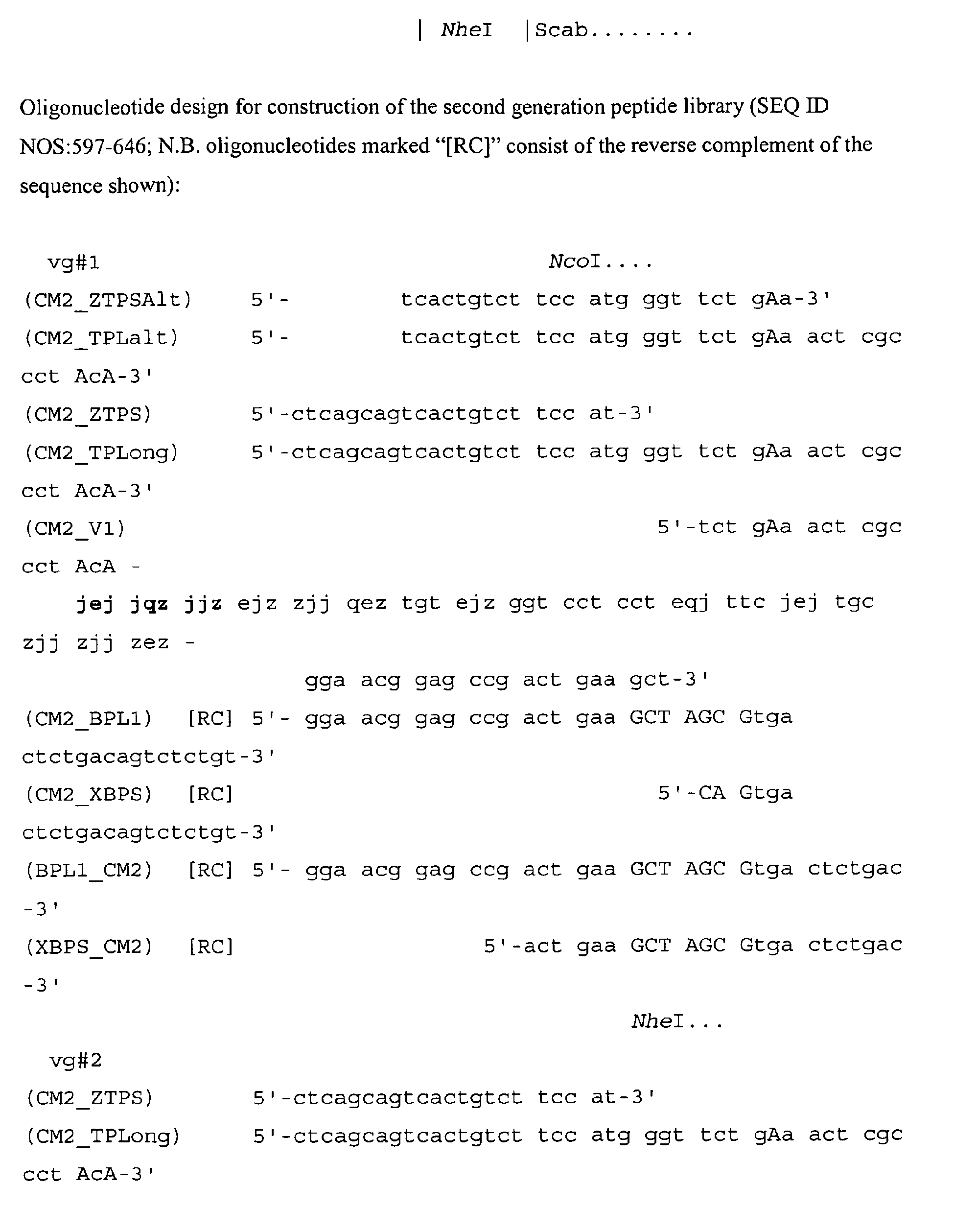


- The phage vector DY3P82 was digested with NheI and NcoI, cleaned and treated with alkaline phosphatase. The 10 templates, CM2-V1 through CM2-V7, plus CM2-V8vg, CM2-V9vg and CM2-V 10vg, were amplified separately, using the primer pairs listed in Table 5 below.
Table 5. Template Sense Antisense CM2_V1 CM2_TPLONG CM2_BPL1 CM2_V2 CM2_TPLONG CM2_BPL1 CM2_V3 CM2_TPLONG CM2_BPL1 CM2_V4 CM2_TPLONG CM2_BPL1 CM2_V5 CM2_TPLONG CM2_BPL1 CM2_V6 CM2_TPLONG CM2_BPL1 CM2_V7 CM2_TPLONG CM2_BPL1 CM2_V8vg CM2_ZTPSALT L20BOTAMP CM2_V9vg CM2_ZTPSALT L20BOTAMP CM2_V10vg CM2_ZTPSALT L20BOTAMP - Using a BIAcore machine, the binding constants were determined for the peptide dimers (shown in
FIGS. 13A-13C ) binding to immobilized cMet-Fc. - Three densities of cMet-Fc (R&D Systems) were cross- linked to the dextran surface of a CM5 sensor chip by the standard amine coupling procedure (3 :M solution diluted 1:100, 1:50, or 1:20 with 50 mM acetate, pH 5.5). Flow
cell 1 was activated and then blocked to serve as a reference subtraction.
Final immobilization levels achieved:


- Experiments were performed in PBST buffer (5.5 mM phosphate, pH 7.65, 0.15 M NaCl) + 0.05% (v/v) Tween-20). Peptide dimers were dissolved in deionized H2O to 1 mg/mL solutions. Dimers were diluted to 50 nM in PBS. Serial dilutions were performed to produce 25, 12.5, 6.25, and 3.125 nM solutions. All samples were injected in duplicate. For association, dimers were injected at 30 :L/minute for 3 minutes using the kinject program. Following a 10-minute dissociation, any remaining peptide was stripped from the cMet surface with two quickinjects of 4M MgCl2 for 2 minutes at 50 :L/minute. Sensorgrams were analyzed using BIAevaluation software 3.1. The heterodimer, Ac-GSPEMCMMFPFLYPCNHHAPGGGK{PnA06-Glut-K[Ac-GSFFPCWRIDRFGYCHANAPGGGKJJ-Glut]-NH2}-NH2 (SEQ ID NO514 linked to SEQ ID NO:515), exhibits a KD of 0.79 nM.
- It is known in the art that compounds that contain maleimide and other groups that can react with thiols react with thiols on serum proteins, especially serum albumin, when the compounds are injected. The adducts have serum life times similar to serum albumin, more than 14 days in humans for example.
- Methods are available that allow for the direct synthesis of maleimide-labeled linear peptides encompassed by the present invention (Holmes, D. et al., 2000. Bioconjug. Chem., 11:439-444.).
- Peptides that include disulfides can be derivatized with maleimide in one of several ways. For example, a third cysteine can be added at the carboxy terminus. The added cysteine is protectted with protecting group that is orthogonal to the type of groups used for the cysteines that are to form the disulfide. The disulfide is formed by selectively deprotecting the intended cysteines and oxidizing the peptide. The final cysteine is then deprotected and the peptide reacted with a large molar excess of a bismaleimide. The resulting compound has one of the maleimides free to react with serum albumin or other thiol-containing serum proteins.
- Alternatively, a cyclic peptide of the present invention is synthesized with a lysine-containing C-terminal extention, such as -GGGK (SEQ ID NO:513). Lysines of the cMet-binding motif are protected with ivDde and the C-terminal lysine is deprotected. This lysine is reacted with a maleimide-containing compound, such as N-[e-maleimidocaproyloxy]succinimide ester (Pierce Biotechnology, Rockford, IL) or N-[a-Maleimidoacetoxy]succinimide ester (Pierce Biotechnology).
- Polypeptides having a molecular weight less than 50-60 kDa are rapidly excreted. Many small molecules, such as fatty acids, bind to serum albumin. Attaching a fatty acid or other serum albumin binding moiety to a peptide causes it to bind non-covalently to serum albumin and can greatly prolong serum residence. Fatty acids attached to peptides of the present invention should contain at least 12 carbons, preferably at least 14 carbons and, more preferably at least 16 carbons. The fatty acid could be straight-chain or branched. The fatty acid could be saturated or unsaturated. Palmate (CH3-(CH2)14-CO- is a preferred fatty acid. This binding in serum can reduce the rate of excretion (Knudsen, L. et al., 2000. J. Med. Chem., 43:1664-1669). Using methods known in the art, serum-albumin-binding moieties can be conjugated to any one of the peptides or multimeric polypeptide binding constructs herein disclosed. The serum-albumin-binding moiety can be joined to the cMet-binding peptide through a linker. The linker can be peptidic or otherwise, such as PEG. Linkers of zero to about thirty atoms are preferred. It is preferred that the linker be hydrophilic. The serum-albumin-binding moiety can be conjugated to the cMet-binding peptide or construct at either end or though a side group of an appended amino acid. Suitable side groups include lysine and cysteine. Such compounds also can comprise, for example, chelators for radionuclides, or other detectable labels or therapeutic constructs, as discussed herein. A cMet peptide or construct joined to a serum-albumin-binding moiety will bind cMet.
- Attachment of PEG to proteins and peptides enhances the serum residence of these molecules. Attachment of PEG (linear or branched) to a cMet-binding peptide or multimeric polypeptide construct is expected give substantial enhancement of serum residence time. The molecular weight of the PEG be at least 10 kDa, more preferably at least 20 kDa, and most preferably 30 kDa or more. The PEG can be attached at the N- or C-terminus. Methods of attaching PEG to peptides are well known in the art. PEG can be attached to reactive side groups such as lysine or cysteine.
- Proteins comprising serum albumin (SA) and other proteins have enhanced serum residence times. The amino-acid sequence of human SA (hSA) is shown in Table 10. Table 11 shows a fusion protein comprising of (SEQ ID NO:604), mature hSA, and SEQ ID NO:605. The cMet-binding peptides are separated from mature hSA by linkers that are rich in glycine to allow flexible spacing. One need not use all of hSA to obtain an injectable protein that will have an enhanced serum residence time. Chemical groups, such as maleimide and alpha bromo carboxylates, react with the unpaired cysteine (residue 34) to form stable adducts. Thus, one can attach a single chelator to hSA fusion proteins so that the adduct will bind a radionuclide. One can prepare a chelator with a maleimide group and couple that to hSA or an hSA derivative. Alternatively, hSA or an hSA derivative can be reacted with a bismaleimide and a chelator carrying a reactive thiol could be reacted with the bismaleimide-derivatized hSA.
- Construction of genes that encode a given amino-acid sequence are known in the art. Expression of HSA fusions in Saccharomyces cerevisiae is known in the art.
- Conventional radioimmune cancer therapy is plagued by two problems. The generally attainable targeting ratio (ratio of administered dose localizing to tumor versus administered dose circulating in blood or ratio of administered dose localizing to tumor versus administered dose migrating to bone marrow) is low. Also, the absolute dose of radiation or therapeutic agent delivered to the tumor is insufficient in many cases to elicit a significant tumor response. Improvement in targeting ratio or absolute dose to tumor would be of great importance for cancer therapy.
- The present invention provides methods of increasing active agent localization at a target cell site of a mammalian recipient. The methods include, for example, a) administering to a recipient a fusion protein comprising a targeting moiety and a member of a ligand-anti-ligand binding pair; b) thereafter administering to the recipient a clearing agent capable of directing the clearance of circulating fusion protein via hepatocyte receptors of the recipient, wherein the clearing agent incorporates a member of the ligand-anti-ligand binding pair; and c) subsequently administering to the recipient an active agent comprising a ligand/anti-ligand binding pair member.
- It is known in the art that hexoses, particularly the hexoses galactose, glucose, mannose, mannose-6-phosphate, N-acetylglucosamine, pentamannosyl phosphate, N-acetylgalactosamine, thioglycosides of galactose, and mixtures thereof are effective in causing hepatic clearance. Binding of sugars to hepatic receptors is not, however, the only means of directing a molecule to the liver.
- Clearance of carcinoembryonic antigen (CEA) from the circulation is by binding to Kupffer cells in the liver. We have shown that CEA binding to Kupffer cells occurs via a peptide sequence YPELPK representing amino acids 107-112 of the CEA sequence. This peptide sequence is located in the region between the N-terminal and the first immunoglobulin like loop domain. Using native CEA and peptides containing this sequence complexed with a heterobifunctional crosslinking agent and ligand blotting with biotinylated CEA and NCA we have shown binding to an 80kD protein on the Kupffer cell surface. This binding protein may be important in the development of hepatic metastases. (Thomas, P. et al., 1992. Biochem. Biophys. Res. Commun., 188:671-677)
- To use YPELPK (SEQ ID NO:655) as a clearance agent, one fuses this sequence via a linker to a moiety that binds the fusion protein (Ab). For example, if the Ab has affinity for DOTA/Re, one would make a derivative having YPELPK attached to DOTA/Re; for example, rvYPELPKpsGGG-DOTA. 'rvYPELPKps' is a fragment of CEA which includes the YPELPK sequence identified by Thomas et al. (supra). Any convenient point on DOTA can be use for attachment. RVYPELPKPSGGG-DOTA/cold Re (SEQ ID NO:656) would then be used as a clearing agent. The Fab corresponding to the fusion Ab would have affinity for the clearing agent of Kd < 100 nM, preferably Kd < 10 nM, and most preferably Kd < 1 nM.
- The therapeutic agent would contain DOTA/185Re. In a preferred embodiment, the therapeutic agent would contain two or more DOTA moieties so that the Ab immobilized on the tumor would bind the bis-DOTA compound with high avidity. The two DOTA moieties would preferably be connected with a hydrophilic linker of ten to thirty units of PEG. PEG is a preferred linker because it is not degraded, promotes solubility. Ten to thirty units of PEG is not sufficient to give the bis DOTA compound a very long serum residence time. A half life of 30 minutes to 10 hours is acceptable. The serum half life should be longer than the radioactive half life of the radionuclide used so that most of the radiation is delivered to the tumor or to the external environment.
- In one embodiment, a "fusion protein" of the present invention comprises at least one cMet-binding peptide fused to the amino terminus or the carboxy terminus of either the light chain (LC) or the heavy chain (HC) of a human antibody. Optionally and preferably, two or more cMet-binding peptides are fused to the antibody. The antibody is picked to have high affinity for a small molecule that can be made radioactive or have a toxin attached. Preferably, the affinity of the Fab corresponding to the Ab has affinity for the small molecule with Kd less than 100 nM, more preferably less than 10 nM, and most preferably less than 1 nM. The small molecule could be a chelator capable of binding a useful radioactive atom, many of which are listed herein. The small molecule could be a peptide having one or more tyrosines to which radioactive iodine can be attached without greatly affecting the binding property of the peptide.
- Any cMet-binding peptide (CMBP) of the present invention can be fused to either end of either chain of an antibody that is capable of binding a small radioactive compound. Useful embodiments include:
- 1) CMBP#1::link::LC / HC,
- 2) LC::link::
CMBP# 1 / HC, - 3) LC / CMBP#1::link::HC,
- 4) LC / HC::link::
CMBP# 1, - 5) CMBP#1::link1::LC::link2::
CMBP# 2 / HC, - 6) LC / CMBP#1::link1::HC::link2::
CMBP# 2, - 7) CMBP#1::link1::LC / CMBP#2::link2::HC,
- 8) CMBP#1::link1::LC / HC::link2::
CMBP# 2, - 9) LC::link1::
CMBP# 1 / CMBP#2::link2::HC, - 10) LC::link1 ::
CMBP# 1 / HC::link2::CMBP# 2, - 11) eMBP#1::link1::LC::link2::
eMBP# 2 / CMBP#3::link3::HC, - 12) CMBP#1::link1::LC::link2::
CMBP# 2 / HC::link3::CMBP# 3, - 13) CMBP#3::link3::LC / CMBP#1::link1::HC::link2::
CMBP# 2, - 14) LC::link3::
CMBP# 3 / CMBP#1::link1::HC::link2::CMBP# 2, and - 15) CMBP#1::link1::LC::link2::
CMBP# 2 / CMBP#3::link3::HC::link4::CMBP# 4. - Since antibodies are dimeric, each fusion protein will present two copies of each of the fused peptides. In case (15), there will be eight CMBPs present and binding to cMet-displaying cells should be highly avid. It is possible that tumor penetration will be aided by moderate cMet affinity in each of the CMBPs rather than maximal affinity.
- The fusion protein is produced in eukaryotic cells so that the constant parts of the HC will be glycosylated. Preferably, the cells are mammalian cells, such as CHO cells.
- The fusion proteins are injected into a patient and time is allowed for the fusion protein to accumulate at the tumor. A clearing agent is injected so that fusion protein that has not become immobilized at the tumor will be cleared. In previous pretargeting methods, the antibody combining site has been used to target to the tumor and biotin/avidin or biotin/streptavidin has been used to attach the radioactive or toxic agent to the immobilized antibody. The biotin/avidin or streptavidin binding is essentially irreversible. Here we fuse a target-binding peptide to the antibody which is picked to bind a radioactive or toxic agent. Because the fusion protein contains 2, 4, 6, or 8 CMBPs, binding of the fusion protein to the tumor is very avid. A clearing agent that will cause fusion protein not immobilized at the tumor to clear can be administered between 2 and 48 hours of the injection of the fusion protein. Because the clearance agent is monomeric in the moiety that binds the antibody, complexes of clearance agent and immobilized fusion protein will not have very long life times. Within 4 to 48 hours of injecting clearance agent, the immobilized antibody will have lost any clearance agent that binds there. The active agent is, preferably, dimeric in the moiety that binds the fusion protein. The active agent is injected between 2 and - 48 hours of injection of clearance agent.
- The spacer length requirements for the binding of a biotinylated derivative of a cMet binding peptide, SEQ ID NO:514, to cMet expressing MDA-MB-231 cells were determined. In order to decide the spacer length to be placed in between peptide and biotin, derivatives were synthesized with no spacer, a single spacer, J, and two spacers, JJ. These three different derivatives of cMet-binding peptide SEQ ID NO:514 and a control peptide that does not bind to cMet, were tested as tetrameric complexes with neutravidin HRP for their ability to bind cMet expressing MB-231 cells. All three tetrameric complexes of cMet-binding peptides bound to the MB231 cells as compared to control peptide; however, the peptide with the JJ spacer exhibited the best KD (12.62 nM). This suggests that inclusion of two spacers (JJ) between the cMet-binding peptide and the biotin is better than one or no spacer.
Cell Culture: MDA-MB231 cells were obtained from ATCC and grown as monolayer culture in their recommended media plus 1 mL/L pen/strep (InVitrogen, Carlsbad, CA). Cells were split the day before the assay, 35000 cells were added to each well of a 96-well plate. - Complexes of control peptide, and the SEQ ID NO:514 derivatives described above, with neutravidin-HRP, were prepared as described above and tested for their ability to bind MDA-MB-231 cells. During the peptide/neutravidin-HRP complex preparation, a 7.5-fold excess of biotinylated peptides over neutravidin-HRP was used to make sure that all four biotin binding sites on neutravidin were occupied. After complex formation, the excess of free biotinylated peptides was removed using soft release avidin-sepharose to avoid any competition between free biotinylated peptides and neutravidin HRP-complexed biotinylated peptides. The experiment was performed at several different concentrations of peptide/neutravidin-HRP, from 0.28 nM to 33.33 nM, to generate saturation binding curves for derivatives without a J spacer and with a single J spacer (
FIG. 14 ), and 0.28 nM to 16.65 nM to generate a saturation binding curve for the derivative with the JJ spacer (FIG. 14 ). In order to draw the saturation binding curve, the background binding of the control peptide/neutravidin HRP complex was subtracted from the binding of the SEQ ID NO:514 derivatives in complex with neutravidin-HRP for each concentration tested. Therefore, absorbance on the Y-axis ofFIG. 14 is differential absorbance (cMet-binding peptide minus control peptide) and not the absolute absorbance. Analysis of the saturation binding data in FIG. using Graph Pad Prism software (version 3.0) yielded a KD of 12.62 nM (+/-3.16) for the tetrameric derivative with the JJ spacer, 155.4 nM (+/- 86.56) for the tetrameric derivative with the J spacer and 123.8 nM (+/- 37.71) for the tetrameric derivative without a spacer peptide complexes. These binding constants are, as expected, lower than that measured by FP for the related monodentate peptide SEQ ID NO:514 (880 nM).
Results: It is evident fromFIG. 14 that the derivative with the JJ spacer showed much better binding to cMet on MDA-MB-231 cells than either of the other two derivatives, with a KD of 12.62 nM after subtracting binding of control peptide as background binding (n = 1). This suggests that a certain minimum spacer length may be required to be able to reach multiple different binding sites on cells and thus achieve multimeric binding. This minimum spacer length could depend on the spacing between different target molecules on cells. As was the case where the binding target was KDR, the neutravidin-HRP assay with biotinylated peptides identified with phage display was useful for identifying peptides capable of binding to an immobilized target even when the affinity of the monomeric binding sequence is too low for an ELISA-type assay (with washing steps after binding) to work well.Table 6. cMet-binding peptide sequences CLASS I TN6: SEQ ID NO: Isolate Sequence SEQ ID NO:001 571-C05, GSWIICWWDNCGSSAP SEQ ID NO:002 465-A06, GSYYDCREFQCNKPAP SEQ ID NO:003 465-D09, GSSHLCNPEFCHFTAP SEQ ID NO:004 569-H10, GSMLMCELWWCRFLAP SEQ ID NO:005 470-E11, GSLIFCPYGECMMYAP SEQ ID NO:006 452-F01, GSEYSCRTSRCIFSAP SEQ ID NO:007 569-C03, GSFILCWWTFCDTNAP SEQ ID NO:008 574-H03, GSSTICPGTACVDHAP SEQ ID NO:009 567-C08, GSLIICWWSWCDKQAP SEQ ID NO:010 561-C08, GSFNICPYQWCTLWAP - Consensus Motif:
G-S-X1-X2-X3-C-X4-X5-X6-X7-C-X8-X9-X10-A-P-G-G-K;
where X1 is F, L, S, W, Y, or M; X2 is I, Y, H, T, or N; X3 is I, L, D, M, F, or S, preferably I; X4 is P, R, W, N, or E, preferably W or P; X5 is W, Y, E, P, L, T, or G; X6 is S, T, D, F, E, W, G, or Q; X7 is F, W, N, Q, E, R, or A; X8 is G, N, H, R, M, I, D, V, or T; X9 is S, K, F, M, T, D, or L; and X10 is S, P, T, L, Y, N, H, Q, or W.CLASS II TN8: SEQ ID NO: Isolate Sequence SEQ ID NO:011 573-F04, AGGFACGPPWDICWMFGT SEQ ID NO:012 570-E07, AGAWNCEYPTFICEWQGA SEQ ID NO:013 456-E04, AGNWICNLSEMRCYPKGT SEQ ID NO:014 434-E12, AGDGWCMAWPEICEWLGT SEQ ID NO:015 489-A04, AGLYLCDLSIMYCFFQGT SEQ ID NO:016 484-D08, AGWWSCQWELNVCIWQGT SEQ ID NO:017 482-D02, AGYYHCIDDFPQCKWMGT SEQ ID NO:018 437-A09, AGWFECEFGFWGCNWLGT SEQ ID NO:019 352-E04, AGTVYCSWESSECWWVGT SEQ ID NO:020 376-E05, AGVWICRVWDDECFFQGT SEQ ID NO:021 482-A12, AGDHYCWEEWWFCWDSGT SEQ ID NO:022 423-C11, AGVLQCIGFEWFCDIWGT SEQ ID NO:023 499-C09, AGVIVCNLSMMYCLYPGT SEQ ID NO:024 457-A09, AGYPECKDNYHWCEWKGT SEQ ID NO:025 573-E07, AGWTWCDLSMMSCIFHGT SEQ ID NO:026 465-F08, AGVTNCNLSTMFCFLHGT SEQ ID NO:027 465-E09, AGTLSCSEEYKSCQLQGT SEQ ID NO:028 444-B08, AGTIRCNLAMMVCMFEGT SEQ ID NO:029 465-E11, AGQYLCTQAALGCPEWGT SEQ ID NO:030 465-D12, AGQMWCAEKNSKCYQWGT SEQ ID NO:031 470-A02, AGQAVCEWGPFWCQMQGT SEQ ID NO:032 465-C01, AGPYSCHSESHDCKLMGT SEQ ID NO:033 448-H02, AGPLFCFEWPSLCHWGGT SEQ ID NO:034 465-D01, AGNLPCHWNMSVCDHQGT SEQ ID NO:035 571-C11, AGMDFCEGFWFLCIGNAT SEQ ID NO:036 465-B11, AGLLGCWDMPMECTGEGT SEQ ID NO:037 442-E08, AGKYMCEGFEWFCEMWGT SEQ ID NO:038 465-C11, AGKTVCQKWESVCSGMGT SEQ ID NO:039 465-F10, AGKQWCVVWEETCDQLGT SEQ ID NO:040 471-A11, AGIWFCNNEEKSCWAYGT SEQ ID NO:041 465-C07, AGHTICQHKALGCPANGT SEQ ID NO:042 465-D04, AGHFECPKHQYMCDMPGT SEQ ID NO:043 445-E04, AGGNWCSFYEELCEWLGT SEQ ID NO:044 465-B06, AGGHWCLELKHLCPPYGT SEQ ID NO:045 470-C02, AGFWDCGWMMQDCHMHGT SEQ ID NO:046 458-B05, ADAWMCEYFQWNCGDKGT SEQ ID NO:047 545-E08, GDGFLCRWENGWCEFWDP - A-G-X1-X2-X3-C-X4-X5-X6-X7-X8-X9-C-X10-X11-X12-G-T-G-G-G-K; where
- X1 is any amino acid other than C, preferably G, V, W, T, K, Q;
- X2 is any amino acid other than C, preferably W, Y, L, F, T;
- X3 is any amino acid other than C, preferably W, E, F, I, L, S
- X4 is any amino acid other than C, preferably E, N, Q;
- X5 is any amino acid other than C, preferably W, L, E;
- X6 is any amino acid other than C, preferably E, S, Y;
- X7 is any amino acid other than C, preferably E, M, P;
- X8 is any amino acid other than C, preferably M, S, W;
- X9 is any amino acid other than C, preferably F, L, V;
- X10 is any amino acid other than C, preferably E, D, W;
- X11 is any amino acid other than C, preferably W, F, M; and
- X12 is any amino acid other than C, preferably Q, W, L.
- G-X1-X2-X3-C-X4-G-P-P-X5-F-X6-C-X7-X8-X9-X10-X11-X12-P-T-E,
where: - X1 is any amino acid other than C, preferably S, R, I, D, or N;
- X2 is any amino acid other than C, preferably W, L, F, V, or I;
- X3 is any amino acid other than C, preferably H, Y, L, Q, N, or V;
- X4 is any amino acid other than C, preferably S, K, or L;
- X5 is any amino acid other than C, preferably T, S, A, or W;
- X6 is any amino acid other than C, preferably E or S;
- X7 is any amino acid other than C, preferably W;
- X8 is any amino acid other than C, preferably W, S, or L;
- X9 is any amino acid other than C, preferably Y or F;
- X10 is any amino acid other than C, preferably D, G, V, or E;
- X11 is any amino acid other than C, preferably T, P, M, or S; and
- X12 is any amino acid other than C, preferably E or G.
- X1 is any amino acid other than C, preferably S;
- X2 is any amino acid other than C, preferably H, Y, or N;
- X3 is any amino acid other than C, preferably S or G;
- X4 is any amino acid other than C, preferably T;
- X5 is any amino acid other than C, preferably E;
- X6 is any amino acid other than C, preferably W;
- X7 is any amino acid other than C, preferably Y;
- X8 is any amino acid other than C, preferably G, D, A, E, or S;
- X9 is any amino acid other than C, preferably T or S; and
- X10 is any amino acid other than C, preferably E or D.
- While this invention has been particularly shown and described with references to preferred embodiments thereof, it will be understood by those skilled in the art that various changes in form and details may be made therein without departing from the scope of the invention encompassed by the appended claims.


| TN9 #1: | ||
| SEQ ID NO: | Isolate | Sequence |
| SEQ ID NO:048 | 325-H05, | AGSIQCKGPPWFSCAMYGT |
| SEQ ID NO:049 | 330-F05, | AGYYGCKGPPTFECQWMGT |
| SEQ ID NO:050 | 333-F09, | AGQFKCAGPPSFACWMTGT |
| SEQ ID NO:051 | 336-G04, | AGWFQCKGPPSFECERHGT |
| SEQ ID NO:052 | 334-G06, | AGWTHCIGPPTFECIPMGT |
| SEQ ID NO:053 | 330-B07, | AGSFACKGPPTFACVEFGT |
| SEQ ID NO:054 | 330-C10, | AGNYFCAGSPSFSCYFMGT |
| SEQ ID NO:055 | 331-G04, | AGSWHCAGPPSFECWEFGT |
| SEQ ID NO:056 | 548-F06, | AGWISCAGPPTFACWPGGT |
| SEQ ID NO:057 | 538-F08, | AGFVNCKGPPTFECILTGT |
| SEQ ID NO:058 | 547-H07, | AGDWICHGPPMFECEWVGT |
| SEQ ID NO:059 | 323-A11, | AGYTSCVGPPSFECTPYGT |
| SEQ ID NO:060 | 333-H03, | AGYFECKGPPTFECWLSGT |
| SEQ ID NO:061 | 329-D02, | AGHAWCSGPPRFECWPPGT |
| SEQ ID NO:062 | 550-C09, | AGHYWCAGPPTFICMGPGT |
| SEQ ID NO:063 | 548-E08, | AGETTCLGWPTFVCVDYGT |
| SEQ ID NO:064 | 332-A05, | AGHGTCRGWPTFECIYFGT |
| SEQ ID NO:065 | 330-C01, | AGDWHCQGPPAFMCWMIGT |
| SEQ ID NO:066 | 545-A09, | AGLPKCSGPPWFSCYYGGT |
| SEQ ID NO:067 | 334-C08, | AGGWECTGPPWFQCGYYGT |
| SEQ ID NO:068 | 333-C05, | AGDIVCTGHPYFECWSWGT |
| SEQ ID NO:069 | 551-B02, | AGTWHCAGPPWFTCYMDGT |
| SEQ ID NO:070 | 551-G12, | AGSWECTGPPSFHCQWYGT |
| SEQ ID NO:071 | 330-G09, | AGHWICVGPPTFSCQWHGT |
| SEQ ID NO:072 | 331-F01, | AGEWWCHGPPEFLCYWTGT |
| SEQ ID NO:073 | 274-B07, | AGETVCYWLNGWFCVDDGT |
| SEQ ID NO:074 | 335-D11, | AGSIQCVGPPSFECTPYGT |
| SEQ ID NO:075 | 336-D07, | AGYSVCKGYPSFECAFFGT |
| SEQ ID NO:076 | 332-C03, | AGVNSCLGPPTFECYQMGT |
| SEQ ID NO:077 | 331-D03, | AGYWHCKGPPHFACEFHGT |
| SEQ ID NO:078 | 331-G06, | AGNWICTGPPSFGCWYHGT |
| SEQ ID NO:079 | 552-G03, | AGYWSCAGPPMFMCTWQGT |
| SEQ ID NO:080 | 552-G11, | AGYWDCKGPPHFFCEWHGT |
| SEQ ID NO:081 | 550-G08, | AGYFHCSGSPWFQCDYYGT |
| SEQ ID NO:082 | 550-G12, | AGWYNCSGENFWNCKWIGT |
| SEQ ID NO:083 | 552-A01, | AGWSDCLGPPQFTCVHWGT |
| SEQ ID NO:084 | 548-C06, | AGTMYCLGPPTFICQQYGT |
| SEQ ID NO:085 | 545-B12, | AGSYWCSGPPTFMCRYEGT |
| SEQ ID NO:086 | 549-F06, | AGSTDCRGHPTFECWGWGT |
| SEQ ID NO:087 | 552-F01, | AGSSPCKGWPTFECYFYGT |
| SEQ ID NO:088 | 547-H12, | AGSIACTGWPYFSCIDLGT |
| SEQ ID NO:089 | 550-F11, | AGQFYCSGPPTFQCIMIGT |
| SEQ ID NO:090 | 548-D08, | AGPWKCTGPPTFSCIQFGT |
| SEQ ID NO:091 | 549-D02, | AGNYWCSGPPSFICHAVGT |
| SEQ ID NO:092 | 552-F02, | AGMTLCAGPPTFECYEVGT |
| SEQ ID NO:093 | 545-E04, | AGETKCSGPPYFYCWMEGT |
| SEQ ID NO:094 | 545-E05, | AGETFCVGNPSFECWSWGT |
| SEQ ID NO:095 | 547-H03, | AGETFCSGWPTFECMQWGT |
| SEQ ID NO:096 | 552-G09, | AGEIFCVGPPTFTCMWTGT |
| SEQ ID NO:097 | 550-A08, | AGDFICQGPPSFVCTNIGT |
| SEQ ID NO:098 | 550-G07, | AGAFFCSGPPTFMCSLYGT |
| SEQ ID NO:099 | 551-A05, | AGWGWCSGPPMFMCTEYGT |
| SEQ ID NO:100 | 548-C10, | GSEFECTGWPEFRCYEYAP |
| SEQ ID NO:101 | 465-C10, | GSILYCINRNDPQCPYTAP |
G-X1-X2-X3-C-X4-G-X5-P-X6-F-X7-C-X8-X9-X10-G-T; where:
X1 is any amino acid other than C, preferably E, S, Y, or W;
X2 is any amino acid other than C, preferably W, T, or F;
X3 is any amino acid other than C, preferably W, H, or F;
X4 is any amino acid other than C, preferably A,K, S, or T;
X5 is any amino acid other than C, preferably P or W;
X6 is any amino acid other than C, preferably T or S;
X7 is any amino acid other than C, preferably E or S;
X8 is any amino acid other than C, preferably W, Y, or I;
X9 is any amino acid other than C, preferably W, Y, M, or E; and
X10 is any amino acid other than C, preferably Y.
| CLASS IV | ||
| TN9 #2: | ||
| SEQ ID NO: | Isolate | Sequence |
| SEQ ID NO:102 | 605-G10, | SETRPTEAGDLICSGPPTFICTLYHTEPTE |
| SEQ ID NO:103 | 593-C01, | SETRPTQAVRSQCSGPPTFECWYFGTEPTE |
| SEQ ID NO:104 | 592-C01, | SETRPTEGGSWYCSGPPAFECWWYGTEPTE |
| SEQ ID NO:105 | 591-E01, | SETRPTVASRWHCNGPPTFECWRYGTEPTE |
| SEQ ID NO:106 | 590-E01, | SETRPTEAGTFHCSGPPTFECWSYGPKPTE |
| SEQ ID NO:107 | 589-BO1, | SETRPTEAGSLWCMGPPWFCCVIYGTQPTE |
| SEQ ID NO:108 | 607-A02, | SETRPTEAGILHCSGPPTFECWWNYTEPTE |
| SEQ ID NO:109 | 590-F01, | SETRPTESGRVHCPGPPWFRCARNGTEPTE |
| SEQ ID NO:110 | 589-CO1, | SETRPTAAGRILCTGPPWFSCAMYGTEPTE |
| SEQ ID NO:111 | 606-B11, | SETRPTEAADWLCSGPPTFECWWFGTEPTE |
| SEQ ID NO:112 | 593-E01, | SETRPTQVGRWQCDGPPTFACRSYGTEPTE |
| SEQ ID NO:113 | 592-F12, | SETRPTEAGSTKCSGPPTFECWWFDTEPTE |
| SEQ ID NO:114 | 590-F07, | SETRPTVAGSWHCSGPPTFECWWYGTEPTE |
| SEQ ID NO:115 | 588-D02, | SETRPTEAGRNHCKGPPGFRCAMTDTEPTE |
| SEQ ID NO:116 | 607-H09, | SETRPTETDFVYCRGPPTFECWWYGTEPTE |
| SEQ ID NO:117 | 590-H01, | SETRPTSSGSRHCKGPPTFECWGYGTEPTE |
| SEQ ID NO:118 | 589-F01, | SETRPTEAGSWRCSGPPTFECWWYETSPTE |
| SEQ ID NO:119 | 608-F11, | SETRPTDAIRSYCSGPPTFECWWFGTEPTE |
| SEQ ID NO:120 | 606-D11, | SETRPTEAGSWNCSGPPAFECWWYGSEPTE |
| SEQ ID NO:121 | 604-D04, | SETRPTEAGSWQCSGPPTFECWSFGTEPTE |
| SEQ ID NO:122 | 602-A11, | SETRPTEAGSWHCNGPPTFECWWYDMEPTE |
| SEQ ID NO:123 | 593-F02, | SETRPTEAGRVSCLGPPTFECWWFVPEPTE |
| SEQ ID NO:124 | 591-H05, | SETRPTDAGSWRCAGPPTFECWWFGTEPTE |
| SEQ ID NO:125 | 590-H06, | SETRPTEPVTWQCTGPPTFECWWLGTEPTE |
| SEQ ID NO:126 | 588-F10, | SETRPTDAVSTHCNGPPTFECYIYGTEPTE |
| SEQ ID NO:127 | 608-G03, | SETRPTVAESWYCVGPPSFECWWYGTEPTE |
| SEQ ID NO:128 | 604-D09, | SETRPTEAGSWNCSGPPTFECWSYQTEPTE |
| SEQ ID NO:129 | 602-A12, | SETRPTEAGSGHCNGPPTFKCWWYDMEPTE |
| SEQ ID NO:130 | 592-G11, | SETRPTDQDSWQCSGPPTFECWWYGTEPTE |
| SEQ ID NO:131 | 588-G01, | SETRPTESTQVQCAGPPSFACWMTGTEPTE |
| SEQ ID NO:132 | 606-E05, | SETRPTEVESWHCSGPPTFECWWYGTEPTE |
| SEQ ID NO:133 | 594-C07, | SETRPTEAGSFHCSGPPTFECWLYWTDPTE |
| SEQ ID NO:134 | 592-H01, | SETRPTEAGQFGCKGPPPFECKLMGRVPTE |
| SEQ ID NO:135 | 605-C05, | SETRPTDTVTWHCNGPPTFECWWYGTEPTE |
| SEQ ID NO:136 | 594-E08, | SETRPTEADRWHCDGPPTFECWWYGTEPTE |
| SEQ ID NO:137 | 593-B11, | SETRPTEAGSIQCVGPPWFSCRMYVTEPTE |
| SEQ ID NO:138 | 590-C01, | SETRPTVSGSWQCVGPPTFECWSYGTEPTE |
| SEQ ID NO:139 | 612-G11, | SETRPTENGSWHCNGPPTFECWWYGTEPTE |
| SEQ ID NO:140 | 612-E08, | SETRPTEAGSWHCSGPPIFECWWYDMEPTE |
| SEQ ID NO:141 | 612-A02, | SETRPTVDGGWHCNGPPTFECWMYGTEPTE |
| SEQ ID NO:142 | 611-G01, | SETRPTDAGTWNCTGPPSFECWWFGTEPTE |
| SEQ ID NO:143 | 610-G04, | SETRPTWDGKWHCSGPPTFECWWYGTEPTE |
| SEQ ID NO:144 | 610-E06, | SETRPTEAGSWRCSGPPTFECWWYYTEPTE |
| SEQ ID NO:145 | 610-C06, | SETRPTEAGNWLCSGPPTFECWWYVTGPTE |
| SEQ ID NO:146 | 610-A04, | SETRPTEGGNWHCSGPPTFECWLYGTEPTE |
| SEQ ID NO:147 | 612-D02, | SETRPTEAGGWHCSGPPTFECWWFNMEPTE |
| SEQ ID NO:148 | 612-A12, | SETRPTEVISWHCSGPPTFECYRYGTEPTE |
| SEQ ID NO:149 | 611-D03, | SETRPTEVGSWHCNGPPTFECWWYGTEPTE |
| SEQ ID NO:150 | 610-G10, | SETRPTLASTWYCSGPPTFECWWYGTEPTE |
| SEQ ID NO:151 | 610-A11, | SETRPTEAGGWYCKGPPTFECWWDGTEPTE |
| SEQ ID NO:152 | 612-H02, | SETRPTEAGGWFCSGPPTFECWWYDTVPTE |
| SEQ ID NO:153 | 612-B01, | SETRPTEAATWQCSGPPTFECWGYGTEPTE |
| SEQ ID NO:154 | 610-C12, | SETRPTEAGDYVCVGPPTFECYLMDAEPTE |
| SEQ ID NO:155 | 610-B01, | SETRPTEAGGWYCSGPPSFECWSYGTEPTE |
| SEQ ID NO:156 | 612-H04, | SETRPTESSSWHCSGPPTFECWRFGTEPTE |
| SEQ ID NO:157 | 612-B09, | SETRPTEAGSWYCSGPPTFECWWYYAEPTE |
| SEQ ID NO:158 | 611-G07, | SETRPTLAGNWQCSGPPTFECWWYGTEPTE |
| SEQ ID NO:159 | 611-E10, | SETRPTEAGSWHCNGPPTFECWQYGTEPTE |
| SEQ ID NO:160 | 610-H02, | SETRPTEAGSWECHGPPSFECWWYGTEPTE |
| SEQ ID NO:161 | 610-D03, | SETRPTEAGSWRCSGPPTFECWWYDAEPTE |
| SEQ ID NO:162 | 610-BO3, | SETRPTEAGSWNCAGPPTFECWWYGTEPTE |
| SEQ ID NO:163 | 612-H05, | SETRPTEAGSFYCSGPPTFECWQYVPEPTE |
| SEQ ID NO:164 | 612-F05, | SETRPTEAGSWMCSGPPTFECWQYFTEPTE |
| SEQ ID NO:165 | 612-B10, | SETRPTEAGSLHCSGPPTFECWWWETEPTE |
| SEQ ID NO:166 | 611-E11, | SETRPTEEGVWHCNGPPTFECWWYGTEPTE |
| SEQ ID NO:167 | 610-F08, | SETRPTEAGRWNCSGPPTFECWWYSTEPTE |
| SEQ ID NO:168 | 610-D05, | SETRPTEAGSWRCSGPPTFECWWFGTEPTE |
| SEQ ID NO:169 | 610-B04, | SETRPTQAVSSYCSGPPTFECWSFGTEPTE |
| SEQ ID NO:170 | 612-B12, | SETRPTEAGRSYCSGPPTFECWWYATEPTE |
| SEQ ID NO:171 | 611-H01, | SETRPTWAKVHCAGPPTFECWTYGTEPTE |
| SEQ ID NO:172 | 610-H05, | SETRPTEPGSWHCSGPPTFVCWWWGTEPTE |
| SEQ ID NO:173 | 610-F10, | SETRPTEAGRWHCSGPPTFECWWHDTEPTE |
| SEQ ID NO:174 | 612-H07, | SETRPTEAGSWQCTGPPTFECWGYVEEPTE |
| SEQ ID NO:175 | 612-G09, | SETRPTEAGSWQCGGPPTFECWWYYTGPTE |
| SEQ ID NO:176 | 612-F08, | SETRPTEAGSWYCTGPPTFECWLYETYPTE |
| SEQ ID NO:177 | 611-H08, | SETRPTAAWSGSCSGPPSFECWNYGTEPTE |
| SEQ ID NO:178 | 610-E01, | SETRPTEAGSWQCSGPPTFACWWYGTEPTE |
| SEQ ID NO:179 | 610-B09, | SETRPTEAGILHCSGPPTFECWWEVMEPTE |
| SEQ ID NO:180 | 612-E07, | SETRPTEAGRVACSGPPTFECWSYDEEPTE |
| SEQ ID NO:181 | 612-C11, | SETRPTEAGNWECQGPPTFECWWFGTEPTE |
| SEQ ID NO:182 | 610-E04, | SETRPTLASNGYCNGPPTFECWHYGTEPTE |
| SEQ ID NO:183 | 610-B12, | SETRPTEAGSFHCSGPPTFECIWYGSEPTE |
| SEQ ID NO:184 | 616-B11, | SETRPTEAGSWYCSGPPTFACWWDGTEPTE |
| SEQ ID NO:185 | 615-H08, | SETRPTQGDNWNCSGPPTFECWWYGTEPTE |
| SEQ ID NO:186 | 615-B11, | SETRPTEAGRWHCNGPPTFECWRYDYDPTE |
| SEQ ID NO:187 | 614-C07, | SETRPTEAYSWECTGPPMFECWWYGTEPTE |
| SEQ ID NO:188 | 613-H12, | SETRPTEWDWHCSGPPQFECWWYGTEPTE |
| SEQ ID NO:189 | 613-F02, | SETRPTEAGSWNCSGPPTFECWWYGSEPTE |
| SEQ ID NO:190 | 613-D05, | SETRPTASGSWHCSGPPTFECWIFGTEPTE |
| SEQ ID NO:191 | 612-H12, | SETRPTEAGAWYCMGPPTFECWWYDRGPTE |
| SEQ ID NO:192 | 616-D05, | SETRPTEAGGLHCSGPPTFECWWYDTEPTE |
| SEQ ID NO:193 | 615-C01, | SETRPTVGGSWDCKGPPTFECWSYGTEPTE |
| SEQ ID NO:194 | 614-E09, | SETRPTEAGAWSCLGPPTFECWWYGTEPTE |
| SEQ ID NO:195 | 614-A03, | SETRPTEAGSLHCSGPPTFECWWFDTEPTE |
| SEQ ID NO:196 | 616-C02, | SETRPTAGRSWECSGPPTFECWVFGTEPTE |
| SEQ ID NO:197 | 615-C04, | SETRPTDNGSWHCNGPPTFECWWYGTEPTE |
| SEQ ID NO:198 | 614-C12, | SETRPTEAGSWQCKGPPTFECWWYGTEPTE |
| SEQ ID NO:199 | 615-C11, | SETRPTEVGNYKCSGPPTFECWWYGTEPTE |
| SEQ ID NO:200 | 614-H08, | SETRPTEAGSWHCVGPPTFECWGYVTEPTE |
| SEQ ID NO:201 | 614-E11, | SETRPTEAGSFVCKGPPTFECYWFGQDPTE |
| SEQ ID NO:202 | 616-E10, | SETRPTEAGSWHCSGPPTFECWWYGPDPTE |
| SEQ ID NO:203 | 615-D02, | SETRPTEAERWHCSGPPTFECWWYGTEPTE |
| SEQ ID NO:204 | 614-F04, | SETRPTEAGSWHCSGPPTFECWFYVKEPTE |
| SEQ ID NO:205 | 614-D06, | SETRPTEAGSWDCSGPPTFECWWFGTEPTE |
| SEQ ID NO:206 | 614-B08, | SETRPTEPAGWECRGPPSFECLWYGTEPTE |
| SEQ ID NO:207 | 613-H01, | SETRPTDAGPWNCTGPPSFECWWYGTEPTE |
| SEQ ID NO:208 | 613-E04, | SETRPTEARGWHCSGPPTFECWLWGTEPTE |
| SEQ ID NO:209 | 613-B08, | SETRPTEAGRWNCSGPPTFECWQYEMDPTE |
| SEQ ID NO:210 | 615-D04, | SETRPTEAGSWYCSGPPTFECFWYDTEPTE |
| SEQ ID NO:211 | 615-A05, | SETRPTESGSWHCSGPPTFECWWFGTEPTE |
| SEQ ID NO:212 | 614-E04, | SETRPTEAGSWLCTGPPTFECWWFDTDPTE |
| SEQ ID NO:213 | 613-E06, | SETRPTEPSHWHCVGPPTFACWWYVTDPTE |
| SEQ ID NO:214 | 613-C05, | SETRPTEAGSWYCSGPPMFECYLFVTEPTE |
| SEQ ID NO:215 | 616-C07, | SETRPTEAVNWHCLGPPSFECWQFGTEPTE |
| SEQ ID NO:216 | 615-G02, | SETRPTEAGSWHCSGPPTFECWWYGTDPTE |
| SEQ ID NO:217 | 615-E06, | SETRPTEAGSWHCSGPPTFECWSFVSLPTE |
| SEQ ID NO:218 | 615-A08, | SETRPTEGSEWSCIGPPSFECWWYGTEPTE |
| SEQ ID NO:219 | 614-G01, | SETRPTEDGYWNCSGPPTFECWWHGTEPTE |
| SEQ ID NO:220 | 613-D01, | SETRPTEAGSWSCSGPPTFECWPYYTEPTE |
| SEQ ID NO:221 | 614-G02, | SETRPTEAGSWYCSGPPTFECWWYWPEPTE |
| SEQ ID NO:222 | 614-E06, | SETRPTDDGRWSCAGPPTFECWRYGTEPTE |
| SEQ ID NO:223 | 620-E11, | SETRPTEGGSWSCGGPPTFECWWFGTEPTE |
| SEQ ID NO:224 | 620-A11, | SETRPTVTGSWYCSGPPTFECWWYGTEPTE |
| SEQ ID NO:225 | 618-F04, | SETRPTEASSWYCTGPPAFECWWYGTEPTE |
| SEQ ID NO:226 | 617-G06, | SETRPTEAGSWLCSGPPTFECWWYGTEPTE |
| SEQ ID NO:227 | 616-G06, | SETRPTESVRWYCSGPPTFECWWYGTEPTE |
| SEQ ID NO:228 | 620-F10, | SETRPTEAGRLVCSGPPTFMCRTYATDPTE |
| SEQ ID NO:229 | 619-G04, | SETRPTEAGSWECTGPPWFVCRQYAIEPTE |
| SEQ ID NO:230 | 618-F12, | SETRPTEAGYLYCSGPPTFECWWYDTMPTE |
| SEQ ID NO:231 | 618-B06, | SETRPTEAGSWHCSGPPTFECWWFGTEPTE |
| SEQ ID NO:232 | 617-E09, | SETRPTEAGNWHCLGPPTFECWWYGTEPTE |
| SEQ ID NO:233 | 616-F10, | SETRPTEAGSWHCSGPPTFECWWYDTEPTE |
| SEQ ID NO:234 | 620-B11, | SETRPTESGGWYCSGPPAFECWWYGTEPTE |
| SEQ ID NO:235 | 619-G07, | SETRPTVAGAVSCSGPPTFECWWYGTEPTE |
| SEQ ID NO:236 | 619-E11, | SETRPTEAGRWYCSGPPTFECWWFLPDPTE |
| SEQ ID NO:237 | 619-B12, | SETRPTEAGGWHCSGPPSFECWWFDTVPTE |
| SEQ ID NO:238 | 618-G11, | SETRPTGVGGWYCSGPPSFECWLYGTEPTE |
| SEQ ID NO:239 | 618-B11, | SETRPTQADYLHCSGPPTFECFWYGTEPTE |
| SEQ ID NO:240 | 617-F01, | SETRPTGDGNWHCNGPPTFECWRFGTEPTE |
| SEQ ID NO:241 | 617-B01, | SETRPTEASNYHCIGPPTFECFWYGTEPTE |
| SEQ ID NO:242 | 616-G12, | SETRPTEAGDWLCKGPPTFECWWQVTDPTE |
| SEQ ID NO:243 | 620-G01, | SETRPTEAGSWHCNGPPTFECWWYSSDPTE |
| SEQ ID NO:244 | 620-C10, | SETRPTEDGGWRCSGPPTFECWWYGTEPTE |
| SEQ ID NO:245 | 619-G09, | SETRPTEAGRIECKGPPWFSCVIYGTEPTE |
| SEQ ID NO:246 | 619-F06, | SETRPTGGGSWNCSGPPTFECWWYGTEPTE |
| SEQ ID NO:247 | 618-C03, | SETRPTEAGSLYCSGPPTFECWWYITHPTE |
| SEQ ID NO:248 | 617-F02, | SETRPTEAGRWHCSGPPRFECWWYDTEPTE |
| SEQ ID NO:249 | 616-H01, | SETRPTEYGSWHCSGPPTFECWYHGTEPTE |
| SEQ ID NO:250 | 618-D01, | SETRPTEAGNWHCSGPPSFECWWYATEPTE |
| SEQ ID NO:251 | 617-F03, | SETRPTEQGSWHCKGPPTFECWSYGTEPTE |
| SEQ ID NO:252 | 616-H03, | SETRPTDAANYHCSGPPTFECWWYGTEPTE |
| SEQ ID NO:253 | 616-G02, | SETRPTEAGSWYCSGPPMFECWWLAEEPTE |
| SEQ ID NO:254 | 620-G09, | SETRPTEAGGWYCSGPPAFECWWYATEPTE |
| SEQ ID NO:255 | 620-D12, | SETRPTEAGIWSCSGPPTFECWWYESSPTE |
| SEQ ID NO:256 | 619-A09, | SETRPTEEGLRVCSGPPTFECWWYGTEPTE |
| SEQ ID NO:257 | 618-D06, | SETRPTEAGSWLCFGPPTFECWSFGTEPTE |
| SEQ ID NO:258 | 617-H12, | SETRPTVAGSWDCSGPPTFECWWYGTEPTE |
| SEQ ID NO:259 | 616-H05, | SETRPTKADNWHCSGPPTFECWWYGTEPTE |
| SEQ ID NO:260 | 619-H10, | SETRPTEAGIVYCSGPPTFECWWFGTEPTE |
| SEQ ID NO:261 | 619-D03, | SETRPTEAGYWHCLGPPTFECWWYVKEPTE |
| SEQ ID NO:262 | 618-D12, | SETRPTEPGLLHCSGPPTFECWWYGTEPTE |
| SEQ ID NO:263 | 620-E04, | SETRPTEASSWYCSGPPSFECWWYGTEPTE |
| SEQ ID NO:264 | 620-A05, | SETRPTEAGSWHCLGPPTFECWWYVKEPTE |
| SEQ ID NO:265 | 619-D04, | SETRPTEAGIILCKGPPWFSCDIYDTGPTE |
| SEQ ID NO:266 | 618-A11, | SETRPTAAGNWHCSGPPTFECWAYGTEPTE |
| SEQ ID NO:267 | 617-D07, | SETRPTVGGSWYCSGPPTFECWSYGTEPTE |
| SEQ ID NO:268 | 627-A10, | SETRPTEDGWLDCKGPPTFECWWYGTEPTE |
| SEQ ID NO:269 | 626-H02, | SETRPTEDGNWHCSGPPTFECWSYGTEPTE |
| SEQ ID NO:270 | 626-F06, | SETRPTEAGSWHCSGPPTFECWYYWPEPTE |
| SEQ ID NO:271 | 624-D02, | SETRPTEAGSLYCSGPPMFECWWYDWYPTE |
| SEQ ID NO:272 | 622-D09, | SETRPTEAGGWYCMGPPAFECWWYASEPTE |
| SEQ ID NO:273 | 621-F11, | SETRPTNAGSWYCSGPPTFECWWYGTEPTE |
| SEQ ID NO:274 | 621-B11, | SETRPTEASRWHCNGPPTFECWWYGTEPTE |
| SEQ ID NO:275 | 627-B03, | SETRPTEAGSFVCSGPPTFECWWYNTGPTE |
| SEQ ID NO:276 | 626-H03, | SETRPTEAGSWHCSGPPTFECWSYGTEPTE |
| SEQ ID NO:277 | 626-F07, | SETRPTESDIWLCSGPPTFECWWYGTEPTE |
| SEQ ID NO:278 | 626-D02, | SETRPTDADPWHCSGPPTFECWWFGTEPTE |
| SEQ ID NO:279 | 625-B03, | SETRPTEAGVVLCSGPPTFECWWYDTEPTE |
| SEQ ID NO:280 | 622-D10, | SETRPTEVGSVHCSGPPTFECWWFGTEPTE |
| SEQ ID NO:281 | 621-G02, | SETRPTEAGRWLCSGPPTFECWEYDTEPTE |
| SEQ ID NO:282 | 621-E04, | SETRPTDAGWLQCSGPPTFECWWYGTEPTE |
| SEQ ID NO:283 | 621-B12, | SETRPTEASRRHCNGPPTFECWRYGTEPTE |
| SEQ ID NO:284 | 626-H04, | SETRPTEAGRWYCSGPPTFECWLFVEEPTE |
| SEQ ID NO:285 | 626-F11, | SETRPTAADSWQCSGPPTFECWSFGTEPTE |
| SEQ ID NO:286 | 626-D03, | SETRPTEAGSWHCGGPPTFECWMYVTEPTE |
| SEQ ID NO:287 | 626-A02, | SETRPTDDGSWYCSGPPTFECWWYGTEPTE |
| SEQ ID NO:288 | 623-E07, | SETRPTEAGYWHCLGPPTFECWWYDMEPTE |
| SEQ ID NO:289 | 622-G09, | SETRPTEAGILRCSGPPTFECWYYETEPTE |
| SEQ ID NO:290 | 622-E05, | SETRPTEDVSVHCAGPPTFECWLYGTEPTE |
| SEQ ID NO:291 | 622-B12, | SETRPTEEGVFQCVGPPTFECWWYGTEPTE |
| SEQ ID NO:292 | 621-G07, | SETRPTEDGGFFCSGPPTFECWWYGTEPTE |
| SEQ ID NO:293 | 621-E07, | SETRPTEPGSWHCSGPPTFECWWYGTEPTE |
| SEQ ID NO:294 | 621-C01, | SETRPTEAGSWHCSGPPTFECWWYDRAPTE |
| SEQ ID NO:295 | 626-A05, | SETRPTEAGTWYCSGPPTFECWYYATEPTE |
| SEQ ID NO:296 | 623-G02, | SETRPTEAGSLYCSGPPAFECYWYGTVPTE |
| SEQ ID NO:297 | 622-H11, | SETRPTDPGVLHCSGPPTFECWWFGTEPTE |
| SEQ ID NO:298 | 622-C04, | SETRPTEAGTWYCLGPPTFECWSFWQDPTE |
| SEQ ID NO:299 | 621-G11, | SETRPTEAGRWGCSGPPTFECWWYVAEPTE |
| SEQ ID NO:300 | 621-C07, | SETRPTEAGIWHCAGPPTFICWLYETEPTE |
| SEQ ID NO:301 | 627-C03, | SETRPTEAGSWHCSGPPSFECWQYSTEPTE |
| SEQ ID NO:302 | 626-D12, | SETRPTEAGSWQCSGPPTFECWVYETEPTE |
| SEQ ID NO:303 | 626-A06, | SETRPTEAGSWYCSGPPTFECWWYDVGPTE |
| SEQ ID NO:304 | 623-H02, | SETRPTDEVSWECRGPPTFECWWYGTEPTE |
| SEQ ID NO:305 | 623-B05, | SETRPTEGGSWVCSGPPTFECWWYGTEPTE |
| SEQ ID NO:306 | 622-E10, | SETRPTEYGSWYCSGPPTFECWWLGTEPTE |
| SEQ ID NO:307 | 622-C06, | SETRPTEAGVWLCSGPPTFECWWYDTDPTE |
| SEQ ID NO:308 | 621-H03, | SETRPTMAGSYYCSGPPTFECWVYGTEPTE |
| SEQ ID NO:309 | 621-E11, | SETRPTEAGYVQCYGPPSFVCHPMVPDPTE |
| SEQ ID NO:310 | 621-C08, | SETRPTEDGFVLCKGPPWFSCEMYGTEPTE |
| SEQ ID NO:311 | 627-C04, | SETRPTEAGGWNCSGPPTFECWWYVTEPTE |
| SEQ ID NO:312 | 626-A07, | SETRPTEDGSWECFGPPTFECWSYGTEPTE |
| SEQ ID NO:313 | 623-H08, | SETRPTDAVSYVCKGPPTFECWWYGTEPTE |
| SEQ ID NO:314 | 622-F05, | SETRPTEARSWHCSGPPTFECWWYGTEPTE |
| SEQ ID NO:315 | 627-A04, | SETRPTASVSWHCSGPPTFECWSYGTEPTE |
| SEQ ID NO:316 | 626-G05, | SETRPTEAGSWYCSGPPTFECWYYDMDPTE |
| SEQ ID NO:317 | 623-H11, | SETRPTEAGSWLCSGPPTFECWWFGTEPTE |
| SEQ ID NO:318 | 622-F11, | SETRPTGDGSWYCSGPPTFECWWLGTEPTE |
| SEQ ID NO:319 | 621-F03, | SETRPTEAGSWYCSGPPTFECWWYFLDPTE |
| SEQ ID NO:320 | 626-F01, | SETRPTEAGGWYCSGPPTFECWWFATEPTE |
| SEQ ID NO:321 | 621-F04, | SETRPTEAGDLDCLGPPTFICRIYGTEPTE |
| SEQ ID NO:322 | 630-F06, | SETRPTEAGSWQCVGPPTFECWSFGTEPTE |
| SEQ ID NO:323 | 630-A03, | SETRPTEADSWYCSGPPTFECWLFGTEPTE |
| SEQ ID NO:324 | 629-F10, | SETRPTQADSWYCSGPPTFECWWWGTEPTE |
| SEQ ID NO:325 | 629-D11, | SETRPTEAFSWDCSGPPTFECWWFGTEPTE |
| SEQ ID NO:326 | 629-B06, | SETRPTEAGSWQCSGPPVFECWWYDTEPTE |
| SEQ ID NO:327 | 628-H01, | SETRPTEAGNVQCSGPPTFECWWFDTEPTE |
| SEQ ID NO:328 | 628-F03, | SETRPTEAGSWCSGPPRFECWAFVTEPTE |
| SEQ ID NO:329 | 627-G02, | SETRPTEDGTLHCSGPPTFACWWYGTEPTE |
| SEQ ID NO:330 | 629-E01, | SETRPTDAEVWVCNGPPTFECWWYGTEPTE |
| SEQ ID NO:331 | 628-H09, | SETRPTEDVTFHCSGPPTFECWLYGTEPTE |
| SEQ ID NO:332 | 628-A05, | SETRPTSDFDWHCKGPPTFECWSYGTEPTE |
| SEQ ID NO:333 | 627-G04, | SETRPTEADSWYCSGPPTFECWWYVPEPTE |
| SEQ ID NO:334 | 630-A05, | SETRPTDDGNWYCSGPPTFECWWYGTEPTE |
| SEQ ID NO:335 | 629-E03, | SETRPTEAGSWYCSGPPTFECWRYDTDPTE |
| SEQ ID NO:336 | 629-C02, | SETRPTEAGPWSCSGPPTFECWWFDTEPTE |
| SEQ ID NO:337 | 628-H10, | SETRPTEAGMFLCSGPPAFECWWYDTEPTE |
| SEQ ID NO:338 | 628-F12, | SETRPTEAGSLYCSGPPTFECWLYDVEPTE |
| SEQ ID NO:339 | 627-D12, | SETRPTEAGQWNCSGPPTFECWWYDIEPTE |
| SEQ ID NO:340 | 630-G02, | SETRPTEAGSWYCSGPPTFECWWFETEPTE |
| SEQ ID NO:341 | 629-E06, | SETRPTEAGSFVCSGPPTFECWGYVTEPTE |
| SEQ ID NO:342 | 628-D07, | SETRPTQDGTWFCSGPPTFECWWYGTEPTE |
| SEQ ID NO:343 | 627-E06, | SETRPTEGDSWHCAGPPTFECWWYGTEPTE |
| SEQ ID NO:344 | 629-E07, | SETRPTEAGSWSCSGPPTFECWSYGTEPTE |
| SEQ ID NO:345 | 629-C11, | SETRPTEAGRIQCSGPPTFECWWYDEEPTE |
| SEQ ID NO:346 | 629-A03, | SETRPTEAGTIVCKGPPWFSCEIYETEPTE |
| SEQ ID NO:347 | 628-A12, | SETRPTEAGDWYCSGPPAFECWEYLGEPTE |
| SEQ ID NO:348 | 627-E08, | SETRPTEAGSWFCSGPPSFECWSYVTEPTE |
| SEQ ID NO:349 | 629-E08, | SETRPTEAGSWHCSGPPAFECWWYDNEPTE |
| SEQ ID NO:350 | 629-B02, | SETRPTEAGRWTCSGPPTFECWWYVSDPTE |
| SEQ ID NO:351 | 628-E06, | SETRPTEAGEWYCGGPPTFECWWFDTAPTE |
| SEQ ID NO:352 | 627-G09, | SETRPTEAGSWHCSGPPSFECWWFDTGPTE |
| SEQ ID NO:353 | 631-A11, | SETRPTEAGSFICSGPPTFECWWYGTEPTE |
| SEQ ID NO:354 | 630-C10, | SETRPTEDVRWYCSGPPTFECWWFGTEPTE |
| SEQ ID NO:355 | 628-B08, | SETRPTEAGSWYCSGPPTFECWWYVPEPTE |
| SEQ ID NO:356 | 629-F03, | SETRPTEAGNWLCSGPPAFECWWFVAEPTE |
| SEQ ID NO:357 | 632-A09, | SETRPTEAGSWYCSGPPTFECWWYGTEPTE |
| SEQ ID NO:358 | 632-G07, | SETRPTEAGDWLCAGPPTFECWWWGTDPTE |
| SEQ ID NO:359 | 631-F12, | SETRPTEAGSWHCVGPPTFECWWFDTEPTE |
| SEQ ID NO:360 | 633-A02, | SETRPTEAGEWSCSGPPTFECWWWDMEPTE |
| SEQ ID NO:361 | 633-B06, | SETRPTYYVSWYCSGPPTFECWSYGTEPTE |
| SEQ ID NO:362 | 632-D11, | SETRPTEDGSWYCSGPPTFECWWYGTEPTE |
| SEQ ID NO:363 | 631-D10, | SETRPTEDGTWYCSGPPTFECWWYGTEPTE |
| SEQ ID NO:364 | 633-F09, | SETRPTETDSWVCSGPPTFECWWYGTEPTE |
T-X1-X2-X3-X4-X5-X6-C-X7-G-P-P-X8-F-X9-C-X10-X11-X12-G, where:
X1 is any amino acid other than C, preferably E, D, or V;
X2 is any amino acid other than C, preferably A, D, G, S, or V;
X3 is any amino acid other than C, preferably G, V, D, or S;
X4 is any amino acid other than C, preferably S, N, R, T, or G;
X5 is any amino acid other than C, preferably W;
X6 is any amino acid other than C, preferably H or Q;
X7 is any amino acid other than C, preferably S, N, or K;
X8 is any amino acid other than C, preferably T;
X9 is any amino acid other than C, preferably E;
X10 is any amino acid other than C, preferably W;
X11 is any amino acid other than C, preferably W or S; and
X12 is any amino acid other than C, preferably Y or F.
| CLASS V | ||
| TN10: | ||
| SEQ ID NO: | Isolate | Sequence |
| SEQ ID NO:365 | 545-C02, | GSWRFCGGEYSFQVCQDVAP |
| SEQ ID NO:366 | 546-E02, | GSHHTCLDGFAGWRCTEVAP |
| SEQ ID NO:367 | 545-C11, | GSFAPCGWPSFAIDCIAEAP |
| SEQ ID NO:368 | 549-G01, | GSTKVCHEKWNQLFCHNQAP |
| SEQ ID NO:369 | 548-F07, | GSPEMCMMFPFLYPCNHHAP |
| SEQ ID NO:370 | 551-H10, | GSFFPCWRIDRFGYCHANAP |
S-X1-X2-X3-C-X4-X5-X6-X7-X8-X9-X10-X11-C-X12-X13-X14-A-P, where
X1 is one of W, H, F, T, or P;
X2 is one of R, H, A, K, E, or F;
X3 is one of F, T, P, V, or M;
X4 is one of F, L, H, M, or W;
X5 is one of G, D, W, E, M, or R;
X6 is one of E, G, P, K, F, or I;
X7 is one of Y, F, S, W, P, or D;
X8 is one of S, A, F, N, or R;
X9 is one of F, G, A, Q, or L;
X10 is one of Q, W, I, L, Y, or G;
X11 is one of V, R, D, F, P, or Y;
X12 is one of Q, T, I, H, or N;
X13 is one of D, E, A, N, or H; and
X14 is one of V, E, Q, H, or N
| CLASS VI | ||
| TN11 #1: | ||
| SEQ ID NO: | Isolate | Sequence |
| SEQ ID NO:371 | 443-H10, | GSQQICDRKEYRFQACLSDAP |
| SEQ ID NO:372 | 557-A12, | GSTMSCWRWGRDAYSCNQMAP |
| SEQ ID NO:373 | 465-A03, | GSSQICAVYLDDTHNCERHAP |
| SEQ ID NO:374 | 446-E12, | GSSHCNQMITPWQNCGMRAP |
| SEQ ID NO:375 | 445-E06, | GSSARCDELINDFHSCLVMAP |
| SEQ ID NO:376 | 452-A03, | GSRFHCWQGDLMQTYCMPMAP |
| SEQ ID NO:377 | 465-C06, | GSQNNCEYGSRGSSFCLAMAP |
| SEQ ID NO:378 | 441-H01, | GSMNMCDTTDEISPTCHPSAP |
| SEQ ID NO:379 | 443-D04, | GSMLGCLFEHQNKYDCYVLAP |
| SEQ ID NO:380 | 445-G12, | GSLYRCLGEASPTPPCAYEAP |
| SEQ ID NO:381 | 442-E03, | GSGMGCHQVNISTGDCAEDAP |
| SEQ ID NO:382 | 453-A05, | GSGDPCSPGPSINGHCSVMAP |
| SEQ ID NO:383 | 445-E07, | GSFWNCTTDLGAMSDCGFFAP |
| SEQ ID NO:384 | 451-B12, | GSFTACNKTSTTRQPCNPYAP |
| SEQ ID NO:385 | 465-B07, | GSELFCFYHHQGYEGCDVLAP |
| SEQ ID NO:386 | 451-C06, | GSDMNCTVLAQDQIFCFREAP |
| SEQ ID NO:387 | 445-E11, | GSAGWCYTMNYVDQLCTYMAP |
S-X1-X2-X3-C-X4-X5-X6-X7-X8-X9-X10-X11-X12-C-X13-X14-X15-A-P,
where
X1 is any amino acid other than C, preferably S, F, G, M, or Q;
X2 is any amino acid other than C, preferably M, L, N, or Q;
X3 is any amino acid other than C, preferably N, G, H, I, or R;
X4 is any amino acid other than C, preferably D, L, N, T, or W;
X5 is any amino acid other than C, preferably Q, T, R, V, or Y;
X6 is any amino acid other than C, preferably G, E, L, M, or T;
X7 is any amino acid other than C, preferably A, D, H, I, L, N, or S;
X8 is any amino acid other than C, preferably Q, R, S, T, or Y;
X9 is any amino acid other than C, preferably D, G, I, or P;
X10 is any amino acid other than C, preferably T, F, or Q;
X11 is any amino acid other than C, preferably Q, F, H, P, S, or Y;
X12 is any amino acid other than C, preferably D, F, N, P, or S;
X13 is any amino acid other than C, preferably L, A, G, N, or S;
X14 is any amino acid other than C, preferably V, P, R, or Y; and
X15 is any amino acid other than C, preferably M, D, E, or L.
| CLASS | ||
| TN11 # | ||
| 2 | ||
| SEQ ID NO: | Isolate | Sequence |
| SEQ ID NO:388 | 593-G11, | SETRPTEAGMCACRGPPAFVCQWYGSEPTE |
| SEQ ID NO:389 | 631-E12, | SETRPTEAGSCHCSGPPTFECWSYVTEPTE |
| CLASS VIII | ||
| TN12: | ||
| SEQ ID NO: | Isolate | Sequence |
| SEQ ID NO:390 | 546-G02, | GDYDYCDFDLETYIPECHSYDP |
| SEQ ID NO:391 | 333-C03, | GDDFHCEFIDDYQSEICYFNDP |
| SEQ ID NO:392 | 549-G05, | GDLLVCKFDDKFWTETCEWADP |
| SEQ ID NO:393 | 546-B01, | GDSYNCSWDSKTFEVTCLYADP |
| SEQ ID NO:394 | 551-D02, | GDASWCDENSPAAWFYCELWDP |
| SEQ ID NO:395 | 334-F05, | GDLLGCGYQEKGGEYKCRFNDP |
| SEQ ID NO:396 | 330-G02, | GDPWWCFEKDSFIPFACWHHDP |
| SEQ ID NO:397 | 316-F08, | GDYYQCQFSKDMYSERCWPYDP |
| SEQ ID NO:398 | 332-H09, | GDNRFCSWVYNVDDWWCVDNDP |
| SEQ ID NO:399 | 545-H12, | GDYSECFFEPDSFEVKCYDRDP |
| SEQ ID NO:400 | 548-G05, | GDYRMCQISDMWGNYECSSDDP |
| SEQ ID NO:401 | 547-C09, | GDPDECQLNRETFEVWCPWHDP |
| SEQ ID NO:402 | 545-F04, | GDHRKCEISAKTHEVTCYDNDP |
| SEQ ID NO:403 | 552-F06, | GDHLTCEFRDDGWKEHCWWSDP |
| SEQ ID NO:404 | 531-E11, | GDASMCYDGLALRWDQCWPHDP |
D-X1-X2-X3-C-X4-X5-X-6-X7-X8-X9-X10-X11-X12-X13-C-X14-X15-X16-D-P,
where
X1 is any amino acid other than C, preferably Y, A, H, L, or P;
X2 is any amino acid other than C, preferably L, R, S, D, or Y;
X3 is any amino acid other than C, preferably E, M, or W;
X4 is any amino acid other than C, preferably E, Q, D, F, or S;
X5 is any amino acid other than C, preferably F, I, W, or E;
X6 is any amino acid other than C, preferably D, S, or N;
X7 is any amino acid other than C, preferably D, S, or L;
X8 is any amino acid other than C, preferably D, K, or E;
X9 is any amino acid other than C, preferably T, F, or G;
X10 is any amino acid other than C, preferably F, W, Y, or G;
X11 is any amino acid other than C, preferably E, S, or W;
X12 is any amino acid other than C, preferably E, V, F, or Y;
X13 is any amino acid other than C, preferably T, E, K, or V;
X14 is any amino acid other than C, preferably W or E;
X15 is any amino acid other than C, preferably D, W, F, P, or S; and
X16 is any amino acid other than C, preferably N, I, or A.
| CLASS IX | ||
| TN9 #3: | ||
| SEQ ID NO: | Isolate | Sequence |
| SEQ ID NO:405 | 606-B08, | SETRPTEAGSCHCSGPPTFQCWCYEVEPTE |
| SEQ ID NO:406 | 602-G12, | SETRPTEAGSCHCSGPPTFECWCYGTEPTE |
| SEQ ID NO:407 | 603-E09, | SETRPTGESDCHCSGPPTFECYCYGTEPTE |
| SEQ ID NO:408 | 606-C12, | SETRPTESGNCYCSGPPWFECWCYGTEPTE |
| SEQ ID NO:409 | 603-H03, | SETRPTEAGACRCSGPPTFECYCYDMAPTE |
| SEQ ID NO:410 | 604-G01, | SETRPTEAGSCYCSGPPRFECWCYETEPTE |
| SEQ ID NO:411 | 602-G04, | SETRPTEAGSCHCSGPPSFECWCFGTEPTE |
| SEQ ID NO:412 | 611-G11, | SETRPTVSVSCSCGGPPTFECWCFGTEPTE |
| SEQ ID NO:413 | 611-F02, | SETRPTEAGSCHCNGPPTFECFCFGTEPTE |
| SEQ ID NO:414 | 610-G02, | SETRPTEAGSCYCGGPPSFECWCYGTEPTE |
| SEQ ID NO:415 | 614-E08, | SETRPTEAGSCHCSGPPTFECWCYGSNPTE |
| SEQ ID NO:416 | 615-A01, | SETRPTEAGSCHCSGPPAFECWCYRAEPTE |
| SEQ ID NO:417 | 617-H02, | SETRPTEAGSCDCSGPPTFECWCFGTEPTE |
| SEQ ID NO:418 | 616-F12, | SETRPTEAGKCHCGGPPSFECWCYATEPTE |
| SEQ ID NO:419 | 620-G06, | SETRPTEAGKCHCSGPPTFECTCYHTDPTE |
| SEQ ID NO:420 | 627-B04, | SETRPTEAGFCQCSGPPAFECWCYDTEPTE |
| SEQ ID NO:421 | 627-B06, | SETRPTEAVSCECKGPPTFECWCFGTEPTE |
| SEQ ID NO:422 | 626-H05, | SETRPTEAGDCHCSGPPTFECWCYGTEPTE |
| SEQ ID NO:423 | 626-D11, | SETRPTEAGACDCIGPPTFECWCYDTYPTE |
| SEQ ID NO:424 | 626-E05, | SETRPTEAGNCLCSGPPTFECACYHSEPTE |
| SEQ ID NO:425 | 621-D01, | SETRPTEAGSCHCSGPPTFQCWCYSTEPTE |
| SEQ ID NO:426 | 622-A10, | SETRPTEAGICHCSGPPTFECWCYATEPTE |
| SEQ ID NO:427 | 630-D09, | SETRPTEEGSCHCSGPPTFECWCFGTEPTE |
| SEQ ID NO:428 | 628-D01, | SETRPTEAGICNCSGPPTFECWCYSMGPTE |
| SEQ ID NO:429 | 628-F11, | SETRPTQGGNCHCSGPPTFECWCYGTEPTE |
| SEQ ID NO:430 | 628-D04, | SETRPTEAGSCNCSGPPTFECYCYTLDPTE |
| SEQ ID NO:431 | 630-G01, | SETRPTDNGSCQCSGPPTFECWCFGTEPTE |
| SEQ ID NO:432 | 627-G06, | SETRPTESGSCHCSGPPTFECWCYGTEPTE |
| SEQ ID NO:433 | 630-G05, | SETRPTEAGSCNCSGPPSFECWCYVTEPTE |
| SEQ ID NO:434 | 630-C03, | SETRPTEGGSCYCGGPPTFECWCYGTEPTE |
| SEQ ID NO:435 | 627-G07, | SETRPTEAGRCHCSGPPTFECWCYVQEPTE |
| SEQ ID NO:436 | 630-H10, | SETRPTESGSCLCSGPPQFECWCYGTEPTE |
| SEQ ID NO:437 | 628-B01, | SETRPTETDSCHCIGPPTFECWCYGTEPTE |
| SEQ ID NO:438 | 630-F01, | SETRPTEAGFCRCSGPPTFECWCYDTEPTE |
| SEQ ID NO:439 | 629-D01, | SETRPTEHGSCNCYGPPTFECWCYGTEPTE |
| SEQ ID NO:440 | 633-G02, | SETRPTALGGCLCSGPPTFECWCYGTEPTE |
| SEQ ID NO:441 | 631-F07, | SETRPTEGGSCECSGPPTFECWCYGTEPTE |
| SEQ ID NO:442 | 633-G08, | SETRPTEEGSCHCSGPPAFECWCYGTEPTE |
| SEQ ID NO:443 | 632-H07, | SETRPTEAGTCYCSGPPTFECWCYGTEPTE |
| SEQ ID NO:444 | 631-D03, | SETRPTEDGSCHCSGPPRFECWCYGTEPTE |
| SEQ ID NO:445 | 633-G12, | SETRPTEAGSCHCSGPPTFECWCYSTEPTE |
| SEQ ID NO:446 | 633-H03, | SETRPTEAGSCYCSGPPTFECWCYAEEPTE |
| SEQ ID NO:447 | 632-F05, | SETRPTEAGSCHCSGPPTFECWCFEPEPTE |
where
where:
X1 is any amino acid other than C, preferably E;
X2 is any amino acid other than C, preferably A, S, E, or G;
X3 is any amino acid other than C, preferably G;
X4 is any amino acid other than C, preferably S;
X5 is any amino acid other than C, preferably H;
X6 is any amino acid other than C, preferably S;
X7 is any amino acid other than C, preferably T;
X8 is any amino acid other than C, preferably W, Y, or F; and
X9 is any amino acid other than C, preferably Y or F.
| CLASS X | ||
| SEQ ID NO: | Isolate | Sequence |
| SEQ ID NO:448 | 606-E11, | SEYPTWVSKEFHECAGELVAMQGGSGTE |
| CLASS XI | ||
| Linear #1: | ||
| SEQ ID NO: | Isolate | Sequence |
| SEQ ID NO:449 | 525-A07, | AQQASRFTFTDGDSYWWFEDF |
| SEQ ID NO:450 | 528-F05, | AQIQGIQKTEQGEFYWFNWFPA |
| SEQ ID NO:451 | 524-E09, | AQREVEEPYWYLDFLSSWRMHE |
| SEQ ID NO:452 | 96-H12 , | AQRPEAHYKLAMSYPIIPRTKT |
| SEQ ID NO:453 | 118-A08, | AQRWSSPGMSQSFVLEWKWNDN |
| SEQ ID NO:454 | 94-E08 , | AQYDTWVFQFIHEVPGELVAMQ |
| SEQ ID NO:455 | 119-F06, | AQMYQTPDGVIGKFVDWMFN |
| SEQ ID NO:456 | 95-A11 , | AQVGSPMLPSWFSFEANWSS |
| SEQ ID NO:457 | 94-H04 , | AQNAWPPPMLWSIYWDYGREG |
| SEQ ID NO:458 | 94-F07, | AQPYYELQDADMLLVVALLSTG |
| SEQ ID NO:459 | 103-G08, | AQVGTAEAIMFSDVEDTGVHKF |
| SEQ ID NO:460 | 118-C07, | AQFPLEFDVPNFSYHWLVSFNP |
| SEQ ID NO:461 | 104-C09, | AQDLKPWTAGWEPPWLWTDRGP |
| SEQ ID NO:462 | 117-F08, | AQHQYGQMMVLHIQYDMGEFIP |
| SEQ ID NO:463 | 76-D09, | AQSPYIFPIDDSGRQIFVIQWG |
| SEQ ID NO:464 | 93-C08, | AQVPDWLSAVVIEKLIEYGMMV |
| SEQ ID NO:465 | 92-B05, | AQFDRYWHFAWMDVSFSSGQSG |
| SEQ ID NO:466 | 116-H02, | AQKETWEFFDIVYGSGWKFNSP |
| SEQ ID NO:467 | 02-B08, | AQHSVQRQMDVWMPVQFMAGFT |
| SEQ ID NO:468 | 117-F03, | AQEWQTWTWNMIEVISENKTP |
| SEQ ID NO:469 | 127-A07, | AQGFELWVDHTRNFFIAISP |
| SEQ ID NO:470 | 94-B08 , | AQAYEWWADESIFNHGYYWGHQ |
| SEQ ID NO:471 | 115-G02, | AQDPGFSKHSMGHGYPSKMNWG |
| SEQ ID NO:472 | 130-E10, | AQEWEREYFVDGFWGSWFGIPH |
| SEQ ID NO:473 | 136-D01, | AQMGHHWDVQWDYKLFHVARGD |
| SEQ ID NO:474 | 15-D02 , | AQELFQILEKQMWSDFMEWATP |
| SEQ ID NO:475 | 79-B02 , | AQHWDYDSGSDFWFPVFFLEHH |
| SEQ ID NO:476 | 94-A06 , | AQHGYLSPLKQYQMSHVEFWTY |
| SEQ ID NO:477 | 94-G02 , | AQFSGLVMYGRTHEVQWTFGSM |
| SEQ ID NO:478 | 75-B12 , | AQAEWVITSEEFYWKMADFGPP |
| SEQ ID NO:479 | 117-F04, | AQWPHDGLVHWGEVIMLRF |
| SEQ ID NO:480 | 151-B08, | AQWNQWDEFMWFLNPPPIGLMW |
| SEQ ID NO:481 | 117-E09, | AQDNTADQMFNGFHVLAMYMV |
| SEQ ID NO:482 | 93-B10, | AQSDHDHAHWGVKHWPFRRYQ |
| SEQ ID NO:483 | 98-F05 , | AQLFQYLWHDDPQGAFFQLSMW |
| SEQ ID NO:484 | 118-B12, | AQHWTLTLIQMPFAFNFEPRM |
| SEQ ID NO:485 | 27-D10, | AQVGESLDDGWTFFSDKWFDFF |
| SEQ ID NO:486 | 122-D07, | AQFMYEKEHYVMSISLPGLWFY |
| SEQ ID NO:487 | 149-E06, | AQHMDPAEWDWFIRIYSPVVNP |
| SEQ ID NO:488 | 166-H04, | AQMWHRVHDPGYTFEVTWLWDN |
| SEQ ID NO:489 | 96-D06 , | AQWNWDMGFMWTTDSAQVQPSM |
| SEQ ID NO:490 | 103-C04, | AQKTWFLEADLFQMFQEVTWQF |
| SEQ ID NO:491 | 527-E08, | AQWGAVDNDWYDWEMEQIWMFE |
| SEQ ID NO:492 | 524-H02, | AQVEDMATVHFKFNPATHEVIW |
| SEQ ID NO:493 | 523-A04, | AQRDYLFYWNDGSYQPWQVFVG |
| SEQ ID NO:494 | 524-D07, | AQQWMFQIHQSMAWPYEWIDSY |
| SEQ ID NO:495 | 522-H03, | AQGIAWQLEWSYMPQSPPSFDR |
| SEQ ID NO:496 | 527-A10, | AQGGRYPFYDTDWFKWEMYVL |
| CLASS XII | ||
| Linear #2 | ||
| SEQ ID NO: | Isolate | Sequence |
| SEQ ID NO:497 | 594-F01, | SEEDTWLFWQIIEVPVGQVLMQGGSGTE |
| SEQ ID NO:498 | 592-E11, | SEYDTLLFQRTGEWGKLGSMQGGSGTE |
| SEQ ID NO:499 | 591-G09, | SEYDTWVFQFMLEVPGSWMARLGGSGTE |
| SEQ ID NO:500 | 601-G11, | SEYDTWIFQFYREVPGVPGAMQGGSGTE |
| SEQ ID NO:501 | 592-G01, | SEVDTGVQLLTHEGPGELVAMQGGSGTE |
| SEQ ID NO:502 | 591-H01, | SESDTWVFQLIHEVPASWAMQGGSGTE |
| SEQ ID NO:503 | 592-G05, | SEYDTWVFQFRHGVKAQLVAMRGGSGTE |
| SEQ ID NO:504 | 606-D12, | SEYDSRVFQYAPEVAGQVEAMQGGSGTE |
| SEQ ID NO:505 | 592-B01, | SEDESRVVQFQHEVSGELVAMQGGSGTE |
| SEQ ID NO:506 | 591-A06, | SEQDTFVFMYNGEVSGDMVAMQGGSGTE |
| SEQ ID NO:507 | 588-H01, | SEYDTWVFQFRRQVPGVLETMLGGSGTE |
| SEQ ID NO:508 | 589-A01, | SEQETLVFAVIDGDPGELVAMQGGSGTE |
| SEQ ID NO:509 | 619-F10, | SEYDTWVFQFIHVARGEMEGTLGGSGTE |
| SEQ ID NO:510 | 592-B01, | SEDESRVVQFQHEVSGELVAMQGGSGTE |
| SEQ ID NO:511 | 591-A06, | SEQDTFVFMYNGEVSGDMVAMQGGSGTE |
| CLASS I | |||||
| SEQ ID NO: | Isolate | Protein | WC | HGF | HGF |
| ELISA | ELISA | 100ng/mL | 500ng/mL | ||
| SEQ ID NO:001 | 571-C05 | 4.9 | 1.30 | 102% | 74% |
| SEQ ID NO:002 | 465-A06 | 4.4 | 1.33 | 56% | 32% |
| SEQ ID NO:003 | 465-D09 | 3.2 | 1.30 | 90% | 70% |
| SEQ ID NO:004 | 569-H10 | 3.4 | 1.27 | 98% | 83% |
| SEQ ID NO:005 | 470-E11 | 3.5 | 1.33 | 55% | 127% |
| SEQ ID NO:006 | 452-F01 | 3.2 | 1.33 | 117% | 110% |
| SEQ ID NO:007 | 569-C03 | 3.4 | 1.30 | 95% | 89% |
| SEQ ID NO:008 | 574-H03 | 3.2 | 1.27 | 88% | 18% |
| SEQ ID NO:009 | 567-C08 | 3.8 | 1.27 | 85% | 94% |
| SEQ ID NO:010 | 561-C08 | 3.0 | 1.37 | 92% | 96% |
| CLASS II | |||||
| SEQ ID NO: | Isolate | Protein | WC | HGF | HGF |
| ELISA | ELISA | 100ng/mL | 500ng/mL | ||
| SEQ ID NO:011 | 573-F04 | 5.6 | 1.30 | 76% | 71% |
| SEQ ID NO:012 | 570-E07 | 4.5 | 1.27 | 81% | 71% |
| SEQ ID NO:013 | 456-E04 | 3.9 | 1.40 | 82% | 81% |
| SEQ ID NO:014 | 434-E12 | 4.8 | 1.33 | 117% | 41% |
| SEQ ID NO:015 | 489-A04 | 4.3 | 1.33 | 30% | 13% |
| SEQ ID NO:016 | 484-D08 | 4.1 | 1.33 | 105% | 90% |
| SEQ ID NO:017 | 482-D02 | 3.9 | 1.37 | 66% | 44% |
| SEQ ID NO:018 | 437-A09 | 3.9 | 1.13 | 89% | 78% |
| SEQ ID NO:019 | 352-E04 | 3.9 | 1.37 | 88% | 74% |
| SEQ ID NO:020 | 376-E05 | 3.7 | 1.37 | 122% | 121% |
| SEQ ID NO:021 | 482-A12 | 3.5 | 1.37 | 98% | 79% |
| SEQ ID NO:022 | 423-C11 | 3.4 | 1.40 | 132% | 75% |
| SEQ ID NO:023 | 499-C09 | 3.2 | 1.33 | 91% | 70% |
| SEQ ID NO:024 | 457-A09 | 14.5 | 1.30 | 27% | 67% |
| SEQ ID NO:025 | 573-E07 | 3.2 | 1.37 | 77% | 82% |
| SEQ ID NO:026 | 465-F08 | 3.8 | 1.30 | 68% | 116% |
| SEQ ID NO:027 | 465-E09 | 3.6 | 1.30 | 60% | 77% |
| SEQ ID NO:028 | 444-B08 | 3.6 | 1.43 | 111% | 93% |
| SEQ ID NO:029 | 465-E11 | 4.3 | 1.23 | 33% | 124% |
| SEQ ID NO:030 | 465-D12 | 3.2 | 1.27 | 34% | 0% |
| SEQ ID NO:031 | 470-A02 | 3.2 | 1.30 | 78% | 62% |
| SEQ ID NO:032 | 465-C01 | 3.2 | 1.27 | 267% | 23% |
| SEQ ID NO:033 | 448-H02 | 3.8 | 1.43 | 113% | 92% |
| SEQ ID NO:034 | 465-D01 | 3.3 | 1.30 | 235% | 134% |
| SEQ ID NO:035 | 571-C11 | 3.5 | 1.23 | 107% | 72% |
| SEQ ID NO:036 | 465-B11 | 3.6 | 1.27 | 97% | 89% |
| SEQ ID NO:037 | 442-E08 | 4.1 | 1.43 | 81% | 75% |
| SEQ ID NO:038 | 465-C11 | 3.1 | 1.30 | 41% | 4% |
| SEQ ID NO:039 | 465-F10 | 3.7 | 1.33 | 61% | 42% |
| SEQ ID NO:040 | 471-A11 | 3.0 | 1.37 | 85% | 80% |
| SEQ ID NO:041 | 465-C07 | 3.1 | 1.27 | 102% | 138% |
| SEQ ID NO:042 | 465-D04 | 3.1 | 1.23 | 77% | 31% |
| SEQ ID NO:043 | 445-E04 | 4.2 | 1.37 | 127% | 102% |
| SEQ ID NO:044 | 465-B06 | 4.1 | 1.23 | 89% | 57% |
| SEQ ID NO:045 | 470-C02 | 3.9 | 1.33 | 340% | 227% |
| SEQ ID NO:046 | 458-B05 | 4.5 | 1.33 | 201% | 247% |
| SEQ ID NO:047 | 545-E08 | 4.7 | 1.30 | 81% | 57% |
| CLASS III | |||||
| SEQ ID NO: | Isolate | Protein | WC | HGF | HGF |
| ELISA | ELISA | 100ng/mL | 500ng/mL | ||
| SEQ ID NO:048 | 325-H05 | 15.9 | 1.47 | 41% | 32% |
| SEQ ID NO:049 | 330-F05 | 13.8 | 1.33 | 51% | 27% |
| SEQ ID NO:050 | 333-F09 | 14.8 | 1.43 | 52% | 32% |
| SEQ ID NO:051 | 336-G04 | 5.4 | 1.33 | 46% | 23% |
| SEQ ID NO:052 | 334-G06 | 8.0 | 1.30 | 56% | 43% |
| SEQ ID NO:053 | 330-B07 | 18.1 | 1.27 | 58% | 40% |
| SEQ ID NO:054 | 330-C10 | 13.4 | 1.33 | 48% | 25% |
| SEQ ID NO:055 | 331-G04 | 18.3 | 1.47 | 56% | 36% |
| SEQ ID NO:056 | 548-F06 | 14.3 | 1.23 | 76% | 18% |
| SEQ ID NO:057 | 538-F08 | 12.3 | 1.23 | 55% | 43% |
| SEQ ID NO:058 | 547-H07 | 15.9 | 1.17 | 60% | 45% |
| SEQ ID NO:059 | 323-A11 | 21.2 | 1.43 | 41% | 18% |
| SEQ ID NO:060 | 333-H03 | 8.1 | 1.43 | 55% | 37% |
| SEQ ID NO:061 | 329-D02 | 3.2 | 1.27 | 53% | 31% |
| SEQ ID NO:062 | 550-C09 | 10.2 | 1.40 | 25% | 25% |
| SEQ ID NO:063 | 548-E08 | 5.3 | 1.27 | 102% | 50% |
| SEQ ID NO:064 | 332-A05 | 6.0 | 1.40 | 40% | 21% |
| SEQ ID NO:065 | 330-C01 | 4.7 | 1.30 | 58% | 43% |
| SEQ ID NO:066 | 545-A09 | 13.5 | 1.30 | 44% | 22% |
| SEQ ID NO:067 | 334-C08 | 8.0 | 1.47 | 70% | 57% |
| SEQ ID NO:068 | 333-C05 | 6.3 | 1.33 | 83% | 66% |
| SEQ ID NO:069 | 551-B02 | 9.0 | 1.30 | 69% | 43% |
| SEQ ID NO:070 | 551-G12 | 3.9 | 1.37 | 88% | 46% |
| SEQ ID NO:071 | 330-G09 | 13.5 | 1.40 | 42% | 26% |
| SEQ ID NO:072 | 331-F01 | 12.6 | 1.47 | 77% | 73% |
| SEQ ID NO:073 | 274-B07 | 7.8 | 1.10 | 342% | 296% |
| SEQ ID NO:074 | 335-D11 | 6.7 | 1.37 | 56% | 37% |
| SEQ ID NO:075 | 336-D07 | 5.8 | 1.33 | 44% | 37% |
| SEQ ID NO:076 | 332-C03 | 5.7 | 1.20 | 37% | 95% |
| SEQ ID NO:077 | 331-D03 | 5.5 | 1.40 | 64% | 55% |
| SEQ ID NO:078 | 331-G06 | 4.7 | 1.40 | 59% | 51% |
| SEQ ID NO:079 | 552-G03 | 10.7 | 1.27 | 101% | 83% |
| SEQ ID NO:080 | 552-G11 | 7.4 | 1.23 | 55% | 41% |
| SEQ ID NO:081 | 550-G08 | 9.1 | 1.40 | 79% | 58% |
| SEQ ID NO:082 | 550-G12 | 14.3 | 1.43 | 61% | 79% |
| SEQ ID NO:083 | 552-A01 | 3.9 | 1.33 | 76% | 81% |
| SEQ ID NO:084 | 548-C06 | 13.0 | 1.23 | 94% | 77% |
| SEQ ID NO:085 | 545-B12 | 17.1 | 1.27 | 51% | 42% |
| SEQ ID NO:086 | 549-F06 | 5.2 | 1.30 | 96% | 40% |
| SEQ ID NO:087 | 552-F01 | 4.8 | 1.30 | 56% | 37% |
| SEQ ID NO:088 | 547-H12 | 5.6 | 1.10 | 92% | 81% |
| SEQ ID NO:089 | 550-F11 | 12.4 | 1.23 | 58% | 23% |
| SEQ ID NO:090 | 548-D08 | 19.5 | 1.23 | 97% | 62% |
| SEQ ID NO:091 | 549-D02 | 8.9 | 1.27 | 47% | 36% |
| SEQ ID NO:092 | 552-F02 | 12.3 | 1.23 | 60% | 40% |
| SEQ ID NO:093 | 545-E04 | 16.3 | 1.23 | 48% | 17% |
| SEQ ID NO:094 | 545-E05 | 10.3 | 1.27 | 70% | 32% |
| SEQ ID NO:095 | 547-H03 | 16.2 | 1.23 | 109% | 53% |
| SEQ ID NO:096 | 552-G09 | 9.7 | 1.27 | 98% | 68% |
| SEQ ID N0:097 | 550-A08 | 8.4 | 1.27 | 52% | 51% |
| SEQ ID NO:098 | 550-G07 | 6.2 | 1.27 | 63% | 36% |
| SEQ ID NO:099 | 551-A05 | 4.0 | 1.30 | 68% | 42% |
| SEQ ID NO:100 | 548-C10 | 8.4 | 1.20 | 69% | 57% |
| SEQ ID NO:101 | 465-C10 | 3.0 | 1.27 | 95% | 71% |
| CLASS V | |||||
| SEQ ID NO: | Isolate | Protein | WC | HGF | HGF |
| ELISA | ELISA | 100ng/mL | 500ng/mL | ||
| SEQ ID NO:365 | 545-C02 | 26.3 | 1.33 | 54% | 31% |
| SEQ ID NO:366 | 546-E02 | 10.4 | 1.33 | 74% | 54% |
| SEQ ID NO:367 | 545-C11 | 7.7 | 1.30 | 77% | 50% |
| SEQ ID NO:368 | 549-G01 | 7.0 | 1.27 | 62% | 18% |
| SEQ ID NO:369 | 548-F07 | 27.5 | 2.43 | 54% | 37% |
| SEQ ID NO:370 | 551-H10 | 13.3 | 1.87 | 88% | 49% |
| CLASS VI | |||||
| SEQ ID NO: | Isolate | Protein | WC | HGF | HGF |
| ELISA | ELISA | 100ng/mL | 500ng/mL | ||
| SEQ ID NO:371 | 443-H10 | 3.4 | 1.40 | 124% | 143% |
| SEQ ID NO:372 | 557-A12 | 4.6 | 1.37 | 87% | 62% |
| SEQ ID NO:373 | 465-A03 | 4.0 | 1.30 | 33% | 17% |
| SEQ ID NO:374 | 446-E12 | 3.3 | 1.37 | 73% | 83% |
| SEQ ID NO:375 | 445-E06 | 4.3 | 1.33 | 83% | 73% |
| SEQ ID NO:376 | 452-A03 | 3.0 | 1.30 | 140% | 112% |
| SEQ ID NO:377 | 465-C06 | 6.4 | 1.23 | 184% | 104% |
| SEQ ID NO:378 | 441-H01 | 3.6 | 1.40 | 91% | 69% |
| SEQ ID NO:379 | 443-D04 | 3.2 | 1.43 | 69% | 73% |
| SEQ ID NO:380 | 445-G12 | 4.0 | 1.37 | 85% | 52% |
| SEQ ID NO:381 | 442-E03 | 3.9 | 1.43 | 130% | 81% |
| SEQ ID NO:382 | 453-A05 | 4.5 | 1.33 | 51% | 28% |
| SEQ ID NO:383 | 445-E07 | 3.1 | 1.37 | 82% | 64% |
| SEQ ID NO:384 | 451-B12 | 3.1 | 1.37 | 61% | 27% |
| SEQ ID NO:385 | 465-B07 | 4.8 | 1.27 | 111% | 79% |
| SEQ ID NO:386 | 451-C06 | 3.0 | 1.37 | 108% | 86% |
| SEQ ID NO:387 | 445-E11 | 3.7 | 1.43 | 69% | 79% |
| CLASS VIII | |||||
| SEQ ID NO: | Isolate | Protein | WC | HGF | HGF |
| ELISA | ELISA | 100ng/mL | 500ng/mL | ||
| SEQ ID NO:390 | 546-G02 | 16.1 | 1.27 | 32% | 19% |
| SEQ ID NO:391 | 333-C03 | 12.4 | 1.37 | 52% | 43% |
| SEQ ID NO:392 | 549-G05 | 23.7 | 1.47 | 28% | 21% |
| SEQ ID NO:393 | 546-B01 | 8.4 | 1.20 | 95% | 77% |
| SEQ ID NO:394 | 551-D02 | 13.4 | 1.37 | 91% | 70% |
| SEQ ID NO:395 | 334-F05 | 13.5 | 1.40 | 58% | 29% |
| SEQ ID NO:396 | 330-G02 | 7.4 | 1.30 | 37% | 31% |
| SEQ ID NO:397 | 316-F08 | 7.0 | 1.30 | 72% | 38% |
| SEQ ID NO:398 | 332-H09 | 6.2 | 1.30 | 50% | 43% |
| SEQ ID NO:399 | 545-H12 | 11.3 | 1.30 | 74% | 60% |
| SEQ ID NO:400 | 548-G05 | 6.1 | 1.30 | 110% | 47% |
| SEQ ID NO:401 | 547-C09 | 4.3 | 1.23 | 50% | 32% |
| SEQ ID NO:402 | 545-F04 | 5.2 | 1.17 | 143% | 114% |
| SEQ ID NO:403 | 552-F06 | 11.1 | 1.23 | 82% | 32% |
| SEQ ID NO:404 | 531-E11 | 3.4 | 1.30 | 61% | 33% |
| CLASS XI | |||||
| SEQ ID NO: | Isolate | Protein | WC | HGF | HGF |
| ELISA | ELISA | 100ng/mL | 500ng/mL | ||
| SEQ ID NO:449 | 525-A07 | 7.0 | 1.17 | 93% | 88% |
| SEQ ID NO 450 | 528-F05 | 4.3 | 1.10 | 84% | 81% |
| SEQ ID NO 451 | 524-E09 | 8.2 | 1.33 | 100% | 93% |
| SEQ ID NO:452 | 96-H12 | 35.3 | 1.37 | 88% | 64% |
| SEQ ID NO:453 | 118-A08 | 11.3 | 1.30 | 85% | 74% |
| SEQ ID NO:454 | 94-E08 | 8.9 | 1.23 | 102% | 74% |
| SEQ ID NO:455 | 119-F06 | 8.0 | 1.33 | 4% | 27% |
| SEQ ID NO:456 | 95-A11 | 7.0 | 1.30 | 109% | 108% |
| SEQ ID NO:457 | 94-H04 | 7.0 | 1.37 | 150% | 101% |
| SEQ ID NO:458 | 94-F07 | 6.1 | 1.20 | 106% | 104% |
| SEQ ID NO:459 | 103-G08 | 5.7 | 1.33 | 140% | 95% |
| SEQ ID NO:460 | 118-C07 | 5.6 | 1.27 | 100% | 84% |
| SEQ ID NO:461 | 104-C09 | 5.0 | 1.30 | 64% | 50% |
| SEQ ID NO:462 | 117-F08 | 4.5 | 1.27 | 102% | 270% |
| SEQ ID NO:463 | 76-D09 | 4.4 | 1.23 | 79% | 87% |
| SEQ ID NO:464 | 93-C08 | 4.4 | 1.37 | 101% | 96% |
| SEQ ID NO:465 | 92-B05 | 4.3 | 1.20 | 94% | 94% |
| SEQ ID NO:466 | 116-H02 | 4.0 | 1.23 | 84% | 72% |
| SEQ ID NO:467 | 02-B08 | 3.9 | 1.30 | 84% | 96% |
| SEQ ID NO:468 | 117-F03 | 3.8 | 1.40 | 104% | 93% |
| SEQ ID NO:469 | 127-A07 | 3.8 | 1.20 | 101% | 107% |
| SEQ ID NO:470 | 94-B08 | 3.8 | 1.20 | 111% | 121% |
| SEQ ID NO:471 | 115-G02 | 3.7 | 1.27 | 59% | 0% |
| SEQ ID NO:472 | 130-E10 | 3.7 | 1.80 | 100% | 92% |
| SEQ ID NO:473 | 136-D01 | 3.7 | 1.23 | 85% | 149% |
| SEQ ID NO:474 | 15-D02 | 3.6 | 1.23 | 97% | 118% |
| SEQ ID NO:475 | 79-B02 | 3.5 | 1.30 | 102% | 86% |
| SEQ ID NO:476 | 94-A06 | 3.5 | 1.17 | 84% | 96% |
| SEQ ID NO:477 | 94-G02 | 3.5 | 1.30 | 108% | 76% |
| SEQ ID NO:478 | 75-B12 | 3.4 | 1.23 | 95% | 108% |
| SEQ ID NO:479 | 117-F04 | 3.3 | 1.37 | 93% | 91% |
| SEQ ID NO:480 | 151-B08 | 3.3 | 1.23 | 102% | 368% |
| SEQ ID NO:481 | 117-E09 | 3.3 | 1.37 | 109% | 102% |
| SEQ ID NO:482 | 93-B10 | 3.1 | 1.20 | 0% | 0% |
| SEQ ID NO:483 | 98-F05 | 3.1 | 1.23 | 88% | 57% |
| SEQ ID NO:484 | 118-B12 | 3.1 | 1.30 | 98% | 112% |
| SEQ ID NO:485 | 27-D10 | 3.0 | 1.17 | 111% | 131% |
| SEQ ID NO:486 | 122-D07 | 3.0 | 1.63 | 102% | 92% |
| SEQ ID NO:487 | 149-E06 | 3.0 | 1.80 | 80% | 86% |
| SEQ ID NO:488 | 166-H04 | 3.0 | 1.27 | 77% | 85% |
| SEQ ID NO:489 | 96-D06 | 3.0 | 1.37 | 154% | 151% |
| SEQ ID NO:490 | 103-C04 | 3.0 | 1.40 | 73% | 86% |
| SEQ ID NO:491 | 527-E08 | 3.2 | 1.23 | 98% | 95% |
| SEQ ID NO:492 | 524-H02 | 3.2 | 1.53 | 26% | 25% |
| SEQ ID NO:493 | 523-A04 | 5.5 | 1.30 | 133% | 143% |
| SEQ ID NO:494 | 524-D07 | 3.9 | 1.23 | 105% | 104% |
| SEQ ID NO:495 | 522-H03 | 4.5 | 1.17 | 107% | 94% |
| SEQ ID NO:496 | 527-A10 | 3.8 | 1.30 | 84% | 78% |
| Note: Protein ELISAs were measured as fold over background (cMet-Fc vs. TRAIL-Fc) Whole Cell ELISAs were measured as fold over background (3T3 cells expressing human cMet vs. non-expressing 3T3 cells) HGF competition ELISA measured as a % of binding in the absence of HGF. |
| CLASS I | |||
| SEQ ID NO: | Isolate | Kd (human) | Kd (mouse) |
| SEQ ID NO:001 | 571-C05 | 0.20 | 3.50 |
| CLASS III | |||
| SEQ ID NO: | Isolate | Kd (human) | Kd (mouse) |
| SEQ ID NO:048 | 325-H05 | 3.50 | NT |
| SEQ ID NO:051 | 336-G04 | 3.20 | NT |
| SEQ ID NO:052 | 334-G06 | 2.70 | NT |
| SEQ ID NO:053 | 330-B07 | 2.90 | NT |
| SEQ ID NO:055 | 331-G04 | 0.90 | 1.10 |
| SEQ ID NO:056 | 548-F06 | 2.70 | NT |
| SEQ ID NO:059 | 323-A11 | 4.30 | NT |
| SEQ ID NO:061 | 329-D02 | 5.20 | NT |
| SEQ ID NO:067 | 334-C08 | 1.65 | NT |
| SEQ ID NO:068 | 333-C05 | 2.80 | NT |
| SEQ ID NO:071 | 330-G09 | 1.85 | NT |
| SEQ ID NO:072 | 331-F01 | 0.98 | NT |
| SEQ ID NO:074 | 335-D11 | 3.30 | NT |
| SEQ ID NO:078 | 331-G06 | 2.90 | NT |
| CLASS V | |||
| SEQ ID NO: | Isolate | Kd (human) | Kd (mouse) |
| SEQ ID NO:369 | 548-F07 | 0.88 | NB |
| SEQ ID NO:370 | 551-H10 | 0.22 | NB |
| CLASS VIII | |||
| SEQ ID NO: | Isolate | Kd (human) | Kd (mouse) |
| SEQ ID NO:390 | 546-G02 | 1.50 | NT |
| SEQ ID NO:391 | 333-C03 | 1.80 | NT |
| SEQ ID NO:399 | 545-H12 | 1.15 | NB |
| CLASS XI | |||
| SEQ ID NO: | Isolate | Kd (human) | Kd (mouse) |
| SEQ ID NO:449 | 525-A07 | 6.90 | NT |
| SEQ ID NO:450 | 528-F05 | 2.70 | NT |
| SEQ ID NO:451 | 524-E09 | 2.00 | NT |
| SEQ ID NO:452 | 96-H12 | >2.00 | NT |
| SEQ ID NO:453 | 118-A08 | >2.00 | NT |
| SEQ ID NO:454 | 94-E08 | 0.93 | NT |
| SEQ ID NO:456 | 95-A11 | 2.30 | NT |
| SEQ ID NO:458 | 94-F07 | 3.75 | NT |
| SEQ ID NO:459 | 103-G08 | >2.00 | NT |
| SEQ ID NO:461 | 104-C09 | >2.00 | NT |
| SEQ ID NO:462 | 117-F08 | >2.00 | NT |
| SEQ ID NO:463 | 76-D09 | >2.00 | NT |
| SEQ ID NO:464 | 93-C08 | >2.00 | NT |
| SEQ ID NO:466 | 116-H02 | >2.00 | NT |
| SEQ ID NO:467 | 02-B08 | >2.00 | NT |
| SEQ ID NO:469 | 127-A07 | 2.40 | NT |
| SEQ ID NO:472 | 130-E10 | 2.60 | 7.65 |
| SEQ ID NO:475 | 79-B02 | 1.90 | NT |
| SEQ ID NO:479 | 117-F04 | 1.70 | NT |
| SEQ ID NO:492 | 524-H02 | 0.80 | NT |
| Kd values are in µM. NB = no binding, NT = not tested |
| PAIR I | ||
| SEQ ID NO: | Isolate | CLASS |
| SEQ ID NO:472 | 130-E10 | XI |
| SEQ ID NO:370 | 551-H10 | V |
| PAIR II | ||
| SEQ ID NO: | Isolate | CLASS |
| SEQ ID NO:369 | 548-F07 | V |
| SEQ ID NO:370 | 551-H10 | V |
| PAIR III | ||
| SEQ ID NO: | Isolate | CLASS |
| SEQ ID NO:370 | 551-H10 | V |
| SEQ ID NO:399 | 545-H12 | VIII |


Claims (11)
- A polypeptide or multimeric polypeptide comprising an amino acid sequence: Gly-X1-Cys-X2-Cys-X3-Gly-Pro-Pro-X4-Phe-X5-Cys-X6-Cys-X7-X8-X9-X10-Pro (SEQ ID NO: 534), whereinX1 is Ser;X2 is His, Tyr or Asn;X3 is Ser or Gly;X4 is any amino acid other than Cys;X5 is Glu;X6 is Trp;X7 is Tyr;X8 is Gly, Asp, Ala, Glu or Ser;X9 is Thr or Ser; andX10 is Glu or Asp.
- The polypeptide or multimeric polypeptide according to claim 1, comprising the amino acid sequence of SEQ ID NO: 405, 406, 408-411, 413-420, 422-436, or 438-447.
- The polypeptide or multimeric polypeptide according to claim 1, comprising the amino acid sequence of SEQ ID NO: 410.
- The polypeptide or multimeric polypeptide according to claim 1, consisting of the amino acid sequence of SEQ ID NO: 410.
- The polypeptide or multimeric polypeptide according to claim 1, wherein said polypeptide or multimeric polypeptide has a Kd for cMet of less than about 1.0 TM.
- A method of detecting cMet or a complex of cMet and HGF in an animal or human subject comprising:providing a polypeptide or multimeric polypeptide according to claim 1, wherein the polypeptide or multimeric polypeptide is labeled;administering to the subject the labeled polypeptide or multimeric polypeptide; anddetecting the labeled polypeptide or multimeric polypeptide in the subject.
- A method according claim 6, wherein the label is radioactive or paramagnetic.
- A pharmaceutical composition for treating a condition involving activation of cMet, comprising a polypeptide or multimeric polypeptide according to claim 1.
- The composition according to claim 8, wherein the condition is a solid tumor.
- The composition according to claim 9, wherein the tumor is selected from the group consisting of: breast, thyroid, glioblastoma, prostate, malignant mesothelioma, colorectal, heptacellular, heptobiliary, renal, osteosarcoma and cervical tumors.
- A method of purifying cMet or a cMet and HGF complex from a solution containing it, comprising:contacting the solution with at least one polypeptide or multimeric polypeptide according to claim 1; andseparating the polypeptide or multimeric polypeptide from the solution.
Priority Applications (6)
| Application Number | Priority Date | Filing Date | Title |
|---|---|---|---|
| PL08008144T PL1944312T3 (en) | 2003-03-03 | 2004-03-03 | Peptides that specifically bind HGF receptor (CMET) and uses thereof |
| DK10185426.3T DK2284180T3 (en) | 2003-03-03 | 2004-03-03 | Uses of peptides that specifically bind to the HGF receptor (cMET) |
| PL15157779T PL2949658T3 (en) | 2003-03-03 | 2004-03-03 | Peptides that specifically bind HGF receptor (cMet) and uses thereof |
| EP10185426.3A EP2284180B1 (en) | 2003-03-03 | 2004-03-03 | Uses of peptides that specifically bind HGF receptor (cMET) |
| EP15157779.8A EP2949658B1 (en) | 2003-03-03 | 2004-03-03 | Peptides that specifically bind HGF receptor (cMet) and uses thereof |
| CY181100929T CY1120683T1 (en) | 2003-03-03 | 2018-09-06 | PEPTIDES SPECIFICLY BINDED WITH HGF RECEPTORS (cMet) AND THEIR USES |
Applications Claiming Priority (2)
| Application Number | Priority Date | Filing Date | Title |
|---|---|---|---|
| US45158803P | 2003-03-03 | 2003-03-03 | |
| EP04716901A EP1603935A4 (en) | 2003-03-03 | 2004-03-03 | PEPTIDES THAT SPECIFICALLY BIND HGF RECEPTOR (cMet) AND USES THEREOF |
Related Parent Applications (2)
| Application Number | Title | Priority Date | Filing Date |
|---|---|---|---|
| EP04716901A Division EP1603935A4 (en) | 2003-03-03 | 2004-03-03 | PEPTIDES THAT SPECIFICALLY BIND HGF RECEPTOR (cMet) AND USES THEREOF |
| EP04716901.6 Division | 2004-03-03 |
Related Child Applications (5)
| Application Number | Title | Priority Date | Filing Date |
|---|---|---|---|
| EP10185426.3A Division EP2284180B1 (en) | 2003-03-03 | 2004-03-03 | Uses of peptides that specifically bind HGF receptor (cMET) |
| EP10185426.3A Previously-Filed-Application EP2284180B1 (en) | 2003-03-03 | 2004-03-03 | Uses of peptides that specifically bind HGF receptor (cMET) |
| EP15157779.8A Division EP2949658B1 (en) | 2003-03-03 | 2004-03-03 | Peptides that specifically bind HGF receptor (cMet) and uses thereof |
| EP10185256.4 Division-Into | 2010-10-01 | ||
| EP10185426.3 Division-Into | 2010-10-01 |
Publications (2)
| Publication Number | Publication Date |
|---|---|
| EP1944312A1 true EP1944312A1 (en) | 2008-07-16 |
| EP1944312B1 EP1944312B1 (en) | 2012-06-27 |
Family
ID=32962606
Family Applications (4)
| Application Number | Title | Priority Date | Filing Date |
|---|---|---|---|
| EP15157779.8A Expired - Lifetime EP2949658B1 (en) | 2003-03-03 | 2004-03-03 | Peptides that specifically bind HGF receptor (cMet) and uses thereof |
| EP10185426.3A Expired - Lifetime EP2284180B1 (en) | 2003-03-03 | 2004-03-03 | Uses of peptides that specifically bind HGF receptor (cMET) |
| EP08008144A Expired - Lifetime EP1944312B1 (en) | 2003-03-03 | 2004-03-03 | Peptides that specifically bind HGF receptor (CMET) and uses thereof |
| EP04716901A Withdrawn EP1603935A4 (en) | 2003-03-03 | 2004-03-03 | PEPTIDES THAT SPECIFICALLY BIND HGF RECEPTOR (cMet) AND USES THEREOF |
Family Applications Before (2)
| Application Number | Title | Priority Date | Filing Date |
|---|---|---|---|
| EP15157779.8A Expired - Lifetime EP2949658B1 (en) | 2003-03-03 | 2004-03-03 | Peptides that specifically bind HGF receptor (cMet) and uses thereof |
| EP10185426.3A Expired - Lifetime EP2284180B1 (en) | 2003-03-03 | 2004-03-03 | Uses of peptides that specifically bind HGF receptor (cMET) |
Family Applications After (1)
| Application Number | Title | Priority Date | Filing Date |
|---|---|---|---|
| EP04716901A Withdrawn EP1603935A4 (en) | 2003-03-03 | 2004-03-03 | PEPTIDES THAT SPECIFICALLY BIND HGF RECEPTOR (cMet) AND USES THEREOF |
Country Status (13)
| Country | Link |
|---|---|
| US (6) | US20050214859A1 (en) |
| EP (4) | EP2949658B1 (en) |
| JP (4) | JP5466350B2 (en) |
| AU (2) | AU2004217894B2 (en) |
| CA (1) | CA2517939C (en) |
| CY (1) | CY1120683T1 (en) |
| DK (2) | DK2949658T3 (en) |
| ES (3) | ES2557286T3 (en) |
| HU (1) | HUE039154T2 (en) |
| PL (2) | PL1944312T3 (en) |
| PT (1) | PT2949658T (en) |
| SI (1) | SI2949658T1 (en) |
| WO (1) | WO2004078778A2 (en) |
Families Citing this family (56)
| Publication number | Priority date | Publication date | Assignee | Title |
|---|---|---|---|---|
| US7211240B2 (en) | 2002-03-01 | 2007-05-01 | Bracco International B.V. | Multivalent constructs for therapeutic and diagnostic applications |
| US7261876B2 (en) | 2002-03-01 | 2007-08-28 | Bracco International Bv | Multivalent constructs for therapeutic and diagnostic applications |
| AU2003278807A1 (en) | 2002-03-01 | 2004-08-13 | Bracco International B.V. | Kdr and vegf/kdr binding peptides and their use in diagnosis and therapy |
| US8623822B2 (en) | 2002-03-01 | 2014-01-07 | Bracco Suisse Sa | KDR and VEGF/KDR binding peptides and their use in diagnosis and therapy |
| US7794693B2 (en) | 2002-03-01 | 2010-09-14 | Bracco International B.V. | Targeting vector-phospholipid conjugates |
| SI2949658T1 (en) * | 2003-03-03 | 2018-10-30 | Dyax Corp. | Peptides that specifically bind HGF receptor (cMet) and uses thereof |
| NO20034350D0 (en) * | 2003-09-29 | 2003-09-29 | Amersham Health As | Optical imaging of colorectal cancer |
| US20060008844A1 (en) * | 2004-06-17 | 2006-01-12 | Avidia Research Institute | c-Met kinase binding proteins |
| ATE475099T1 (en) | 2004-10-20 | 2010-08-15 | Peas Inst Ab | ASSESSMENT OF THE BIOLOGICAL ACTIVITY OF HGF (HEPATOCYTE GROWTH FACTOR) |
| US7910555B2 (en) | 2006-07-07 | 2011-03-22 | Washington State University Research Foundation | C-Met receptor regulation by angiotensin IV (AT4 ) receptor ligands |
| US8236761B2 (en) | 2006-07-07 | 2012-08-07 | Washington State University Research Foundation | C-Met receptor regulation by angiotensin IV (AT4) receptor ligands |
| WO2008094275A1 (en) * | 2007-01-30 | 2008-08-07 | New York University | Peptides for treatment of conditions associated with nitric oxide |
| CN101848734B (en) * | 2007-05-16 | 2013-08-28 | 通用电气医疗集团股份有限公司 | Labelled IIGF binding peptides for imaging |
| GB0718967D0 (en) * | 2007-09-28 | 2007-11-07 | Ge Healthcare Ltd | Peptide imaging agents |
| GB0718957D0 (en) * | 2007-09-28 | 2007-11-07 | Ge Healthcare Ltd | Optical imaging agents |
| US20100196272A1 (en) | 2009-01-30 | 2010-08-05 | Neoprobe Corporation | Compositions for radiolabeling diethylenetriaminepentaacetic acid (dtpa)-dextran |
| GB0918321D0 (en) | 2009-10-20 | 2009-12-02 | Ge Healthcare As | Cyclic peptide synthesis |
| EP2496948B1 (en) | 2009-11-05 | 2018-01-10 | University of Virginia Patent Foundation | Compositions and methods for detecting plectin-1 as a biomarker for cancer |
| GB201013808D0 (en) * | 2010-08-18 | 2010-09-29 | Ge Healthcare Ltd | Peptide radiotracer compositions |
| GB201103696D0 (en) | 2011-03-04 | 2011-04-20 | Ge Healthcare Ltd | Technetium labelled peptides |
| GB201116733D0 (en) | 2011-09-28 | 2011-11-09 | Ge Healthcare As | Peptide margin imaging agents |
| GB201116862D0 (en) | 2011-09-30 | 2011-11-09 | Ge Healthcare As | Infusion imaging method |
| GB201121914D0 (en) | 2011-12-20 | 2012-02-01 | Ge Healthcare Ltd | Method for patient selection |
| WO2014074907A1 (en) * | 2012-11-09 | 2014-05-15 | Indi Molecular, Inc. | C-met-specific capture agents, compositions, and methods of using and making |
| CA2908198A1 (en) | 2013-04-18 | 2014-10-23 | Armo Biosciences, Inc. | Methods of using interleukin-10 for treating diseases and disorders |
| US9823255B2 (en) | 2013-06-17 | 2017-11-21 | Armo Biosciences, Inc. | Method for assessing protein identity and stability |
| EP3035903B1 (en) | 2013-08-20 | 2018-08-22 | Anutra Medical, Inc. | Syringe fill system and method |
| GB201314936D0 (en) | 2013-08-21 | 2013-10-02 | Ge Healthcare Ltd | Radiolabelling method |
| EP3038642A4 (en) | 2013-08-30 | 2017-01-18 | Armo Biosciences, Inc. | Methods of using interleukin-10 for treating diseases and disorders |
| CA2927592C (en) | 2013-10-28 | 2020-08-18 | Ngm Biopharmaceuticals, Inc. | Fgf-19 variants for treating a fgf-19 dependent cancer or tumor |
| EP3068425B1 (en) | 2013-11-11 | 2021-01-27 | Armo Biosciences, Inc. | Methods of using interleukin-10 for treating diseases and disorders |
| GB201322456D0 (en) | 2013-12-18 | 2014-02-05 | Ge Healthcare Ltd | Radiotracer compositions and methods |
| CN106573072A (en) | 2014-06-02 | 2017-04-19 | 阿尔莫生物科技股份有限公司 | Methods of lowering serum cholesterol |
| USD763433S1 (en) | 2014-06-06 | 2016-08-09 | Anutra Medical, Inc. | Delivery system cassette |
| USD774182S1 (en) | 2014-06-06 | 2016-12-13 | Anutra Medical, Inc. | Anesthetic delivery device |
| USD750768S1 (en) | 2014-06-06 | 2016-03-01 | Anutra Medical, Inc. | Fluid administration syringe |
| US10806803B2 (en) | 2014-07-17 | 2020-10-20 | Ohio State Innovation Foundation | Compositions for targeting macrophages and other CD206 high expressing cells and methods of treating and diagnosis |
| WO2016023895A1 (en) * | 2014-08-11 | 2016-02-18 | Miti Biosystems GmbH | Cyclic peptides expressed by a genetic package |
| WO2016049044A1 (en) * | 2014-09-22 | 2016-03-31 | National Health Research Institutes | Use of 5-methoxytryptophan as diagnostic agent of inflammatory diseases |
| MX2017004838A (en) | 2014-10-14 | 2017-10-16 | Armo Biosciences Inc | Interleukin-15 compositions and uses thereof. |
| ES2941234T3 (en) | 2014-10-22 | 2023-05-19 | Armo Biosciences Inc | Methods of using interleukin-10 for the treatment of diseases and disorders |
| WO2016126615A1 (en) | 2015-02-03 | 2016-08-11 | Armo Biosciences, Inc. | Methods of using interleukin-10 for treating diseases and disorders |
| KR20180020141A (en) | 2015-05-28 | 2018-02-27 | 아르모 바이오사이언시스 인코포레이티드 | PEGylated interleukin-10 for cancer treatment |
| EP3302528A1 (en) | 2015-05-29 | 2018-04-11 | Armo Biosciences, Inc. | Methods of using interleukin-10 for treating diseases and disorders |
| JP6921006B2 (en) | 2015-06-15 | 2021-08-18 | ザ ボード オブ トラスティーズ オブ ザ レランド スタンフォード ジュニア ユニバーシティー | Methods and compositions for treating aging-related symptoms |
| CN108025040A (en) | 2015-08-25 | 2018-05-11 | 阿尔莫生物科技股份有限公司 | The method that disease and illness are treated using interleukin-10 |
| RS63143B1 (en) | 2016-06-01 | 2022-05-31 | Athira Pharma Inc | Compounds |
| WO2018022603A1 (en) | 2016-07-25 | 2018-02-01 | The United States Of America, As Represented By The Secretary, Department Of Health And Human Services | Methods of making 18f-labeled precursors and peptides, labeled c-met binding peptides, and methods of use thereof |
| TW201811817A (en) * | 2016-09-13 | 2018-04-01 | 日商第一三共股份有限公司 | Thrombospondin 1-BINDING PEPTIDE |
| AU2017333442A1 (en) | 2016-09-29 | 2019-04-04 | Aebi Ltd. | Therapeutic multi-targeting constructs and uses thereof |
| GB201804835D0 (en) * | 2018-03-26 | 2018-05-09 | Ge Healthcare As | Formulation and method of preparation |
| JP2022537946A (en) * | 2019-06-14 | 2022-08-31 | エディンバラ モレキュラー イメージング リミテッド | Compounds and methods of using them |
| KR20220131221A (en) | 2019-11-13 | 2022-09-27 | 아뮤닉스 파마슈티컬스, 인크. | Barcoded XTEN Polypeptides and Compositions Thereof, and Methods of Making and Using the Same |
| JP7162853B1 (en) | 2021-07-02 | 2022-10-31 | ペプチスター株式会社 | Method for analyzing carrier-bound peptides for liquid-phase peptide synthesis |
| WO2023133595A2 (en) | 2022-01-10 | 2023-07-13 | Sana Biotechnology, Inc. | Methods of ex vivo dosing and administration of lipid particles or viral vectors and related systems and uses |
| WO2023193015A1 (en) | 2022-04-01 | 2023-10-05 | Sana Biotechnology, Inc. | Cytokine receptor agonist and viral vector combination therapies |
Citations (88)
| Publication number | Priority date | Publication date | Assignee | Title |
|---|---|---|---|---|
| US4093709A (en) | 1975-01-28 | 1978-06-06 | Alza Corporation | Drug delivery devices manufactured from poly(orthoesters) and poly(orthocarbonates) |
| US4131648A (en) | 1975-01-28 | 1978-12-26 | Alza Corporation | Structured orthoester and orthocarbonate drug delivery devices |
| US4180646A (en) | 1975-01-28 | 1979-12-25 | Alza Corporation | Novel orthoester polymers and orthocarbonate polymers |
| US4391797A (en) | 1977-01-05 | 1983-07-05 | The Children's Hospital Medical Center | Systems for the controlled release of macromolecules |
| EP0122624A2 (en) | 1983-04-15 | 1984-10-24 | Schering Aktiengesellschaft | Microparticles and ultrasonic contrast means containing gas bubbles |
| EP0123235A2 (en) | 1983-04-15 | 1984-10-31 | Schering Aktiengesellschaft | Microparticles and ultrasonic contrast means containing gas bubbles |
| WO1986006605A1 (en) | 1985-05-08 | 1986-11-20 | The General Hospital Corporation | Hepatobiliary nmr contrast agents |
| US4718433A (en) | 1983-01-27 | 1988-01-12 | Feinstein Steven B | Contrast agents for ultrasonic imaging |
| US4844882A (en) | 1987-12-29 | 1989-07-04 | Molecular Biosystems, Inc. | Concentrated stabilized microbubble-type ultrasonic imaging agent |
| EP0324938A1 (en) | 1987-12-29 | 1989-07-26 | Molecular Biosystems, Inc. | Concentrated stabilized microbubble-type ultrasonic imaging agent and method of production |
| US4900540A (en) | 1983-06-20 | 1990-02-13 | Trustees Of The University Of Massachusetts | Lipisomes containing gas for ultrasound detection |
| EP0365467A2 (en) | 1988-10-07 | 1990-04-25 | Schering Aktiengesellschaft | Ultrasonic contrast agent consisting of gas bubbles and microparticles containing a fatty acid |
| WO1991003200A1 (en) | 1989-08-28 | 1991-03-21 | The General Hospital Corporation | Hydroxy-aryl metal chelates for diagnostic nmr imaging |
| US5021556A (en) | 1987-07-22 | 1991-06-04 | Neorx Corporation | Method of radiolabeling chelating compounds comprising sulfur atoms with metal radionuclides |
| EP0458745A1 (en) | 1990-05-18 | 1991-11-27 | BRACCO International B.V. | Polymeric gas or air filled microballoons usable as suspensions in liquid carriers for ultrasonic echography |
| US5075099A (en) | 1988-05-31 | 1991-12-24 | Neorx Corporation | Metal radionuclide chelating compounds for improved chelation kinetics |
| EP0474833A1 (en) | 1990-04-02 | 1992-03-18 | Sint Sa | Stable microbubbles suspensions injectable into living organisms. |
| US5118797A (en) | 1989-08-28 | 1992-06-02 | E. R. Squibb & Sons, Inc. | Rhenium tris dioxime complexes |
| US5123414A (en) | 1989-12-22 | 1992-06-23 | Unger Evan C | Liposomes as contrast agents for ultrasonic imaging and methods for preparing the same |
| US5137928A (en) | 1990-04-26 | 1992-08-11 | Hoechst Aktiengesellschaft | Ultrasonic contrast agents, processes for their preparation and the use thereof as diagnostic and therapeutic agents |
| US5173298A (en) | 1990-09-27 | 1992-12-22 | Allergan, Inc. | Nonaqueous fluorinated drug delivery vehicle suspensions |
| US5183653A (en) | 1990-04-13 | 1993-02-02 | E. R. Squibb & Sons, Inc. | Boronic acid adducts of metal dioxime complexes useful in labelling proteins and other amine-containing compounds |
| US5223409A (en) | 1988-09-02 | 1993-06-29 | Protein Engineering Corp. | Directed evolution of novel binding proteins |
| WO1993013809A1 (en) | 1992-01-09 | 1993-07-22 | Holmes, Michael, John | Improvements in contrast agents, consisting of galactose particles |
| US5230882A (en) | 1989-12-22 | 1993-07-27 | Unger Evan C | Liposomes as contrast agents for ultrasonic imaging and methods for preparing the same |
| EP0554213A1 (en) | 1992-01-24 | 1993-08-04 | BRACCO International B.V. | Long-lasting aqueous dispersions or suspensions of pressure-resistant gas-filled microvesicles and methods for the preparation thereof |
| EP0619743A1 (en) | 1992-11-02 | 1994-10-19 | Bracco Int Bv | Stable microbubble suspensions as enhancement agents for ultrasound echography. |
| US5364613A (en) | 1989-04-07 | 1994-11-15 | Sieving Paul F | Polychelants containing macrocyclic chelant moieties |
| US5367080A (en) | 1990-11-08 | 1994-11-22 | Sterling Winthrop Inc. | Complexing agents and targeting radioactive immunoreagents useful in therapeutic and diagnostic imaging compositions and methods |
| WO1995001187A1 (en) | 1993-07-02 | 1995-01-12 | Molecular Biosystems, Inc. | Protein encapsulated insoluble gas microspheres and their preparation and use as ultrasonic imaging agents |
| WO1995003280A1 (en) | 1993-07-19 | 1995-02-02 | Resolution Pharmaceuticals Inc. | Hydrazino-type radionuclide chelators having an n3s configuration |
| US5387409A (en) | 1990-01-18 | 1995-02-07 | Bracco International B.V. | Boronic acid adducts of rhenium dioxime and technetium-99m dioxime complexes containing a biochemically active group |
| WO1995006633A1 (en) | 1993-09-03 | 1995-03-09 | Resolution Pharmaceuticals Inc. | Hydrazino-type n2s2 chelators |
| WO1995021631A1 (en) | 1994-02-15 | 1995-08-17 | Nycomed Imaging As | Improvements in or relating to contrast agents |
| WO1995028179A1 (en) | 1994-04-15 | 1995-10-26 | Metasyn, Inc. | Contrast agents for diagnostic imaging |
| WO1995028967A1 (en) | 1994-04-20 | 1995-11-02 | Nycomed Salutar Inc | Contrast agents |
| JPH07300452A (en) | 1994-03-11 | 1995-11-14 | Nippon Shokubai Co Ltd | Organic sulfide compound and its production |
| EP0682530A1 (en) | 1993-12-15 | 1995-11-22 | Bracco Research S.A. | Gas mixtures useful as ultrasound contrast media |
| US5469854A (en) | 1989-12-22 | 1995-11-28 | Imarx Pharmaceutical Corp. | Methods of preparing gas-filled liposomes |
| US5474756A (en) | 1986-01-23 | 1995-12-12 | Bracco International B.V. | Method for imaging mammalian tissue using 1-substituted-1,4,7-tricarboxymethyl-1,4,7,10-tetraazacyclododecane and analogs |
| WO1996003427A1 (en) | 1994-07-22 | 1996-02-08 | Resolution Pharmaceuticals Inc. | Peptide derived radionuclide chelators |
| WO1996015815A1 (en) | 1994-11-22 | 1996-05-30 | Bracco Research S.A. | Microcapsules, method of making and their use |
| WO1996017628A1 (en) | 1994-12-07 | 1996-06-13 | Institut für Diagnostikforschung GmbH an der Freien Universität Berlin | In-vivo diagnostic process by near infrared radiation |
| US5529766A (en) | 1991-03-28 | 1996-06-25 | Nycomed Imaging As | Contrast agents |
| US5536490A (en) | 1991-03-28 | 1996-07-16 | Nycomed Imaging As | Contrast agents |
| WO1996023526A2 (en) | 1995-02-01 | 1996-08-08 | Epix Medical, Inc. | Diagnostic imaging contrast agents with extended blood retention |
| WO1996023524A1 (en) | 1995-02-02 | 1996-08-08 | Nycomed Imaging A/S | Contrast media for in vivo imaging based on light transmission on reflection |
| US5556939A (en) | 1994-10-13 | 1996-09-17 | Merck Frosst Canada, Inc. | TC or RE radionuclide labelled chelate, hexapeptide complexes useful for diagnostic or therapeutic applications |
| US5556610A (en) | 1992-01-24 | 1996-09-17 | Bracco Research S.A. | Gas mixtures useful as ultrasound contrast media, contrast agents containing the media and method |
| US5558857A (en) | 1991-06-03 | 1996-09-24 | Nycomed Imaging As | Contrast agents |
| US5558856A (en) | 1992-01-09 | 1996-09-24 | Nycomed Imaging As | Microbubble-generating contrast agents for ultrasound and magnetic resonance imaging |
| US5578292A (en) | 1991-11-20 | 1996-11-26 | Bracco International B.V. | Long-lasting aqueous dispersions or suspensions of pressure-resistant gas-filled microvesicles and methods for the preparation thereof |
| EP0744962A1 (en) | 1994-12-16 | 1996-12-04 | Bracco Research S.A. | Method of storage of ultrasonic gas suspensions |
| US5585112A (en) | 1989-12-22 | 1996-12-17 | Imarx Pharmaceutical Corp. | Method of preparing gas and gaseous precursor-filled microspheres |
| US5607661A (en) | 1991-07-05 | 1997-03-04 | Nycomed Imaging As | Aggregates of x-ray microparticles for ultrasound imaging |
| US5608110A (en) | 1993-06-15 | 1997-03-04 | Bracco International B.V. | Heteroatom-bearing ligands and metal complexes thereof |
| WO1997018841A1 (en) | 1994-07-01 | 1997-05-29 | The Board Of Trustees Of The Leland Stanford Junior University | Non-invasive localization of a light-emitting conjugate in a mammal |
| US5637289A (en) | 1992-01-09 | 1997-06-10 | Nycomed Imaging As | Contrast agents, consisting of galactose particles |
| WO1997029783A1 (en) | 1996-02-19 | 1997-08-21 | Nycomed Imaging A/S | Improvements in or relating to contrast agents |
| WO1997036619A2 (en) | 1996-04-01 | 1997-10-09 | Epix Medical, Inc. | Bioactivated diagnostic imaging contrast agents |
| US5688487A (en) | 1991-10-29 | 1997-11-18 | Bracco International B.V. | Diagnostic imaging methods using rhenium and technetium complexes containing a hypoxia-localizing moiety |
| WO1998005364A2 (en) | 1996-08-02 | 1998-02-12 | Marsden, John, Christopher | Improvements in or relating to contrast agents |
| US5720934A (en) | 1992-04-30 | 1998-02-24 | Diatide, Inc. | Technetium-99M labeled peptides for imaging |
| WO1998017324A2 (en) | 1996-10-21 | 1998-04-30 | Marsden, John, Christopher | Improvements in or relating to contrast agents |
| WO1998018496A2 (en) | 1996-10-28 | 1998-05-07 | Nycomed Imaging As | Contrast agents |
| WO1998018497A2 (en) | 1996-10-28 | 1998-05-07 | Nycomed Imaging As | Contrast agents |
| WO1998018495A2 (en) | 1996-10-28 | 1998-05-07 | Marsden, John, Christopher | Improvements in or relating to diagnostic/therapeutic agents |
| WO1998018498A2 (en) | 1996-10-28 | 1998-05-07 | Marsden, John, Christopher | Improvements in or relating to diagnostic/therapeutic agents |
| WO1998018501A2 (en) | 1996-10-28 | 1998-05-07 | Marsden, John, Christopher | Improvements in or relating to diagnostic/therapeutic agents |
| US5759996A (en) | 1994-04-13 | 1998-06-02 | La Jolla Cancer Research Center | Peptides useful for altering αv β3 -mediated binding |
| US5798091A (en) | 1993-07-30 | 1998-08-25 | Alliance Pharmaceutical Corp. | Stabilized gas emulsion containing phospholipid for ultrasound contrast enhancement |
| WO1998046612A1 (en) | 1997-04-11 | 1998-10-22 | Epix Medical, Inc. | Process for synthesizing phosphodiesters |
| WO1998047538A2 (en) | 1997-04-23 | 1998-10-29 | Institut für Diagnostikforschung GmbH an der Freien Universität Berlin | Acid-labile and enzymatically divisible dye compounds for diagnosis with near infrared light and for therapy |
| WO1998052618A1 (en) | 1997-05-20 | 1998-11-26 | Thomas Jefferson University | Radiolabeled thrombus imaging agents |
| WO1998053857A1 (en) | 1997-05-27 | 1998-12-03 | Mallinckrodt Medical, Inc. | Selectively binding ultrasound contrast agents |
| US5849261A (en) | 1991-02-08 | 1998-12-15 | Diatide, Inc. | Radiolabeled vasoactive intestinal peptides for diagnosis and therapy |
| WO1998057666A1 (en) | 1997-06-16 | 1998-12-23 | Nycomed Imaging As | Use of acousto-optical and sonoluminescence contrast agents |
| US5879658A (en) | 1991-02-08 | 1999-03-09 | Diatide, Inc. | Technetium-99m labeled peptides for thrombus imaging |
| WO1999017809A2 (en) | 1997-10-02 | 1999-04-15 | Epix Medical, Inc. | Contrast-enhanced diagnostic imaging method for monitoring interventional therapies |
| US5990263A (en) | 1994-09-05 | 1999-11-23 | Nycomed Imaging Asa | Contrast agents |
| US6025331A (en) | 1996-02-16 | 2000-02-15 | Children's Medical Center Corporation | Pharmaceutical compositions comprising troponin subunits, fragments and analogs thereof and methods of their use to inhibit angiogenesis |
| US6093382A (en) | 1998-05-16 | 2000-07-25 | Bracco Research Usa Inc. | Metal complexes derivatized with folate for use in diagnostic and therapeutic applications |
| US6165458A (en) | 1997-12-26 | 2000-12-26 | Pharmaderm Laboratories Ltd. | Composition and method for dermal and transdermal administration of a cytokine |
| WO2001016135A2 (en) | 1999-09-01 | 2001-03-08 | Molecumetics Ltd. | Reverse-turn mimetics and methods relating thereto |
| US6258378B1 (en) | 1998-02-09 | 2001-07-10 | Bracco Research S.A. | Delivery of biologically active substance to target sites in the body of patients |
| US6274166B1 (en) | 1997-05-29 | 2001-08-14 | Ben Gurion University Of The Negev Research And Development Authority | Transdermal delivery system |
| US6312665B1 (en) | 1998-12-21 | 2001-11-06 | Generex Pharmaceuticals Incorporated | Aerosol formulations for buccal and pulmonary application |
| US20020136721A1 (en) * | 1998-02-17 | 2002-09-26 | Schwall Ralph H. | Hepatocyte growth factor receptor antagonists and uses thereof |
Family Cites Families (75)
| Publication number | Priority date | Publication date | Assignee | Title |
|---|---|---|---|---|
| US5773024A (en) * | 1989-12-22 | 1998-06-30 | Imarx Pharmaceutical Corp. | Container with multi-phase composition for use in diagnostic and therapeutic applications |
| US6146657A (en) * | 1989-12-22 | 2000-11-14 | Imarx Pharmaceutical Corp. | Gas-filled lipid spheres for use in diagnostic and therapeutic applications |
| US5305757A (en) | 1989-12-22 | 1994-04-26 | Unger Evan C | Gas filled liposomes and their use as ultrasonic contrast agents |
| US5871959A (en) * | 1989-12-27 | 1999-02-16 | The United States Of America As Represented By The Department Of Health And Human Services | Method of producing hepatocycte growth factor/scatter factor and related cell lines |
| DE69232718T2 (en) | 1991-02-22 | 2003-04-03 | American Cyanamid Co | Identification of a human receptor tyrosine kinase gene |
| US5861301A (en) | 1992-02-20 | 1999-01-19 | American Cayanamid Company | Recombinant kinase insert domain containing receptor and gene encoding same |
| US5877289A (en) | 1992-03-05 | 1999-03-02 | The Scripps Research Institute | Tissue factor compositions and ligands for the specific coagulation of vasculature |
| US5965132A (en) * | 1992-03-05 | 1999-10-12 | Board Of Regents, The University Of Texas System | Methods and compositions for targeting the vasculature of solid tumors |
| US5316921A (en) * | 1992-05-18 | 1994-05-31 | Genentech, Inc. | Single-chain hepatocyte growth factor variants |
| ATE222603T1 (en) * | 1992-05-18 | 2002-09-15 | Genentech Inc | HEPATOCYT GROWTH FACTOR VARIANT |
| JPH08500246A (en) * | 1992-08-10 | 1996-01-16 | ケンブリッジ ニューロサイエンス インコーポレーテッド | Cell growth inhibitor, preparation and use thereof |
| AU687727B2 (en) | 1992-10-28 | 1998-03-05 | Genentech Inc. | Vascular endothelial cell growth factor antagonists |
| CA2184584C (en) * | 1994-03-08 | 2003-04-08 | Jing-Shan Hu | Vascular endothelial growth factor 2 |
| US5707624A (en) * | 1994-06-03 | 1998-01-13 | The Regents Of The University Of Michigan | Treatment of Kaposi's sarcoma by inhibition of scatter factor |
| US6221839B1 (en) * | 1994-11-14 | 2001-04-24 | Helsinki University Licensing Ltd. Oy | FIt4 ligand and methods of use |
| US6403088B1 (en) * | 1995-08-01 | 2002-06-11 | Helsinki University Licensing, Ltd. | Antibodies reactive with VEGF-C, a ligand for the Flt4 receptor tyrosine kinase (VEGFR-3) |
| US6245530B1 (en) * | 1995-08-01 | 2001-06-12 | Ludwig Institute For Cancer Research | Receptor ligand |
| US6645933B1 (en) * | 1995-08-01 | 2003-11-11 | Helsinki University Licensing Ltd. Oy | Receptor ligand VEGF-C |
| US5686292A (en) * | 1995-06-02 | 1997-11-11 | Genentech, Inc. | Hepatocyte growth factor receptor antagonist antibodies and uses thereof |
| US6521211B1 (en) | 1995-06-07 | 2003-02-18 | Bristol-Myers Squibb Medical Imaging, Inc. | Methods of imaging and treatment with targeted compositions |
| WO1996040285A1 (en) | 1995-06-07 | 1996-12-19 | Imarx Pharmaceutical Corp. | Novel targeted compositions for diagnostic and therapeutic use |
| US6361946B1 (en) * | 1997-02-05 | 2002-03-26 | Licentia Ltd | Vascular endothelial growth factor C (VEGF-C) protein and gene, mutants thereof, and uses thereof |
| EP0848755B2 (en) | 1995-09-08 | 2011-02-09 | Genentech, Inc. | Vegf-related protein |
| WO1997017442A1 (en) | 1995-11-08 | 1997-05-15 | Immunex Corporation | Flk-1 binding protein |
| TW320761B (en) * | 1996-10-03 | 1997-11-21 | Mos Electronics Taiwan Inc | Manufacturing method of high density DRAM with cylindrical stack capacitor |
| US6331289B1 (en) * | 1996-10-28 | 2001-12-18 | Nycomed Imaging As | Targeted diagnostic/therapeutic agents having more than one different vectors |
| NZ335596A (en) * | 1996-10-28 | 2000-10-27 | Nycomed Imaging As | Diagnostic and therapeutic agents comprising microbubbles of a reporter, a surfactant and a vector |
| GB9708265D0 (en) | 1997-04-24 | 1997-06-18 | Nycomed Imaging As | Contrast agents |
| US6261537B1 (en) * | 1996-10-28 | 2001-07-17 | Nycomed Imaging As | Diagnostic/therapeutic agents having microbubbles coupled to one or more vectors |
| US7125714B2 (en) * | 1997-02-05 | 2006-10-24 | Licentia Ltd. | Progenitor cell materials and methods |
| WO1998058053A1 (en) * | 1997-06-18 | 1998-12-23 | Merck & Co., Inc. | Human receptor tyrosine kinase, kdr |
| EP1037994A1 (en) | 1997-12-09 | 2000-09-27 | Children's Medical Center Corporation | Peptide antagonists of vascular endothelial growth factor |
| WO1999040947A2 (en) | 1998-02-11 | 1999-08-19 | Resolution Pharmaceuticals Inc. | Angiogenesis targeting molecules |
| KR20010042288A (en) | 1998-03-31 | 2001-05-25 | 블레어 큐. 퍼거슨 | Pharmaceuticals For The Imaging Of Angiogenic Disorders |
| HUP0104302A3 (en) | 1998-11-06 | 2002-11-28 | Abbott Gmbh & Co Kg | Inhibition of the formation of vascular hyperpermeability |
| EP1169344A1 (en) * | 1999-03-12 | 2002-01-09 | Human Genome Sciences, Inc. | 47 human secreted proteins |
| JP2002542766A (en) * | 1999-03-12 | 2002-12-17 | ヒューマン ジノーム サイエンシーズ, インコーポレイテッド | 50 human secreted proteins |
| US20090087878A9 (en) * | 1999-05-06 | 2009-04-02 | La Rosa Thomas J | Nucleic acid molecules associated with plants |
| GB9928950D0 (en) | 1999-12-07 | 2000-02-02 | Metris Therapeutics Limited | Binding protein |
| AU783683B2 (en) * | 2000-01-18 | 2005-11-24 | Vegenics Limited | VEGF-D/VEGF-C/VEGF peptidomimetic inhibitor |
| US7740841B1 (en) | 2000-01-28 | 2010-06-22 | Sunnybrook Health Science Center | Therapeutic method for reducing angiogenesis |
| CA2399022A1 (en) * | 2000-02-04 | 2001-08-09 | Supratek Pharma Inc. | Ligand for vascular endothelial growth factor receptor |
| CA2400948A1 (en) | 2000-02-25 | 2001-08-30 | Ludwig Institute For Cancer Research | Materials and methods involving hybrid vascular endothelial growth factor dnas and proteins |
| ES2377119T3 (en) | 2000-03-02 | 2012-03-22 | Vegenics Pty Ltd | Procedures for treating cancers that express vascular endothelial growth factor D |
| US20020102260A1 (en) * | 2000-03-02 | 2002-08-01 | Marc Achen | Methods for treating neoplastic disease characterized by vascular endothelial growth factor D expression, for screening for neoplastic disease or metastatic risk, and for maintaining vascularization of tissue |
| US6769080B2 (en) * | 2000-03-09 | 2004-07-27 | Texas Instruments Incorporated | Scan circuit low power adapter with counter |
| US20010031485A1 (en) | 2000-03-22 | 2001-10-18 | Sibtech, Inc. | Recombinant proteins containing Shiga-like toxin and vascular endothelial growth factor fragments |
| AU2001249393A1 (en) | 2000-03-22 | 2001-10-03 | Glaxo Group Limited | Pharmaceutical comprising an agent that blocks the cell cycle and an antibody |
| CA2404528A1 (en) * | 2000-03-31 | 2001-10-04 | Institut Pasteur | Peptides blocking vascular endothelial growth factor (vegf)-mediated angiogenesis, polynucleotides encoding said peptides and methods of use thereof |
| US20050100991A1 (en) * | 2001-04-12 | 2005-05-12 | Human Genome Sciences, Inc. | Albumin fusion proteins |
| AU2001259195A1 (en) | 2000-04-28 | 2001-11-12 | Glaxo Group Limited | Compounds having affinity for the vascular endothelial growth factor receptor-2 (vegfr-2) and associated uses |
| JP2004501091A (en) | 2000-05-03 | 2004-01-15 | ルードヴィッヒ インスティテュート フォー キャンサー リサーチ | Method for activating only vascular endothelial growth factor receptor-3 and use thereof |
| KR100457350B1 (en) * | 2000-06-01 | 2004-11-16 | 이경림 | IgE-dependent histamine-releasing factor(HRF) receptor, HRF-binding peptides and nucleic acids encoding the same, and uses thereof |
| EP1166798A1 (en) | 2000-06-23 | 2002-01-02 | Schering Aktiengesellschaft | Combinations and compositions which interfere with VEGF/VEGF and angiopoietin/ Tie receptor function and their use |
| EP1166799A1 (en) | 2000-06-28 | 2002-01-02 | Schering Aktiengesellschaft | Combinations and compositions which interfere with VEGF/VEGF and angiopoietin/Tie receptor function and their use (II) |
| KR20030036238A (en) | 2000-06-23 | 2003-05-09 | 쉐링 악티엔게젤샤프트 | Combinations and Compositions Which Interfere with VEGF/VEGF and Angiopoietin/Tie Receptor Function and Their Use (II) |
| AU2001276934A1 (en) * | 2000-07-18 | 2002-02-05 | Joslin Diabetes Center Inc. | Methods of modulating fibrosis |
| US20020091082A1 (en) * | 2000-09-13 | 2002-07-11 | Aiello Lloyd P. | Methods of modulating symptoms of hypertension |
| FR2814744B1 (en) * | 2000-10-04 | 2002-11-29 | Commissariat Energie Atomique | CYCLOPEPTIDES, THEIR PREPARATION PROCESS AND THEIR USE AS ANGIOGENESIS INHIBITOR OR ACTIVATOR |
| US20030082103A1 (en) * | 2000-10-11 | 2003-05-01 | Targesome, Inc. | Targeted therapeutic lipid constructs having cell surface targets |
| US7611711B2 (en) | 2001-01-17 | 2009-11-03 | Vegenics Limited | VEGFR-3 inhibitor materials and methods |
| ATE359300T1 (en) | 2001-01-19 | 2007-05-15 | Ludwig Inst Cancer Res | FLT4 (VEGFR-3) AS A TARGET FOR CANCER DETECTION AND ANTI-CANCER TREATMENT |
| EP1385862A4 (en) | 2001-04-13 | 2005-03-02 | Human Genome Sciences Inc | Vascular endothelial growth factor 2 |
| JP2005518185A (en) * | 2001-06-04 | 2005-06-23 | キュラジェン コーポレイション | Novel protein and nucleic acid encoding it |
| US20050069877A1 (en) * | 2001-06-22 | 2005-03-31 | Gandhi Ameena R. | Protein modification and maintenance molecules |
| WO2003028643A2 (en) | 2001-10-01 | 2003-04-10 | Targesome, Inc. | Targeted therapeutic lipid constructs having cell surface targets |
| US6766860B2 (en) * | 2002-02-22 | 2004-07-27 | Globalsantafe Corporation | Multi-activity offshore drilling facility having a support for tubular string |
| US7794693B2 (en) * | 2002-03-01 | 2010-09-14 | Bracco International B.V. | Targeting vector-phospholipid conjugates |
| US7211240B2 (en) * | 2002-03-01 | 2007-05-01 | Bracco International B.V. | Multivalent constructs for therapeutic and diagnostic applications |
| JP2005524710A (en) | 2002-05-06 | 2005-08-18 | ジェネンテック・インコーポレーテッド | Use of VEGF in the treatment of bone defects |
| SI2949658T1 (en) * | 2003-03-03 | 2018-10-30 | Dyax Corp. | Peptides that specifically bind HGF receptor (cMet) and uses thereof |
| WO2007067979A2 (en) * | 2005-12-09 | 2007-06-14 | Bracco International B.V. | Targeting vector-phospholipid conjugates |
| CA2716826C (en) * | 2008-02-25 | 2017-05-09 | Prometheus Laboratories Inc. | Drug selection for breast cancer therapy using antibody-based arrays |
| US9801473B2 (en) | 2012-09-13 | 2017-10-31 | Kids Ii, Inc. | Play yard with removable liner |
| US9820182B2 (en) | 2013-07-12 | 2017-11-14 | Telefonaktiebolaget Lm Ericsson (Publ) | Method for enabling control of data packet flows belonging to different access technologies |
-
2004
- 2004-03-03 SI SI200432448T patent/SI2949658T1/en unknown
- 2004-03-03 DK DK15157779.8T patent/DK2949658T3/en active
- 2004-03-03 EP EP15157779.8A patent/EP2949658B1/en not_active Expired - Lifetime
- 2004-03-03 ES ES10185426.3T patent/ES2557286T3/en not_active Expired - Lifetime
- 2004-03-03 EP EP10185426.3A patent/EP2284180B1/en not_active Expired - Lifetime
- 2004-03-03 EP EP08008144A patent/EP1944312B1/en not_active Expired - Lifetime
- 2004-03-03 EP EP04716901A patent/EP1603935A4/en not_active Withdrawn
- 2004-03-03 WO PCT/US2004/006473 patent/WO2004078778A2/en active Application Filing
- 2004-03-03 CA CA2517939A patent/CA2517939C/en not_active Expired - Lifetime
- 2004-03-03 AU AU2004217894A patent/AU2004217894B2/en not_active Revoked
- 2004-03-03 ES ES08008144T patent/ES2396368T3/en not_active Expired - Lifetime
- 2004-03-03 JP JP2006509035A patent/JP5466350B2/en not_active Expired - Lifetime
- 2004-03-03 ES ES15157779.8T patent/ES2692166T3/en not_active Expired - Lifetime
- 2004-03-03 PL PL08008144T patent/PL1944312T3/en unknown
- 2004-03-03 US US10/792,582 patent/US20050214859A1/en not_active Abandoned
- 2004-03-03 DK DK10185426.3T patent/DK2284180T3/en active
- 2004-03-03 PT PT15157779T patent/PT2949658T/en unknown
- 2004-03-03 PL PL15157779T patent/PL2949658T3/en unknown
- 2004-03-03 HU HUE15157779A patent/HUE039154T2/en unknown
-
2008
- 2008-01-30 US US12/012,011 patent/US8044175B2/en active Active
-
2009
- 2009-12-10 US US12/635,193 patent/US20100260672A1/en not_active Abandoned
-
2010
- 2010-10-01 JP JP2010223922A patent/JP5876644B2/en not_active Expired - Lifetime
- 2010-10-15 AU AU2010235865A patent/AU2010235865B2/en not_active Expired
-
2011
- 2011-07-14 US US13/182,895 patent/US9000124B2/en active Active
- 2011-09-07 US US13/227,213 patent/US20120039802A1/en not_active Abandoned
-
2014
- 2014-03-28 JP JP2014068150A patent/JP6216280B2/en not_active Expired - Lifetime
-
2015
- 2015-02-19 US US14/626,188 patent/US9845340B2/en active Active
-
2016
- 2016-07-11 JP JP2016137140A patent/JP6374913B2/en not_active Expired - Lifetime
-
2018
- 2018-09-06 CY CY181100929T patent/CY1120683T1/en unknown
Patent Citations (125)
| Publication number | Priority date | Publication date | Assignee | Title |
|---|---|---|---|---|
| US4093709A (en) | 1975-01-28 | 1978-06-06 | Alza Corporation | Drug delivery devices manufactured from poly(orthoesters) and poly(orthocarbonates) |
| US4131648A (en) | 1975-01-28 | 1978-12-26 | Alza Corporation | Structured orthoester and orthocarbonate drug delivery devices |
| US4138344A (en) | 1975-01-28 | 1979-02-06 | Alza Corporation | Erodible agent releasing device comprising poly(orthoesters) and poly(orthocarbonates) |
| US4180646A (en) | 1975-01-28 | 1979-12-25 | Alza Corporation | Novel orthoester polymers and orthocarbonate polymers |
| US4391797A (en) | 1977-01-05 | 1983-07-05 | The Children's Hospital Medical Center | Systems for the controlled release of macromolecules |
| US4718433A (en) | 1983-01-27 | 1988-01-12 | Feinstein Steven B | Contrast agents for ultrasonic imaging |
| EP0123235A2 (en) | 1983-04-15 | 1984-10-31 | Schering Aktiengesellschaft | Microparticles and ultrasonic contrast means containing gas bubbles |
| EP0122624A2 (en) | 1983-04-15 | 1984-10-24 | Schering Aktiengesellschaft | Microparticles and ultrasonic contrast means containing gas bubbles |
| US4900540A (en) | 1983-06-20 | 1990-02-13 | Trustees Of The University Of Massachusetts | Lipisomes containing gas for ultrasound detection |
| WO1986006605A1 (en) | 1985-05-08 | 1986-11-20 | The General Hospital Corporation | Hepatobiliary nmr contrast agents |
| US4899755A (en) | 1985-05-08 | 1990-02-13 | The General Hospital Corporation | Hepatobiliary NMR contrast agents |
| US4774958A (en) | 1985-12-05 | 1988-10-04 | Feinstein Steven B | Ultrasonic imaging agent and method of preparation |
| US5846519A (en) | 1986-01-23 | 1998-12-08 | Bracco Diagnostics Inc. | Method for imaging mammalian tissue using 1-substituted-1,4,7-tricarboxymethyl-1,4,7,10-tetraazacyclododecane and analogs |
| US5474756A (en) | 1986-01-23 | 1995-12-12 | Bracco International B.V. | Method for imaging mammalian tissue using 1-substituted-1,4,7-tricarboxymethyl-1,4,7,10-tetraazacyclododecane and analogs |
| US6143274A (en) | 1986-01-23 | 2000-11-07 | Tweedle; Michael F. | Method for imaging and radiopharmaceutical therapy using 1-substituted-4,7,10-tricarboxymethyl-1,4,7,10-tetraazacyclododecane and analogs |
| US5021556A (en) | 1987-07-22 | 1991-06-04 | Neorx Corporation | Method of radiolabeling chelating compounds comprising sulfur atoms with metal radionuclides |
| US4844882A (en) | 1987-12-29 | 1989-07-04 | Molecular Biosystems, Inc. | Concentrated stabilized microbubble-type ultrasonic imaging agent |
| EP0324938A1 (en) | 1987-12-29 | 1989-07-26 | Molecular Biosystems, Inc. | Concentrated stabilized microbubble-type ultrasonic imaging agent and method of production |
| US5075099A (en) | 1988-05-31 | 1991-12-24 | Neorx Corporation | Metal radionuclide chelating compounds for improved chelation kinetics |
| US5223409A (en) | 1988-09-02 | 1993-06-29 | Protein Engineering Corp. | Directed evolution of novel binding proteins |
| EP0365467A2 (en) | 1988-10-07 | 1990-04-25 | Schering Aktiengesellschaft | Ultrasonic contrast agent consisting of gas bubbles and microparticles containing a fatty acid |
| US5364613A (en) | 1989-04-07 | 1994-11-15 | Sieving Paul F | Polychelants containing macrocyclic chelant moieties |
| US5118797A (en) | 1989-08-28 | 1992-06-02 | E. R. Squibb & Sons, Inc. | Rhenium tris dioxime complexes |
| WO1991003200A1 (en) | 1989-08-28 | 1991-03-21 | The General Hospital Corporation | Hydroxy-aryl metal chelates for diagnostic nmr imaging |
| US5123414A (en) | 1989-12-22 | 1992-06-23 | Unger Evan C | Liposomes as contrast agents for ultrasonic imaging and methods for preparing the same |
| US5585112A (en) | 1989-12-22 | 1996-12-17 | Imarx Pharmaceutical Corp. | Method of preparing gas and gaseous precursor-filled microspheres |
| US5469854A (en) | 1989-12-22 | 1995-11-28 | Imarx Pharmaceutical Corp. | Methods of preparing gas-filled liposomes |
| US5230882A (en) | 1989-12-22 | 1993-07-27 | Unger Evan C | Liposomes as contrast agents for ultrasonic imaging and methods for preparing the same |
| US5387409A (en) | 1990-01-18 | 1995-02-07 | Bracco International B.V. | Boronic acid adducts of rhenium dioxime and technetium-99m dioxime complexes containing a biochemically active group |
| US5380519A (en) | 1990-04-02 | 1995-01-10 | Bracco International B.V. | Stable microbubbles suspensions injectable into living organisms |
| US6110443A (en) | 1990-04-02 | 2000-08-29 | Bracco International N.V. | Dry stable formation to produce microbubble suspension for ultrasound |
| US5271928A (en) | 1990-04-02 | 1993-12-21 | Sintetica S.A. | Stable microbubbles suspensions injectable into living organisms |
| US5643553A (en) | 1990-04-02 | 1997-07-01 | Bracco International B.V. | Stable microbubbles suspensions injectable into living organisms |
| EP0474833A1 (en) | 1990-04-02 | 1992-03-18 | Sint Sa | Stable microbubbles suspensions injectable into living organisms. |
| US5658551A (en) | 1990-04-02 | 1997-08-19 | Bracco International B.V. | Stable microbubbles suspensions injectable into living organisms |
| US5567414A (en) | 1990-04-02 | 1996-10-22 | Bracco International B.V. | Stable microbubbles suspensions injectable into living organisms |
| US5531980A (en) | 1990-04-02 | 1996-07-02 | Bracco International Bv | Stable microbubbles suspensions injectable into living organisms |
| US5911972A (en) | 1990-04-02 | 1999-06-15 | Bracco International B.V. | Stable microbubbles suspensions injectable into living organisms |
| US6136293A (en) | 1990-04-02 | 2000-10-24 | Bracco International B.V. | Stable microbubbles suspensions containing saturated lamellar phospholipids |
| US5183653A (en) | 1990-04-13 | 1993-02-02 | E. R. Squibb & Sons, Inc. | Boronic acid adducts of metal dioxime complexes useful in labelling proteins and other amine-containing compounds |
| US5137928A (en) | 1990-04-26 | 1992-08-11 | Hoechst Aktiengesellschaft | Ultrasonic contrast agents, processes for their preparation and the use thereof as diagnostic and therapeutic agents |
| US5840275A (en) | 1990-05-18 | 1998-11-24 | Bracco International B.V. | Ultrasonic contrast agent with polymeric gas or air filled microballoons |
| US6123922A (en) | 1990-05-18 | 2000-09-26 | Bracco International B.V. | Resilient deformable microballoons for echographic imaging |
| US5711933A (en) | 1990-05-18 | 1998-01-27 | Bracco International B.V. | Method of making polymeric gas or air filled microballoons for ultrasonic echography |
| US5863520A (en) | 1990-05-18 | 1999-01-26 | Bracco International B.V. | Method of echographic imaging using polymeric gas or air filled microballoons |
| US6200548B1 (en) | 1990-05-18 | 2001-03-13 | Bracco International B.V. | Gas or air filled polymeric microballoons |
| EP0458745A1 (en) | 1990-05-18 | 1991-11-27 | BRACCO International B.V. | Polymeric gas or air filled microballoons usable as suspensions in liquid carriers for ultrasonic echography |
| US5173298A (en) | 1990-09-27 | 1992-12-22 | Allergan, Inc. | Nonaqueous fluorinated drug delivery vehicle suspensions |
| US5367080A (en) | 1990-11-08 | 1994-11-22 | Sterling Winthrop Inc. | Complexing agents and targeting radioactive immunoreagents useful in therapeutic and diagnostic imaging compositions and methods |
| US5849261A (en) | 1991-02-08 | 1998-12-15 | Diatide, Inc. | Radiolabeled vasoactive intestinal peptides for diagnosis and therapy |
| US5879658A (en) | 1991-02-08 | 1999-03-09 | Diatide, Inc. | Technetium-99m labeled peptides for thrombus imaging |
| US5529766A (en) | 1991-03-28 | 1996-06-25 | Nycomed Imaging As | Contrast agents |
| US5536490A (en) | 1991-03-28 | 1996-07-16 | Nycomed Imaging As | Contrast agents |
| US5529766C1 (en) | 1991-03-28 | 2002-06-04 | Nycomed Imaging As | Contrast agents |
| US5558857A (en) | 1991-06-03 | 1996-09-24 | Nycomed Imaging As | Contrast agents |
| US5607661A (en) | 1991-07-05 | 1997-03-04 | Nycomed Imaging As | Aggregates of x-ray microparticles for ultrasound imaging |
| US5688487A (en) | 1991-10-29 | 1997-11-18 | Bracco International B.V. | Diagnostic imaging methods using rhenium and technetium complexes containing a hypoxia-localizing moiety |
| US5578292A (en) | 1991-11-20 | 1996-11-26 | Bracco International B.V. | Long-lasting aqueous dispersions or suspensions of pressure-resistant gas-filled microvesicles and methods for the preparation thereof |
| US5558856A (en) | 1992-01-09 | 1996-09-24 | Nycomed Imaging As | Microbubble-generating contrast agents for ultrasound and magnetic resonance imaging |
| US5637289A (en) | 1992-01-09 | 1997-06-10 | Nycomed Imaging As | Contrast agents, consisting of galactose particles |
| WO1993013809A1 (en) | 1992-01-09 | 1993-07-22 | Holmes, Michael, John | Improvements in contrast agents, consisting of galactose particles |
| US5413774A (en) | 1992-01-23 | 1995-05-09 | Bracco International B.V. | Long-lasting aqueous dispersions or suspensions of pressure-resistant gas-filled microvesicles and methods for the preparation thereof |
| US5556610A (en) | 1992-01-24 | 1996-09-17 | Bracco Research S.A. | Gas mixtures useful as ultrasound contrast media, contrast agents containing the media and method |
| EP0554213A1 (en) | 1992-01-24 | 1993-08-04 | BRACCO International B.V. | Long-lasting aqueous dispersions or suspensions of pressure-resistant gas-filled microvesicles and methods for the preparation thereof |
| US6187288B1 (en) | 1992-02-11 | 2001-02-13 | Bracco International B.V. | Stable microbubble suspensions as enhancement agents for ultrasonic echography |
| US5720934A (en) | 1992-04-30 | 1998-02-24 | Diatide, Inc. | Technetium-99M labeled peptides for imaging |
| US5597549A (en) | 1992-11-02 | 1997-01-28 | Bracco International B.V. | Stable microbubble suspensions as enhancement agents for ultrasound echography |
| US5686060A (en) | 1992-11-02 | 1997-11-11 | Bracco International Bv | Stable microbubble suspensions comprising saturated phospholipids for ultrasound echography |
| US5445813A (en) | 1992-11-02 | 1995-08-29 | Bracco International B.V. | Stable microbubble suspensions as enhancement agents for ultrasound echography |
| US5908610A (en) | 1992-11-02 | 1999-06-01 | Bracco International B.V. | Stable microbubble suspensions comprising saturated phospholipios for ultrasonic echography |
| EP0619743A1 (en) | 1992-11-02 | 1994-10-19 | Bracco Int Bv | Stable microbubble suspensions as enhancement agents for ultrasound echography. |
| US6183725B1 (en) | 1992-12-16 | 2001-02-06 | Bracco Research S.A. | Gas mixtures useful as ultrasound contrast media, contrast agents containing the media and method |
| US5846518A (en) | 1992-12-16 | 1998-12-08 | Bracco Research S.A. | Gas mixtures useful as ultrasound contrast media contrast agents containing the media and method |
| US5665329A (en) | 1993-06-15 | 1997-09-09 | Bracco International B.V. | Heteroatom-bearing ligands and metal complexes thereof |
| US5627286A (en) | 1993-06-15 | 1997-05-06 | Bracco International B.V. | Heteroatom-bearing ligands and metal complexes thereof |
| US5608110A (en) | 1993-06-15 | 1997-03-04 | Bracco International B.V. | Heteroatom-bearing ligands and metal complexes thereof |
| US5656254A (en) | 1993-06-15 | 1997-08-12 | Bracco International B.V. | Polyaza heteroatom-bearing ligands and metal complexes thereof for imaging or radiotherapy |
| WO1995001187A1 (en) | 1993-07-02 | 1995-01-12 | Molecular Biosystems, Inc. | Protein encapsulated insoluble gas microspheres and their preparation and use as ultrasonic imaging agents |
| WO1995003280A1 (en) | 1993-07-19 | 1995-02-02 | Resolution Pharmaceuticals Inc. | Hydrazino-type radionuclide chelators having an n3s configuration |
| US5798091A (en) | 1993-07-30 | 1998-08-25 | Alliance Pharmaceutical Corp. | Stabilized gas emulsion containing phospholipid for ultrasound contrast enhancement |
| WO1995006633A1 (en) | 1993-09-03 | 1995-03-09 | Resolution Pharmaceuticals Inc. | Hydrazino-type n2s2 chelators |
| EP0682530A1 (en) | 1993-12-15 | 1995-11-22 | Bracco Research S.A. | Gas mixtures useful as ultrasound contrast media |
| WO1995021631A1 (en) | 1994-02-15 | 1995-08-17 | Nycomed Imaging As | Improvements in or relating to contrast agents |
| JPH07300452A (en) | 1994-03-11 | 1995-11-14 | Nippon Shokubai Co Ltd | Organic sulfide compound and its production |
| US5773412A (en) | 1994-04-13 | 1998-06-30 | La Jolla Cancer Research Center | Use of peptides for altering αV β3 -mediated binding |
| US5759996A (en) | 1994-04-13 | 1998-06-02 | La Jolla Cancer Research Center | Peptides useful for altering αv β3 -mediated binding |
| WO1995028179A1 (en) | 1994-04-15 | 1995-10-26 | Metasyn, Inc. | Contrast agents for diagnostic imaging |
| WO1995028967A1 (en) | 1994-04-20 | 1995-11-02 | Nycomed Salutar Inc | Contrast agents |
| WO1997018841A1 (en) | 1994-07-01 | 1997-05-29 | The Board Of Trustees Of The Leland Stanford Junior University | Non-invasive localization of a light-emitting conjugate in a mammal |
| US5780006A (en) | 1994-07-22 | 1998-07-14 | Resolution Pharmaceuticals Inc. | Peptide derived radionuclide chelators |
| US5976495A (en) | 1994-07-22 | 1999-11-02 | Resolution Pharmaceuticals, Inc. | Peptide derived radionuclide chelators |
| US5662885A (en) | 1994-07-22 | 1997-09-02 | Resolution Pharmaceuticals Inc. | Peptide derived radionuclide chelators |
| WO1996003427A1 (en) | 1994-07-22 | 1996-02-08 | Resolution Pharmaceuticals Inc. | Peptide derived radionuclide chelators |
| US5990263A (en) | 1994-09-05 | 1999-11-23 | Nycomed Imaging Asa | Contrast agents |
| US5556939A (en) | 1994-10-13 | 1996-09-17 | Merck Frosst Canada, Inc. | TC or RE radionuclide labelled chelate, hexapeptide complexes useful for diagnostic or therapeutic applications |
| WO1996015815A1 (en) | 1994-11-22 | 1996-05-30 | Bracco Research S.A. | Microcapsules, method of making and their use |
| WO1996017628A1 (en) | 1994-12-07 | 1996-06-13 | Institut für Diagnostikforschung GmbH an der Freien Universität Berlin | In-vivo diagnostic process by near infrared radiation |
| EP0744962A1 (en) | 1994-12-16 | 1996-12-04 | Bracco Research S.A. | Method of storage of ultrasonic gas suspensions |
| WO1996023526A2 (en) | 1995-02-01 | 1996-08-08 | Epix Medical, Inc. | Diagnostic imaging contrast agents with extended blood retention |
| WO1996023524A1 (en) | 1995-02-02 | 1996-08-08 | Nycomed Imaging A/S | Contrast media for in vivo imaging based on light transmission on reflection |
| US6025331A (en) | 1996-02-16 | 2000-02-15 | Children's Medical Center Corporation | Pharmaceutical compositions comprising troponin subunits, fragments and analogs thereof and methods of their use to inhibit angiogenesis |
| EP0881915A1 (en) | 1996-02-19 | 1998-12-09 | Nycomed Imaging As | Improvements in or relating to contrast agents |
| WO1997029783A1 (en) | 1996-02-19 | 1997-08-21 | Nycomed Imaging A/S | Improvements in or relating to contrast agents |
| WO1997036619A2 (en) | 1996-04-01 | 1997-10-09 | Epix Medical, Inc. | Bioactivated diagnostic imaging contrast agents |
| WO1998005364A2 (en) | 1996-08-02 | 1998-02-12 | Marsden, John, Christopher | Improvements in or relating to contrast agents |
| WO1998017324A2 (en) | 1996-10-21 | 1998-04-30 | Marsden, John, Christopher | Improvements in or relating to contrast agents |
| WO1998018497A2 (en) | 1996-10-28 | 1998-05-07 | Nycomed Imaging As | Contrast agents |
| WO1998018496A2 (en) | 1996-10-28 | 1998-05-07 | Nycomed Imaging As | Contrast agents |
| WO1998018501A2 (en) | 1996-10-28 | 1998-05-07 | Marsden, John, Christopher | Improvements in or relating to diagnostic/therapeutic agents |
| WO1998018498A2 (en) | 1996-10-28 | 1998-05-07 | Marsden, John, Christopher | Improvements in or relating to diagnostic/therapeutic agents |
| WO1998018495A2 (en) | 1996-10-28 | 1998-05-07 | Marsden, John, Christopher | Improvements in or relating to diagnostic/therapeutic agents |
| WO1998046612A1 (en) | 1997-04-11 | 1998-10-22 | Epix Medical, Inc. | Process for synthesizing phosphodiesters |
| WO1998047538A2 (en) | 1997-04-23 | 1998-10-29 | Institut für Diagnostikforschung GmbH an der Freien Universität Berlin | Acid-labile and enzymatically divisible dye compounds for diagnosis with near infrared light and for therapy |
| US5886142A (en) | 1997-05-20 | 1999-03-23 | Thomas Jefferson University | Radiolabeled thrombus imaging agents |
| WO1998052618A1 (en) | 1997-05-20 | 1998-11-26 | Thomas Jefferson University | Radiolabeled thrombus imaging agents |
| WO1998053857A1 (en) | 1997-05-27 | 1998-12-03 | Mallinckrodt Medical, Inc. | Selectively binding ultrasound contrast agents |
| US6274166B1 (en) | 1997-05-29 | 2001-08-14 | Ben Gurion University Of The Negev Research And Development Authority | Transdermal delivery system |
| WO1998057666A1 (en) | 1997-06-16 | 1998-12-23 | Nycomed Imaging As | Use of acousto-optical and sonoluminescence contrast agents |
| WO1999017809A2 (en) | 1997-10-02 | 1999-04-15 | Epix Medical, Inc. | Contrast-enhanced diagnostic imaging method for monitoring interventional therapies |
| US6165458A (en) | 1997-12-26 | 2000-12-26 | Pharmaderm Laboratories Ltd. | Composition and method for dermal and transdermal administration of a cytokine |
| US6258378B1 (en) | 1998-02-09 | 2001-07-10 | Bracco Research S.A. | Delivery of biologically active substance to target sites in the body of patients |
| US20020136721A1 (en) * | 1998-02-17 | 2002-09-26 | Schwall Ralph H. | Hepatocyte growth factor receptor antagonists and uses thereof |
| US6093382A (en) | 1998-05-16 | 2000-07-25 | Bracco Research Usa Inc. | Metal complexes derivatized with folate for use in diagnostic and therapeutic applications |
| US6312665B1 (en) | 1998-12-21 | 2001-11-06 | Generex Pharmaceuticals Incorporated | Aerosol formulations for buccal and pulmonary application |
| WO2001016135A2 (en) | 1999-09-01 | 2001-03-08 | Molecumetics Ltd. | Reverse-turn mimetics and methods relating thereto |
Non-Patent Citations (64)
| Title |
|---|
| "Remington: The Science and Practice of Pharmacy", 2000, LIPPINCOTT, WILLIAMS AND WILKINS |
| ALEXANDER, A. ET AL., MAGN. RESON. MED., vol. 40, 1998, pages 298 - 310 |
| ALLCOCK, H., SCIENCE, vol. 193, 1976, pages 1214 - 1219 |
| ALON, R. ET AL., BIOCHEM. BIOPHYS. RES. COMMUN., vol. 170, 1990, pages 1236 - 41 |
| ANDERSON; MILLER, CLIN. CHEM., vol. 34, 1988, pages 1417 - 21 |
| ANDRIEUX, A. ET AL., J BIOL. CHEM., vol. 264, 1989, pages 9258 - 65 |
| BACKES ET AL., JAM. CHEM. SOC., vol. 118, 1996, pages 3055 - 56 |
| BARDELLI A ET AL: "A peptide representing the carboxyl-terminal tail of the met receptor inhibits kinase activity and invasive growth", JOURNAL OF BIOLOGICAL CHEMISTRY, AMERICAN SOCIETY OF BIOLOCHEMICAL BIOLOGISTS, BIRMINGHAM,, US, vol. 274, no. 41, October 1999 (1999-10-01), pages 29274 - 29281, XP002964660, ISSN: 0021-9258 * |
| BARLOS ET AL.: "Fmoc Solid Phase Peptide Synthesis", 2000, OXFORD UNIVERSITY PRESS, article "Convergent Peptide Synthesis", pages: 215 - 228 |
| BLACKWELL ET AL., ANGEW. CHEM., vol. 37, 1998, pages 3281 - 3284 |
| BODANSZKY; BODANSZKY: "The Practice of Peptide Synthesis", 1984, SPRINGER-VERLAG |
| BOTTI, P. ET AL., J AM. CHEM. SOC., vol. 118, 1996, pages 10018 - 10034 |
| CARLSSON ET AL., BIOCHEM. J, vol. 173, 1978, pages 723 - 737 |
| CHAN, D. ET AL., TETRAHEDRON LETT., vol. 39, 1998, pages 2933 - 2936 |
| CHEN, J., J NUCL. MED., vol. 42, 2001, pages 1847 - 1855 |
| CLARK ET AL., CHEM.EUR. J, vol. 5, 1999, pages 782 - 792 |
| CLARK ET AL., J AM. CHEM. SOC., vol. 117, 1995, pages 12364 - 12365 |
| DATE K ET AL: "HGF/NK4 is a specific antagonist for pleiotrophic actions of hepatocyte growth factor.", FEBS LETTERS. 22 DEC 1997, vol. 420, no. 1, 22 December 1997 (1997-12-22), pages 1 - 6, XP002403361, ISSN: 0014-5793 * |
| DAVIS ET AL., BASIC METHODS IN MOLECULAR BIOLOGY, 1986 |
| EDELMAN, R. ET AL., RADIOLOGY, vol. 177, 1990, pages 45 - 50 |
| EVANS, D. ET AL., TETRAHEDRON LETT., vol. 39, 1998, pages 2937 - 2940 |
| FALORNI, M., TETRAHEDRON LETT., vol. 39, 1998, pages 7607 - 7610 |
| FIELDS ET AL.: "Synthetic Peptides, A Users Guide", 1992, W.H. FREEMAN CO., article "Principles and Practice of Solid Phase Synthesis", pages: 77 - 183 |
| GOODRICH, K. ET AL., INVEST. RADIOL., vol. 31, 1996, pages 323 - 32 |
| HANESSIAN ET AL.: "Methods in Molecular Medicine", vol. 23, 1999, HUMANA PRESS INC., article "Synthesis of a Versatile Peptidomimetic Scaffold", pages: 161 - 174 |
| HANESSIAN, S.; MCNAUGHTON-SMITH, G.: "Methods in Molecular Medicine", vol. 23, 1999, HUMANA PRESS INC., article "Synthesis of a Versatile Peptidomimetic Scaffold", pages: 161 - 174 |
| HART ET AL., J. ORG. CHEM., vol. 64, 1999, pages 2998 - 2999 |
| HART, S.; ETZKORN, F., J ORG. CHEM., vol. 64, 1999, pages 2998 - 2999 |
| HELLER, J., BIOMATERIALS, vol. 1, 1980, pages 51 - 57 |
| HILLER, Y. ET AL., BIOCHEM. J, vol. 248, 1987, pages 167 - 171 |
| HOLMES, D. ET AL., BIOCONJUG. CHEM., vol. 11, 2000, pages 439 - 444 |
| HUTCHINSON, MOLEC. BIOTECHNOL., vol. 3, 1995, pages 47 - 54 |
| INMAN, J. ET AL., BIOCONJUG. CHEM., vol. 2, 1991, pages 458 - 463 |
| JOHNSON, C. ET AL., TETRAHEDRON, vol. 54, 1998, pages 4097 - 4106 |
| KAY ET AL.: "Phage Display of Peptides and Proteins: A Laboratory Manual", 1996, ACADEMIC PRESS, INC. |
| KAY ET AL.: "Phage Display ofPeptides and Proteins: A Laboratory Manual", 1996, ACADEMIC PRESS, INC. |
| KNUDSEN, L. ET AL., J MED. CHEM., vol. 43, 2000, pages 1664 - 1669 |
| LAM, P. ET AL., TETRAHEDRON LETT., vol. 39, 1998, pages 2941 - 2944 |
| LANZA, G. ET AL., ULTRASOUND MED. BIOL., vol. 23, 1997, pages 863 - 870 |
| LIU, S.; EDWARDS, D., CHEM REV., vol. 99, 1999, pages 2235 - 2268 |
| MALMBORG ET AL., J IMMUNOL. METHODS, vol. 198, 1996, pages 51 - 7 |
| MAYER ET AL.: "Proceedings of the 15th American Peptide Symposium", 14 June 1995, KLUMER ACADEMIC PUB., article "Peptides, Frontiers of Peptide Science", pages: 291 - 292 |
| MELLOR ET AL.: "Fmoc Solid Phase Peptide Synthesis", OXFORD UNIVERSITY PRESS, article "Synthesis of Modified Peptides", pages: 169 - 176 |
| MELLOR: "Synthesis of Modified Peptides", in Fmoc Solid Phase Synthesis: A Practical Approach", vol. 6, 2000, OXFORD UNIVERSITY PRESS, pages: 169 - 178 |
| MERRIFIELD, J., AM. CHEM. SOC., vol. 85, 1963, pages 2149 - 2154 |
| MILLER ET AL., J AM. CHEM. SOC., vol. 117, 1995, pages 5855 - 5856 |
| MILLER ET AL., J AM. CHEM. SOC., vol. 118, 1996, pages 9606 - 9614 |
| OLDBERG, A. ET AL., J BIOL. CHEM., vol. 263, 1988, pages 19433 - 19436 |
| PARR C ET AL: "Hepatocyte growth factor activators, inhibitors and antagonists and their implication in cancer intervention.", HISTOLOGY AND HISTOPATHOLOGY. JAN 2001, vol. 16, no. 1, January 2001 (2001-01-01), pages 251 - 268, XP008070078, ISSN: 0213-3911 * |
| PERNERSTORFER ET AL., CHEM. COMMUN., vol. 20, 1997, pages 1949 - 50 |
| PLOINS- KY, A. ET AL., J MED. CHEM., vol. 35, 1992, pages 4185 - 4194 |
| PLOW, E. ET AL., BLOOD, vol. 70, 1987, pages 110 - 5 |
| POLYMER, vol. 23, 1982, pages 1693 |
| RIPKA, A. ET AL., BIOORG. MED. CHEM. LETT., vol. 8, 1998, pages 357 - 360 |
| ROBEY, F.; FIELDS, R., ANAL. BIOCHEM., vol. 177, 1989, pages 373 - 377 |
| S.F. ROSEBROUGH, Q. J NUCL. MED., vol. 40, 1996, pages 234 - 251 |
| SCHUCK, CURR. OP. BIOTECHNOL., vol. 8, 1997, pages 498 - 502 |
| SCHUSTER ET AL., ANGEW. CHEM. INT. EDN ENGL., vol. 36, 1997, pages 2036 - 2056 |
| STANKOVA, M.; LEBL, M., MOL. DIVERS., vol. 2, 1996, pages 75 - 80 |
| STEWART ET AL.: "Solid-Phase Peptide Synthesis", 1989, W. H. FREEMAN CO. |
| TAUB, R. ET AL., J BIOL. CHEM., vol. 264, 1989, pages 259 - 65 |
| THOMAS, P. ET AL., BIOCHEM. BIOPHYS. RES. COMMUN., vol. 188, 1992, pages 671 - 677 |
| TWEEDLE ET AL.: "Magnetic Resonance Imaging", vol. 1, 1988, W.B. SAUNDERS CO., pages: 796 - 797 |
| WAHL, F.; MUTTER, M., TETRAHEDRON LETT., vol. 37, 1996, pages 6861 - 6864 |
Also Published As
Similar Documents
| Publication | Publication Date | Title |
|---|---|---|
| US9845340B2 (en) | Peptides that specifically bind HGF receptor (cMet) and uses thereof | |
| US9629934B2 (en) | KDR and VEGF/KDR binding peptides and their use in diagnosis and therapy | |
| US7666979B2 (en) | Methods for preparing multivalent constructs for therapeutic and diagnostic applications and methods of preparing the same | |
| EP2301587A2 (en) | KDR and VEGF/KDR binding peptides and their use in diagnosis and therapy | |
| US9408926B2 (en) | KDR and VEGF/KDR binding peptides and their use in diagnosis and therapy | |
| US20050250700A1 (en) | KDR and VEGF/KDR binding peptides | |
| AU2012241087B2 (en) | Peptides that specifically bind HGF receptor (cMet) and uses thereof | |
| AU2015201413B2 (en) | Peptides that specifically bind HGF receptor (cMet) and uses thereof |
Legal Events
| Date | Code | Title | Description |
|---|---|---|---|
| PUAI | Public reference made under article 153(3) epc to a published international application that has entered the european phase |
Free format text: ORIGINAL CODE: 0009012 |
|
| 17P | Request for examination filed |
Effective date: 20080428 |
|
| AC | Divisional application: reference to earlier application |
Ref document number: 1603935 Country of ref document: EP Kind code of ref document: P |
|
| AK | Designated contracting states |
Kind code of ref document: A1 Designated state(s): AT BE BG CH CY CZ DE DK EE ES FI FR GB GR HU IE IT LI LU MC NL PL PT RO SE SI SK TR |
|
| 17Q | First examination report despatched |
Effective date: 20090130 |
|
| AKX | Designation fees paid |
Designated state(s): AT BE BG CH CY CZ DE DK EE ES FI FR GB GR HU IE IT LI LU MC NL PL PT RO SE SI SK TR |
|
| GRAP | Despatch of communication of intention to grant a patent |
Free format text: ORIGINAL CODE: EPIDOSNIGR1 |
|
| RAP1 | Party data changed (applicant data changed or rights of an application transferred) |
Owner name: DYAX CORPORATION Owner name: BRACCO SUISSE SA |
|
| GRAS | Grant fee paid |
Free format text: ORIGINAL CODE: EPIDOSNIGR3 |
|
| RIN1 | Information on inventor provided before grant (corrected) |
Inventor name: LADNER, ROBERT C. Inventor name: NANJAPPAN, PALANIAPPA Inventor name: SATO, AARON K. Inventor name: DRANSFIELD, DANIEL T. |
|
| GRAA | (expected) grant |
Free format text: ORIGINAL CODE: 0009210 |
|
| AC | Divisional application: reference to earlier application |
Ref document number: 1603935 Country of ref document: EP Kind code of ref document: P |
|
| AK | Designated contracting states |
Kind code of ref document: B1 Designated state(s): AT BE BG CH CY CZ DE DK EE ES FI FR GB GR HU IE IT LI LU MC NL PL PT RO SE SI SK TR |
|
| REG | Reference to a national code |
Ref country code: GB Ref legal event code: FG4D |
|
| REG | Reference to a national code |
Ref country code: CH Ref legal event code: EP |
|
| REG | Reference to a national code |
Ref country code: AT Ref legal event code: REF Ref document number: 564186 Country of ref document: AT Kind code of ref document: T Effective date: 20120715 |
|
| REG | Reference to a national code |
Ref country code: IE Ref legal event code: FG4D |
|
| REG | Reference to a national code |
Ref country code: DE Ref legal event code: R096 Ref document number: 602004038394 Country of ref document: DE Effective date: 20120823 |
|
| REG | Reference to a national code |
Ref country code: NL Ref legal event code: T3 |
|
| PG25 | Lapsed in a contracting state [announced via postgrant information from national office to epo] |
Ref country code: SE Free format text: LAPSE BECAUSE OF FAILURE TO SUBMIT A TRANSLATION OF THE DESCRIPTION OR TO PAY THE FEE WITHIN THE PRESCRIBED TIME-LIMIT Effective date: 20120627 Ref country code: FI Free format text: LAPSE BECAUSE OF FAILURE TO SUBMIT A TRANSLATION OF THE DESCRIPTION OR TO PAY THE FEE WITHIN THE PRESCRIBED TIME-LIMIT Effective date: 20120627 |
|
| REG | Reference to a national code |
Ref country code: AT Ref legal event code: MK05 Ref document number: 564186 Country of ref document: AT Kind code of ref document: T Effective date: 20120627 |
|
| PG25 | Lapsed in a contracting state [announced via postgrant information from national office to epo] |
Ref country code: GR Free format text: LAPSE BECAUSE OF FAILURE TO SUBMIT A TRANSLATION OF THE DESCRIPTION OR TO PAY THE FEE WITHIN THE PRESCRIBED TIME-LIMIT Effective date: 20120928 Ref country code: SI Free format text: LAPSE BECAUSE OF FAILURE TO SUBMIT A TRANSLATION OF THE DESCRIPTION OR TO PAY THE FEE WITHIN THE PRESCRIBED TIME-LIMIT Effective date: 20120627 |
|
| REG | Reference to a national code |
Ref country code: PL Ref legal event code: T3 |
|
| PG25 | Lapsed in a contracting state [announced via postgrant information from national office to epo] |
Ref country code: BE Free format text: LAPSE BECAUSE OF FAILURE TO SUBMIT A TRANSLATION OF THE DESCRIPTION OR TO PAY THE FEE WITHIN THE PRESCRIBED TIME-LIMIT Effective date: 20120627 Ref country code: CZ Free format text: LAPSE BECAUSE OF FAILURE TO SUBMIT A TRANSLATION OF THE DESCRIPTION OR TO PAY THE FEE WITHIN THE PRESCRIBED TIME-LIMIT Effective date: 20120627 Ref country code: CY Free format text: LAPSE BECAUSE OF FAILURE TO SUBMIT A TRANSLATION OF THE DESCRIPTION OR TO PAY THE FEE WITHIN THE PRESCRIBED TIME-LIMIT Effective date: 20120627 Ref country code: AT Free format text: LAPSE BECAUSE OF FAILURE TO SUBMIT A TRANSLATION OF THE DESCRIPTION OR TO PAY THE FEE WITHIN THE PRESCRIBED TIME-LIMIT Effective date: 20120627 Ref country code: EE Free format text: LAPSE BECAUSE OF FAILURE TO SUBMIT A TRANSLATION OF THE DESCRIPTION OR TO PAY THE FEE WITHIN THE PRESCRIBED TIME-LIMIT Effective date: 20120627 Ref country code: SK Free format text: LAPSE BECAUSE OF FAILURE TO SUBMIT A TRANSLATION OF THE DESCRIPTION OR TO PAY THE FEE WITHIN THE PRESCRIBED TIME-LIMIT Effective date: 20120627 Ref country code: RO Free format text: LAPSE BECAUSE OF FAILURE TO SUBMIT A TRANSLATION OF THE DESCRIPTION OR TO PAY THE FEE WITHIN THE PRESCRIBED TIME-LIMIT Effective date: 20120627 |
|
| REG | Reference to a national code |
Ref country code: ES Ref legal event code: FG2A Ref document number: 2396368 Country of ref document: ES Kind code of ref document: T3 Effective date: 20130221 |
|
| PG25 | Lapsed in a contracting state [announced via postgrant information from national office to epo] |
Ref country code: PT Free format text: LAPSE BECAUSE OF FAILURE TO SUBMIT A TRANSLATION OF THE DESCRIPTION OR TO PAY THE FEE WITHIN THE PRESCRIBED TIME-LIMIT Effective date: 20121029 |
|
| PG25 | Lapsed in a contracting state [announced via postgrant information from national office to epo] |
Ref country code: DK Free format text: LAPSE BECAUSE OF FAILURE TO SUBMIT A TRANSLATION OF THE DESCRIPTION OR TO PAY THE FEE WITHIN THE PRESCRIBED TIME-LIMIT Effective date: 20120627 |
|
| PLBE | No opposition filed within time limit |
Free format text: ORIGINAL CODE: 0009261 |
|
| STAA | Information on the status of an ep patent application or granted ep patent |
Free format text: STATUS: NO OPPOSITION FILED WITHIN TIME LIMIT |
|
| 26N | No opposition filed |
Effective date: 20130328 |
|
| REG | Reference to a national code |
Ref country code: DE Ref legal event code: R097 Ref document number: 602004038394 Country of ref document: DE Effective date: 20130328 |
|
| PG25 | Lapsed in a contracting state [announced via postgrant information from national office to epo] |
Ref country code: BG Free format text: LAPSE BECAUSE OF FAILURE TO SUBMIT A TRANSLATION OF THE DESCRIPTION OR TO PAY THE FEE WITHIN THE PRESCRIBED TIME-LIMIT Effective date: 20120927 |
|
| REG | Reference to a national code |
Ref country code: HU Ref legal event code: AG4A Ref document number: E016250 Country of ref document: HU |
|
| PG25 | Lapsed in a contracting state [announced via postgrant information from national office to epo] |
Ref country code: MC Free format text: LAPSE BECAUSE OF NON-PAYMENT OF DUE FEES Effective date: 20130331 |
|
| PG25 | Lapsed in a contracting state [announced via postgrant information from national office to epo] |
Ref country code: PL Free format text: LAPSE BECAUSE OF NON-PAYMENT OF DUE FEES Effective date: 20130303 |
|
| REG | Reference to a national code |
Ref country code: PL Ref legal event code: LAPE |
|
| PG25 | Lapsed in a contracting state [announced via postgrant information from national office to epo] |
Ref country code: TR Free format text: LAPSE BECAUSE OF FAILURE TO SUBMIT A TRANSLATION OF THE DESCRIPTION OR TO PAY THE FEE WITHIN THE PRESCRIBED TIME-LIMIT Effective date: 20120627 |
|
| PG25 | Lapsed in a contracting state [announced via postgrant information from national office to epo] |
Ref country code: LU Free format text: LAPSE BECAUSE OF NON-PAYMENT OF DUE FEES Effective date: 20130303 |
|
| REG | Reference to a national code |
Ref country code: FR Ref legal event code: PLFP Year of fee payment: 13 |
|
| REG | Reference to a national code |
Ref country code: FR Ref legal event code: PLFP Year of fee payment: 14 |
|
| REG | Reference to a national code |
Ref country code: FR Ref legal event code: PLFP Year of fee payment: 15 |
|
| REG | Reference to a national code |
Ref country code: ES Ref legal event code: PC2A Owner name: TAKEDA PHARMACEUTICAL COMPANY LIMITED Effective date: 20210802 |
|
| REG | Reference to a national code |
Ref country code: DE Ref legal event code: R081 Ref document number: 602004038394 Country of ref document: DE Owner name: TAKEDA PHARMACEUTICAL COMPANY LIMITED, OSAKA-S, JP Free format text: FORMER OWNERS: BRACCO SUISSE SA, CADEMPINO, CH; DYAX CORP., CAMBRIDGE, MASS., US Ref country code: DE Ref legal event code: R081 Ref document number: 602004038394 Country of ref document: DE Owner name: BRACCO SUISSE SA, CH Free format text: FORMER OWNERS: BRACCO SUISSE SA, CADEMPINO, CH; DYAX CORP., CAMBRIDGE, MASS., US |
|
| REG | Reference to a national code |
Ref country code: NL Ref legal event code: PD Owner name: BRACCO SUISSE SA; CH Free format text: DETAILS ASSIGNMENT: CHANGE OF OWNER(S), ASSIGNMENT; FORMER OWNER NAME: DYAX CORPORATION Effective date: 20211105 |
|
| REG | Reference to a national code |
Ref country code: HU Ref legal event code: GB9C Owner name: TAKEDA PHARMACEUTICAL COMPANY LIMITED, JP Free format text: FORMER OWNER(S): DYAX CORPORATION, US; BRACCO SUISSE SA, CH Ref country code: HU Ref legal event code: GB9C Owner name: BRACCO SUISSE SA, CH Free format text: FORMER OWNER(S): DYAX CORPORATION, US; BRACCO SUISSE SA, CH |
|
| REG | Reference to a national code |
Ref country code: GB Ref legal event code: 732E Free format text: REGISTERED BETWEEN 20220324 AND 20220330 |
|
| PGFP | Annual fee paid to national office [announced via postgrant information from national office to epo] |
Ref country code: NL Payment date: 20230221 Year of fee payment: 20 |
|
| PGFP | Annual fee paid to national office [announced via postgrant information from national office to epo] |
Ref country code: IE Payment date: 20230222 Year of fee payment: 20 Ref country code: FR Payment date: 20230222 Year of fee payment: 20 |
|
| PGFP | Annual fee paid to national office [announced via postgrant information from national office to epo] |
Ref country code: IT Payment date: 20230221 Year of fee payment: 20 Ref country code: HU Payment date: 20230301 Year of fee payment: 20 Ref country code: GB Payment date: 20230221 Year of fee payment: 20 Ref country code: DE Payment date: 20230221 Year of fee payment: 20 |
|
| P01 | Opt-out of the competence of the unified patent court (upc) registered |
Effective date: 20230530 |
|
| PGFP | Annual fee paid to national office [announced via postgrant information from national office to epo] |
Ref country code: ES Payment date: 20230403 Year of fee payment: 20 Ref country code: CH Payment date: 20230401 Year of fee payment: 20 |
|
| REG | Reference to a national code |
Ref country code: DE Ref legal event code: R071 Ref document number: 602004038394 Country of ref document: DE |
|
| REG | Reference to a national code |
Ref country code: NL Ref legal event code: MK Effective date: 20240302 |
|
| REG | Reference to a national code |
Ref country code: CH Ref legal event code: PL |
|
| REG | Reference to a national code |
Ref country code: ES Ref legal event code: FD2A Effective date: 20240326 |
|
| REG | Reference to a national code |
Ref country code: GB Ref legal event code: PE20 Expiry date: 20240302 |