EP0885615A1 - Protein encapsulated insoluble gas microsperes and their preparation and use as ultrasonic imaging agents - Google Patents
Protein encapsulated insoluble gas microsperes and their preparation and use as ultrasonic imaging agents Download PDFInfo
- Publication number
- EP0885615A1 EP0885615A1 EP98116817A EP98116817A EP0885615A1 EP 0885615 A1 EP0885615 A1 EP 0885615A1 EP 98116817 A EP98116817 A EP 98116817A EP 98116817 A EP98116817 A EP 98116817A EP 0885615 A1 EP0885615 A1 EP 0885615A1
- Authority
- EP
- European Patent Office
- Prior art keywords
- gas
- protein
- microspheres
- solution
- temperature
- Prior art date
- Legal status (The legal status is an assumption and is not a legal conclusion. Google has not performed a legal analysis and makes no representation as to the accuracy of the status listed.)
- Granted
Links
- 102000004169 proteins and genes Human genes 0.000 title claims abstract description 66
- 108090000623 proteins and genes Proteins 0.000 title claims abstract description 66
- 239000012216 imaging agent Substances 0.000 title claims description 10
- 238000002360 preparation method Methods 0.000 title description 2
- 239000004005 microsphere Substances 0.000 claims abstract description 184
- QYSGYZVSCZSLHT-UHFFFAOYSA-N octafluoropropane Chemical compound FC(F)(F)C(F)(F)C(F)(F)F QYSGYZVSCZSLHT-UHFFFAOYSA-N 0.000 claims abstract description 32
- 239000000203 mixture Substances 0.000 claims abstract description 26
- 229960004065 perflutren Drugs 0.000 claims abstract description 25
- 102000008100 Human Serum Albumin Human genes 0.000 claims abstract description 12
- 108091006905 Human Serum Albumin Proteins 0.000 claims abstract description 12
- 239000007864 aqueous solution Substances 0.000 claims abstract description 9
- 238000000034 method Methods 0.000 claims description 84
- 239000000243 solution Substances 0.000 claims description 41
- 239000000725 suspension Substances 0.000 claims description 31
- 238000004925 denaturation Methods 0.000 claims description 30
- 230000036425 denaturation Effects 0.000 claims description 29
- 239000012460 protein solution Substances 0.000 claims description 20
- SFZCNBIFKDRMGX-UHFFFAOYSA-N sulfur hexafluoride Chemical compound FS(F)(F)(F)(F)F SFZCNBIFKDRMGX-UHFFFAOYSA-N 0.000 claims description 17
- 229960000909 sulfur hexafluoride Drugs 0.000 claims description 17
- 238000004519 manufacturing process Methods 0.000 claims description 16
- WMIYKQLTONQJES-UHFFFAOYSA-N hexafluoroethane Chemical compound FC(F)(F)C(F)(F)F WMIYKQLTONQJES-UHFFFAOYSA-N 0.000 claims description 13
- TXEYQDLBPFQVAA-UHFFFAOYSA-N tetrafluoromethane Chemical compound FC(F)(F)F TXEYQDLBPFQVAA-UHFFFAOYSA-N 0.000 claims description 6
- 238000004945 emulsification Methods 0.000 claims description 5
- 238000010008 shearing Methods 0.000 claims description 5
- 238000010438 heat treatment Methods 0.000 claims description 4
- KAVGMUDTWQVPDF-UHFFFAOYSA-N perflubutane Chemical compound FC(F)(F)C(F)(F)C(F)(F)C(F)(F)F KAVGMUDTWQVPDF-UHFFFAOYSA-N 0.000 claims description 3
- 229950003332 perflubutane Drugs 0.000 claims description 3
- 239000000654 additive Substances 0.000 claims description 2
- 230000001804 emulsifying effect Effects 0.000 claims description 2
- 239000007789 gas Substances 0.000 abstract description 171
- QVGXLLKOCUKJST-UHFFFAOYSA-N atomic oxygen Chemical compound [O] QVGXLLKOCUKJST-UHFFFAOYSA-N 0.000 abstract description 16
- 239000001301 oxygen Substances 0.000 abstract description 16
- 229910052760 oxygen Inorganic materials 0.000 abstract description 16
- XLYOFNOQVPJJNP-UHFFFAOYSA-N water Substances O XLYOFNOQVPJJNP-UHFFFAOYSA-N 0.000 abstract description 13
- 239000012298 atmosphere Substances 0.000 abstract description 11
- 238000002156 mixing Methods 0.000 abstract description 3
- 239000003570 air Substances 0.000 description 39
- 102000009027 Albumins Human genes 0.000 description 30
- 108010088751 Albumins Proteins 0.000 description 30
- 230000008569 process Effects 0.000 description 28
- 239000007788 liquid Substances 0.000 description 25
- 238000000527 sonication Methods 0.000 description 23
- 229910018503 SF6 Inorganic materials 0.000 description 21
- XKRFYHLGVUSROY-UHFFFAOYSA-N Argon Chemical compound [Ar] XKRFYHLGVUSROY-UHFFFAOYSA-N 0.000 description 18
- 238000009826 distribution Methods 0.000 description 12
- 238000002604 ultrasonography Methods 0.000 description 11
- 229910052786 argon Inorganic materials 0.000 description 10
- 230000015572 biosynthetic process Effects 0.000 description 10
- 230000000694 effects Effects 0.000 description 9
- IJGRMHOSHXDMSA-UHFFFAOYSA-N Atomic nitrogen Chemical compound N#N IJGRMHOSHXDMSA-UHFFFAOYSA-N 0.000 description 8
- 239000002245 particle Substances 0.000 description 8
- 239000000872 buffer Substances 0.000 description 6
- 239000000084 colloidal system Substances 0.000 description 6
- LOKCTEFSRHRXRJ-UHFFFAOYSA-I dipotassium trisodium dihydrogen phosphate hydrogen phosphate dichloride Chemical compound P(=O)(O)(O)[O-].[K+].P(=O)(O)([O-])[O-].[Na+].[Na+].[Cl-].[K+].[Cl-].[Na+] LOKCTEFSRHRXRJ-UHFFFAOYSA-I 0.000 description 6
- 238000001727 in vivo Methods 0.000 description 6
- 239000000463 material Substances 0.000 description 6
- 239000002953 phosphate buffered saline Substances 0.000 description 6
- 230000006835 compression Effects 0.000 description 5
- 238000007906 compression Methods 0.000 description 5
- WRQGPGZATPOHHX-UHFFFAOYSA-N ethyl 2-oxohexanoate Chemical compound CCCCC(=O)C(=O)OCC WRQGPGZATPOHHX-UHFFFAOYSA-N 0.000 description 5
- 239000011521 glass Substances 0.000 description 5
- FAPWRFPIFSIZLT-UHFFFAOYSA-M Sodium chloride Chemical compound [Na+].[Cl-] FAPWRFPIFSIZLT-UHFFFAOYSA-M 0.000 description 4
- 210000004369 blood Anatomy 0.000 description 4
- 239000008280 blood Substances 0.000 description 4
- 239000003795 chemical substances by application Substances 0.000 description 4
- 230000007423 decrease Effects 0.000 description 4
- 238000003801 milling Methods 0.000 description 4
- 229910052757 nitrogen Inorganic materials 0.000 description 4
- 238000012545 processing Methods 0.000 description 4
- 239000000126 substance Substances 0.000 description 4
- 239000007983 Tris buffer Substances 0.000 description 3
- 239000007900 aqueous suspension Substances 0.000 description 3
- 230000008859 change Effects 0.000 description 3
- 238000010382 chemical cross-linking Methods 0.000 description 3
- 238000004581 coalescence Methods 0.000 description 3
- 150000001875 compounds Chemical class 0.000 description 3
- 238000011109 contamination Methods 0.000 description 3
- 239000002872 contrast media Substances 0.000 description 3
- 238000009792 diffusion process Methods 0.000 description 3
- 238000010790 dilution Methods 0.000 description 3
- 239000012895 dilution Substances 0.000 description 3
- 239000006260 foam Substances 0.000 description 3
- 108010074605 gamma-Globulins Proteins 0.000 description 3
- 238000003505 heat denaturation Methods 0.000 description 3
- 238000003384 imaging method Methods 0.000 description 3
- 230000002427 irreversible effect Effects 0.000 description 3
- 238000005259 measurement Methods 0.000 description 3
- 239000003094 microcapsule Substances 0.000 description 3
- 230000002107 myocardial effect Effects 0.000 description 3
- 230000003287 optical effect Effects 0.000 description 3
- 238000011084 recovery Methods 0.000 description 3
- 239000002904 solvent Substances 0.000 description 3
- CURLTUGMZLYLDI-UHFFFAOYSA-N Carbon dioxide Chemical compound O=C=O CURLTUGMZLYLDI-UHFFFAOYSA-N 0.000 description 2
- LFQSCWFLJHTTHZ-UHFFFAOYSA-N Ethanol Chemical compound CCO LFQSCWFLJHTTHZ-UHFFFAOYSA-N 0.000 description 2
- 238000004458 analytical method Methods 0.000 description 2
- 239000002518 antifoaming agent Substances 0.000 description 2
- 108010054176 apotransferrin Proteins 0.000 description 2
- 239000003431 cross linking reagent Substances 0.000 description 2
- 230000006378 damage Effects 0.000 description 2
- 238000000151 deposition Methods 0.000 description 2
- 239000003599 detergent Substances 0.000 description 2
- 229940079593 drug Drugs 0.000 description 2
- 239000003814 drug Substances 0.000 description 2
- 239000002961 echo contrast media Substances 0.000 description 2
- 238000005516 engineering process Methods 0.000 description 2
- 230000002209 hydrophobic effect Effects 0.000 description 2
- 239000012528 membrane Substances 0.000 description 2
- 230000004048 modification Effects 0.000 description 2
- 238000012986 modification Methods 0.000 description 2
- 210000000056 organ Anatomy 0.000 description 2
- 238000004806 packaging method and process Methods 0.000 description 2
- 239000012071 phase Substances 0.000 description 2
- 229960005480 sodium caprylate Drugs 0.000 description 2
- BYKRNSHANADUFY-UHFFFAOYSA-M sodium octanoate Chemical compound [Na+].CCCCCCCC([O-])=O BYKRNSHANADUFY-UHFFFAOYSA-M 0.000 description 2
- 239000007787 solid Substances 0.000 description 2
- 229910001220 stainless steel Inorganic materials 0.000 description 2
- 239000010935 stainless steel Substances 0.000 description 2
- QIVBCDIJIAJPQS-UHFFFAOYSA-M tryptophanate Chemical compound C1=CC=C2C(CC(N)C([O-])=O)=CNC2=C1 QIVBCDIJIAJPQS-UHFFFAOYSA-M 0.000 description 2
- 108010056388 Albunex Proteins 0.000 description 1
- 239000004971 Cross linker Substances 0.000 description 1
- YCKRFDGAMUMZLT-UHFFFAOYSA-N Fluorine atom Chemical compound [F] YCKRFDGAMUMZLT-UHFFFAOYSA-N 0.000 description 1
- 102000008192 Lactoglobulins Human genes 0.000 description 1
- 108010060630 Lactoglobulins Proteins 0.000 description 1
- 102000016943 Muramidase Human genes 0.000 description 1
- 108010014251 Muramidase Proteins 0.000 description 1
- 241000238367 Mya arenaria Species 0.000 description 1
- 108010062010 N-Acetylmuramoyl-L-alanine Amidase Proteins 0.000 description 1
- 239000002202 Polyethylene glycol Substances 0.000 description 1
- 102000016941 Rho Guanine Nucleotide Exchange Factors Human genes 0.000 description 1
- 108010053823 Rho Guanine Nucleotide Exchange Factors Proteins 0.000 description 1
- 229910000831 Steel Inorganic materials 0.000 description 1
- 108010046334 Urease Proteins 0.000 description 1
- 238000002835 absorbance Methods 0.000 description 1
- 239000002253 acid Substances 0.000 description 1
- 230000002411 adverse Effects 0.000 description 1
- 150000001413 amino acids Chemical class 0.000 description 1
- 239000012736 aqueous medium Substances 0.000 description 1
- 239000008346 aqueous phase Substances 0.000 description 1
- 238000010923 batch production Methods 0.000 description 1
- 239000000560 biocompatible material Substances 0.000 description 1
- 230000005540 biological transmission Effects 0.000 description 1
- 238000009835 boiling Methods 0.000 description 1
- RJCQBQGAPKAMLL-UHFFFAOYSA-N bromotrifluoromethane Chemical compound FC(F)(F)Br RJCQBQGAPKAMLL-UHFFFAOYSA-N 0.000 description 1
- 239000001569 carbon dioxide Substances 0.000 description 1
- 229910002092 carbon dioxide Inorganic materials 0.000 description 1
- 230000003247 decreasing effect Effects 0.000 description 1
- 238000007872 degassing Methods 0.000 description 1
- 239000003398 denaturant Substances 0.000 description 1
- 230000001419 dependent effect Effects 0.000 description 1
- 230000008021 deposition Effects 0.000 description 1
- 238000001514 detection method Methods 0.000 description 1
- 238000011161 development Methods 0.000 description 1
- 230000003292 diminished effect Effects 0.000 description 1
- 239000006185 dispersion Substances 0.000 description 1
- 238000005538 encapsulation Methods 0.000 description 1
- 238000011156 evaluation Methods 0.000 description 1
- 230000001747 exhibiting effect Effects 0.000 description 1
- 238000002474 experimental method Methods 0.000 description 1
- 238000011049 filling Methods 0.000 description 1
- 238000001914 filtration Methods 0.000 description 1
- 238000005188 flotation Methods 0.000 description 1
- 229910052731 fluorine Inorganic materials 0.000 description 1
- 239000011737 fluorine Substances 0.000 description 1
- 238000005194 fractionation Methods 0.000 description 1
- 238000004817 gas chromatography Methods 0.000 description 1
- 229920001519 homopolymer Polymers 0.000 description 1
- 210000004276 hyalin Anatomy 0.000 description 1
- 125000001165 hydrophobic group Chemical group 0.000 description 1
- 230000002163 immunogen Effects 0.000 description 1
- 230000004941 influx Effects 0.000 description 1
- 238000002347 injection Methods 0.000 description 1
- 239000007924 injection Substances 0.000 description 1
- 239000002198 insoluble material Substances 0.000 description 1
- 239000006194 liquid suspension Substances 0.000 description 1
- 239000004325 lysozyme Substances 0.000 description 1
- 229960000274 lysozyme Drugs 0.000 description 1
- 235000010335 lysozyme Nutrition 0.000 description 1
- 239000002609 medium Substances 0.000 description 1
- 230000005499 meniscus Effects 0.000 description 1
- 231100000324 minimal toxicity Toxicity 0.000 description 1
- 238000012544 monitoring process Methods 0.000 description 1
- 239000007764 o/w emulsion Substances 0.000 description 1
- 230000036961 partial effect Effects 0.000 description 1
- 230000010412 perfusion Effects 0.000 description 1
- 230000002093 peripheral effect Effects 0.000 description 1
- 239000002798 polar solvent Substances 0.000 description 1
- 229920001223 polyethylene glycol Polymers 0.000 description 1
- 229920005862 polyol Polymers 0.000 description 1
- 150000003077 polyols Chemical class 0.000 description 1
- 239000011148 porous material Substances 0.000 description 1
- 230000010349 pulsation Effects 0.000 description 1
- 238000010791 quenching Methods 0.000 description 1
- 230000000171 quenching effect Effects 0.000 description 1
- 230000008707 rearrangement Effects 0.000 description 1
- 230000009467 reduction Effects 0.000 description 1
- 230000002441 reversible effect Effects 0.000 description 1
- 238000005070 sampling Methods 0.000 description 1
- 239000012047 saturated solution Substances 0.000 description 1
- 238000013341 scale-up Methods 0.000 description 1
- 238000000926 separation method Methods 0.000 description 1
- 239000011780 sodium chloride Substances 0.000 description 1
- 239000003381 stabilizer Substances 0.000 description 1
- 230000003068 static effect Effects 0.000 description 1
- 239000010959 steel Substances 0.000 description 1
- 238000003756 stirring Methods 0.000 description 1
- 238000003860 storage Methods 0.000 description 1
- 238000005728 strengthening Methods 0.000 description 1
- 239000004094 surface-active agent Substances 0.000 description 1
- 230000004083 survival effect Effects 0.000 description 1
- 230000026676 system process Effects 0.000 description 1
- 230000008685 targeting Effects 0.000 description 1
- 238000012360 testing method Methods 0.000 description 1
- 230000001052 transient effect Effects 0.000 description 1
- 238000012285 ultrasound imaging Methods 0.000 description 1
- 238000011144 upstream manufacturing Methods 0.000 description 1
- 229920003176 water-insoluble polymer Polymers 0.000 description 1
Images


Classifications
-
- A—HUMAN NECESSITIES
- A61—MEDICAL OR VETERINARY SCIENCE; HYGIENE
- A61K—PREPARATIONS FOR MEDICAL, DENTAL OR TOILETRY PURPOSES
- A61K49/00—Preparations for testing in vivo
-
- B—PERFORMING OPERATIONS; TRANSPORTING
- B01—PHYSICAL OR CHEMICAL PROCESSES OR APPARATUS IN GENERAL
- B01J—CHEMICAL OR PHYSICAL PROCESSES, e.g. CATALYSIS OR COLLOID CHEMISTRY; THEIR RELEVANT APPARATUS
- B01J13/00—Colloid chemistry, e.g. the production of colloidal materials or their solutions, not otherwise provided for; Making microcapsules or microballoons
- B01J13/02—Making microcapsules or microballoons
-
- A—HUMAN NECESSITIES
- A61—MEDICAL OR VETERINARY SCIENCE; HYGIENE
- A61K—PREPARATIONS FOR MEDICAL, DENTAL OR TOILETRY PURPOSES
- A61K49/00—Preparations for testing in vivo
- A61K49/22—Echographic preparations; Ultrasound imaging preparations ; Optoacoustic imaging preparations
- A61K49/222—Echographic preparations; Ultrasound imaging preparations ; Optoacoustic imaging preparations characterised by a special physical form, e.g. emulsions, liposomes
- A61K49/223—Microbubbles, hollow microspheres, free gas bubbles, gas microspheres
-
- B—PERFORMING OPERATIONS; TRANSPORTING
- B01—PHYSICAL OR CHEMICAL PROCESSES OR APPARATUS IN GENERAL
- B01J—CHEMICAL OR PHYSICAL PROCESSES, e.g. CATALYSIS OR COLLOID CHEMISTRY; THEIR RELEVANT APPARATUS
- B01J13/00—Colloid chemistry, e.g. the production of colloidal materials or their solutions, not otherwise provided for; Making microcapsules or microballoons
- B01J13/02—Making microcapsules or microballoons
- B01J13/04—Making microcapsules or microballoons by physical processes, e.g. drying, spraying
Definitions
- This invention relates to ultrasonic imaging agents composed of proteinaceous microspheres encapsulating insoluble gases and methods for their production and use.
- Diagnostic ultrasonic imaging is based on the principle that waves of sound energy can be focused upon an area of interest and reflected in such a way as to produce an image thereof.
- An ultrasonic scanner is placed on a body surface overlying the area to be imaged, and ultrasonic energy in the form of sound waves are directed toward that area. The scanner detects reflected sound waves and translates the data into video images.
- the amount of energy reflected depends upon the velocity of the transmission and the acoustic properties of the substance. Changes in the substance's acoustic properties (e.g., variations in acoustic impedance) are most prominent at the interfaces of different acoustic densities, such as liquid-solid or liquid-gas. Consequently, when ultrasonic energy is directed through tissue, organ structures generate sound reflection signals for detection by the ultrasonic scanner. These signals can be intensified by the proper use of a contrast agent.
- Ultrasound imaging agents of particular importance employ the use of gas because of its efficiency as a reflector of ultrasound. Resonant gas bubbles scatter sound a thousand times more efficiently than a solid particle of the same size.
- Ophir and Parker describe two types of gas-containing imaging agents as being: (1) free air bubbles and (2) encapsulated air bubbles ( Ultrasound in Medicine and Biology 15 (4) : 319-333, 1989).
- free gas bubbles of the appropriate size are too short-lived to be effective for most in vivo applications (Meltzer, et al., Ultrasound in Medicine and Biology 6 :263-269, 1980).
- Ophir and Parker point out that the development of encapsulated gas bubbles was an attempt to overcome this problem.
- microspheres The second major class of gas-containing ultrasound contrast agents described by Ophir and Parker are the encapsulated microbubbles, hereinafter referred to as "microspheres".
- the gas bubble is surrounded by a shell composed of a protein or other biocompatible material.
- a current commercial microsphere contrast agent is ALBUNEX® (Molecular Biosystems, Inc., San Diego, CA) which is composed of human serum albumin encapsulated air microspheres. See U.S. Patent Nos. 4,572,203 and 4,844,882.
- Air microspheres have been shown to quickly lose echogenicity when subjected to pressures of 150 mm Hg, such as would be encountered during injection and circulation in vivo (deJong, N. et al., Ultrasound Med. Biol. 19 :279-288, 1993).
- Present encapsulating technology has yet to produce a material suitable as an ultrasound contrast agent that will survive long enough in vivo for most desired applications.
- an agent capable of imaging the myocardial wall must withstand transient pressures pulses of at least 250 mm Hg (about 5 psig).
- microsphere shells or membranes are too fragile or brittle under pressure, resulting in rapid collapse in vivo .
- Erbel and Zotz (U.S. Patent No. 5,190,982) describe a cross-linked polymeric microcapsule in which air is entrapped.
- Schneider et al. indicates that the pressure resistance of microspheres can be improved by having at least a portion of the gas that is encapsulated be a gas which has a S gas /MW gas ⁇ 0.0031, where S gas is the water solubility of the gas in liters/liter and MW gas is the average molecular weight of the gas in daltons.
- Table 1 of the reference lists N 2 , SF 6 , CBrF 3 and CF 4 as meeting this criterion. The reference teaches that these microspheres may be made in either of two ways.
- the first is a two-step method in which air-containing microspheres are prepared by a known method and the air is replaced with the insoluble gas by a gas-exchange method, e.g., by incubating the air-filled microspheres under an atmosphere of the insoluble gas for an adequate time.
- the second is a one-step method in which microspheres are made by the method of EPA 324,938 (See Example 1 of the reference) using an insoluble gas instead of air.
- the gas was passed over a solution of a shell-forming material (e.g., a solution of albumin) while a sonicator horn was lowered into the vessel and then removed.
- a shell-forming material e.g., a solution of albumin
- Suslick reported that ultrasound-associated cavitation was suitable as a method of production of microspheres only in the presence of oxygen.
- Suslick et al. (Proc. Natl. Acad. Sci. 88:7708-7710, 1991; J. Am. Chem. Soc. 112:7807-7809, 1990) reported that oxygen participates in the cavitation-induced intermolecular rearrangement of disulfide bonds needed for a stable protein shell. Suslick states "We find that microcapsule formation is strongly inhibited by the absence of 0 2 ".
- the current invention relates to the unexpected finding that high concentrations of proteinaceous microspheres, entrapping relatively insoluble gas, can be made by ultrasound or mechanical cavitational processes in the presence of the insoluble gas without the presence of oxygen.
- Such microspheres exhibit surprising and greatly improved stability and elasticity to applied pressure, with better or equivalent echogenicity.
- the proteinaceous shell prevents coalescence and resists expansion due to diffusion of dissolved atmospheric gases from the surrounding environment.
- the current invention relates to a novel procedure for the production of protein-shelled microspheres which employs mechanical energy in the form of shear forces. These forces are responsible for mechanically shearing a liquid-gas mixture to form a microbubble suspension, and also for causing hydrodynamic cavitation which releases energy. This energy can be absorbed by the surrounding liquid to effect localized protein denaturation and deposition at a gas-liquid interface to form discrete microspheres. Hydrodynamic cavitation can be distinguished from ultrasonic (acoustic) cavitation on the basis of the way in which each produces pressure variations on liquid systems which lead to the release of energy.
- Microbubble suspensions produced by mechanical shear forces are used per se as contrast agents or formed into microspheres by further processing.
- PCT Publication No. WO 92/05806 describes the preparation of a microbubble suspension, which they refer to as a "foam", of a filmogenic protein which is prepared by whipping a protein solution containing viscosifiers into a coarse foam at a constant temperature below that which would denature the protein.
- the resultant foam is then sheared mechanically to form bubbles of a desired range which are stabilized by the presence of the viscosifiers.
- the bubbles may then be further processed into microspheres by heat denaturation or by the addition of cross-linkers to harden the protein film surrounding the bubbles.
- European Patent Application Pub. No. 0 450 745 A1 describes a process for making microspheres by forming an oil-in-water emulsion by mechanical shearing and simultaneously or subsequently adding a water-insoluble polymer which deposits at the interface. The hydrophobic phase is then evaporated to form air or gas filled microspheres.
- the present invention also relates to an improved method for making microspheres from heat-denaturable protein by subjecting a protein solution to mechanical shear forces. Such forces create a suspension of microbubbles that are simultaneously or subsequently encapsulated by a discrete shell. Because of the nature of cavitational heating, the denaturation of the protein is localized and forms the shell by depositing at the liquid-gas interface. This new method is easier to scale-up and leads to improved product yields as compared to the prior acoustic-based methods of making microspheres.
- One aspect of the invention is a method of making encapsulated gas microspheres useful as an ultrasonic imaging agent comprising subjecting a mixture of an aqueous solution of a filmogenic protein and a pharmacologically acceptable water insoluble gas to ultrasonic or mechanical cavitation in the absence of oxygen.
- Another aspect of the invention is a method of making encapsulated gas microspheres useful as an ultrasonic imaging agent comprising subjecting a mixture of an aqueous solution of a filmogenic protein and a pharmacologically acceptable insoluble gas to ultrasonic or mechanical cavitation in an apparatus that is closed to the atmosphere.
- Another aspect of the invention is an ultrasonic imaging agent composition
- an ultrasonic imaging agent composition comprising an aqueous suspension of microspheres of a gas encapsulated by a heat-insolubilized filmogenic protein wherein the encapsulated gas is entirely a pharmacologically acceptable water insoluble gas.
- Another aspect of the invention is an ultrasonic imaging agent composition
- an ultrasonic imaging agent composition comprising an aqueous suspension of microspheres of perfluoropropane gas encapsulated by a heat-insolubilized filmogenic protein.
- Another aspect of the invention is a method of making encapsulated gas microspheres useful as an ultrasonic imaging agent comprising:
- novel microspheres of the present invention are formed in an aqueous suspension from the ultrasonic or mechanical cavitation of aqueous solutions of filmogenic proteins in the presence of an insoluble gas and in the substantial absence of oxygen, i.e., under anaerobic (closed system) conditions.
- the microspheres are echo reflective and of a size suitable for transpulmonary passage, with a mean diameter less than 10 microns and greater than 0.1 microns.
- the size distribution may be altered by fractionation into larger or smaller microsphere populations.
- the microspheres may or may not be concentrated by removal of excess aqueous phase, or collected and resuspended in a second aqueous solution.
- gas used to make these novel microspheres need only be pharmacologically acceptable and insoluble in the aqueous media in which they are placed (i.e., initially the medium in which they are made and, when used, in the blood). Solubility in water is a close approximation of solubility in such media.
- gas refers to any compound which is a gas or capable of forming gas at the temperature at which imaging is being performed (typically normal physiological temperature). The gas may be composed of a single compound or a mixture of compounds.
- gases would include, but are not limited to, fluorine-containing gases such as sulfur hexafluoride, perfluoroethane, perfluoropropane, perfluoromethane, and perfluorobutane.
- Solubility of a gas can be defined by determining the Bunsen Coefficient of the gas of interest. This value is the volume of gas which is absorbed by a unit volume of solvent. (see Wen, W-Y, Muccitelli, JA, J. Sol. Chem. 8:225-240 (1979)).
- Gas suitable for use in the present invention should have a Bunsen Coefficient in water at 25°C of less than 0.01 mL/mL of solution. Table 1 gives the Bunsen Coefficients of several gases.
- the diffusivity of the gas is less than 4 x 10 -5 cm 2 /sec at 25°C in water. It should be noted, however, that the diffusivity constant varies in different solvents and at different temperatures, but for purposes of selecting a gas, the gas should meet this criterion.
- Pharmacologically acceptable refers to the property that the selected gas must be biocompatible and have minimal toxicity.
- Perfluoropropane is preferred because it provides an insoluble gas that (1) will not condense at the temperatures of manufacture and use, (2) has no isomeric forms, (3) produces microspheres that exhibit excellent pressure resistance, and (4) is pharmacologically acceptable.
- the gas microbubble is encapsulated by a filmogenic protein shell.
- filmogenic refers to the ability of the protein to form a shell around the entrapped gas, with hydrophilic groups oriented externally and hydrophobic groups oriented internally, upon protein insolubilization (caused by heat denaturation).
- the protein will necessarily have both hydrophilic and hydrophobic amino acids.
- Suitable proteins will include naturally occurring proteins such albumin, gamma-globulin (human), apo-transferrin (human), b-lactoglobulin, and urease. Although naturally occurring proteins are preferred synthetic proteins (homopolymeric or heteropolymeric) which exhibit tertiary structure and are susceptible to heat denaturation may be used.
- albumin and more particularly, human albumin.
- the protein is present in the solution at a concentration in the range of about 0.1 to 10% w/v, preferably about 1 to 5% w/v, and most preferably about 1% w/v.
- Proteins suitable for the present invention, or the resulting microspheres may be chemically modified for the purpose of organ targeting or quenching immunogenic activity (e.g., modification with polyethylene glycol).
- the present invention does not involve addition of chemical crosslinking agents, or additional modifications to the proteins for the purpose of forming the microspheres.
- microspheres of the present invention are formed by insolubilizing portions of a protein in solution as a result of cavitation in the presence of an insoluble gas, without the presence of oxygen (i.e., in a closed system in which air contamination is avoided.)
- Such protein insolubilization is characterized primarily by local protein denaturation and orientation around the gas core, the latter of which may be enhanced in the presence of the insoluble gas.
- the system used to heat-insolubilize the protein for formation of the microspheres of the present invention must be anaerobic, i.e., closed to the atmosphere, and is referred to as a "closed system".
- an "open system” is one which is open to the atmosphere.
- the gas entrapped in microspheres made in such a closed system will necessarily contain only the insoluble gas used for formation. Contamination by atmospheric gases can be monitored using an O 2 electrode to measure the presence of O 2 in the system effluent.
- microspheres are produced which initially contain only the gas used for formation.
- microspheres After formation of the microspheres, exposure to the atmosphere should be avoided during packaging. For example, microspheres should be sealed in vials or other air-tight containers within 5 to 30 seconds from the time they exit from the closed system. Additionally, any head space in the vials should be removed and replaced with the gas used in formation during packaging.
- these protein microspheres exhibit remarkable stability, surviving exposure of 40 psig (>2000 mm Hg, 2.76 bar) at a concentration of about 1.0 x 10 9 microspheres per mL.
- the microspheres also exhibit elasticity in a dilute suspension, exhibiting compression under pressure of 3-10 psig (0.21 to 0.69 bar) and returning to the original volume upon the release of pressure. Additional chemical cross-linking would be disadvantageous because the resultant microspheres would have too rigid a structure to exhibit enhanced pressure stability.
- microspheres of the present invention are resistant to coalescence and diffusion driven expansion.
- the protein shell although elastic when subjected to pressure, is strong enough to resist expansion or tearing by gas diffusion or exchange. The presence of the protein shell prevents coalescence, and maintains the gas in small, individual bubbles for up to several months, similar to air-filled protein microspheres.
- the inability of insoluble gas-filled microspheres to measurably expand by exchange with solvated atmospheric gases is a novel and key property for the use of this material as an ultrasound agent.
- Insoluble gas-entrapped protein microspheres exhibit resistance to collapse upon exposure to degassed aqueous solutions. Unlike free microbubbles or encapsulated microspheres filled with air, insoluble gas-filled microspheres may be added to vacuum degassed water and maintain integrity at high dilution. Air-filled material collapses in blood due to the efflux of the oxygen component of the gas phase. The ability of microspheres filled with an insoluble gas to resist collapse in a partially degassed or pressurized environment increases dramatically the duration of ultrasound contrast in vivo .
- microspheres of the present invention can be produced by ultrasound or mechanical cavitation.
- a process of ultrasound production of air-filled microspheres has been described by Cerny (USP 4,957,656).
- Mechanical cavitation is a preferred process for making the novel insoluble gas-filled microspheres of this invention. It may also be used to make air-filled or soluble gas (e.g., N 2 , H 2 , argon)-filled microspheres.
- air-filled or soluble gas e.g., N 2 , H 2 , argon
- the aqueous solution of heat-denaturable protein is provided at a temperature necessary to achieve incipient denaturation temperature during the subsequent mechanical emulsification of the solution.
- the denaturation temperature of the protein in solution will normally be in the range of 50 to 100°C. It can be obtained from tables of thermal protein denaturation in the literature, or experimentally by any known method. For example, to determine the denaturation temperature experimentally, a protein solution can be heated in a water bath while stirring. The denaturation temperature is the temperature at which insoluble material is first observed.
- the denaturation temperature is affected by the nature, purity and source of the protein, the concentration of protein in the solution, the pH, buffer, ionic strength, the presence of stabilizers and the presence of chemical denaturants or detergents. Therefore, it is necessary to determine the denaturation temperature of the protein in the environment in which it will be employed to make microspheres. If desired, additives such as detergents or polar solvents can be employed to change the temperature at which denaturation takes place.
- Table 2 gives the denaturation temperatures of several naturally occurring proteins which were determined experimentally as described above: PROTEIN CONCENTRATION pH SOLVENT T denaturation Human Serum Albumin, USP Swiss Red Cross (Bern, Switzerland) 50 mg/mL 6.9 0.9% NaCl, 4mM Sodium Caprylate, 4mM Tryptophanate 75°C Human Serum Albumin, USP Swiss Red Cross (Bern, Switzerland 10 mg/mL 6.9 0.9% NaCl, 1 mM Sodium Caprylate, 1 mM Tryptophanate 78°C ⁇ -Lactoglobulin, Sigma (St. Louis, MO) 25 mg/mL 7.6 USP Water 90°C ⁇ -Globin, Sigma (St.
- Each apparatus employed to shear the protein solution/gas mixture will cause a certain amount of additional heating of the protein solution due to the mechanical shear forces exerted on the solution. That heat must be sufficient to cause localized denaturation of the protein at the gas-liquid interface. It is thus important to determine the amount of temperature increase caused by the apparatus so that the temperature at which the protein solution is introduced into the apparatus can be adjusted to achieve such local thermal denaturation. Specifically, the bulk temperature of the liquid in the apparatus must coincide with the incipient denaturation temperature immediately prior to cavitation. The cavitation event generates the additional heat necessary to locally denature the protein. Incipient denaturation temperature is defined as the temperature at which the protein is on the verge of denaturation, but the solution does not contain any denatured protein. This temperature is just below, typically 1 to 5°C below, the denaturation temperature. If necessary, the starting protein solution may be preheated prior to being introduced into the apparatus to a temperature that allows the incipient denaturation temperature to be reached.
- the solution is combined with a suitable gas, for example by introducing the gas into the protein solution prior to or during the emulsification step at a volume to volume ratio in the range of about 5% to 200% gas:liquid, preferably about 20% to 100%.
- a suitable gas for example by introducing the gas into the protein solution prior to or during the emulsification step at a volume to volume ratio in the range of about 5% to 200% gas:liquid, preferably about 20% to 100%.
- the proper gas liquid ratio will depend on the geometry of the apparatus, the physical characteristics of the gas (solubility, density, molecular weight, etc.), and can be adjusted to optimize output.
- the mixture is emulsified and subjected to cavitation under conditions that produce microspheres.
- This is accomplished using an apparatus in which mechanical shearing and hydrodynamic cavitation can be produced, such as high speed mixers, mills, fluidizers and the like.
- a preferred apparatus is a colloid mill which is defined as "a machine consisting of a high-speed rotor and a stator, dispersion or emulsification being effected by the opposing faces.” Advanced Filtration and Separation Technology, p.108-110. Examples of specific milling apparatuses which can be used are as follows:
- the colloid mills When used to make insoluble gas-filled microspheres, the colloid mills should be closed to the atmosphere so as to avoid introducing air into the mixture.
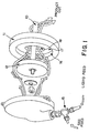
- Figures 1-3 provide further details of several types of mills that may be used in the mechanical cavitation process.
- Fig. 1 depicts the essential elements of a Gaulin mill. These are: a rotating shaft (10) operably connected to a motor (not shown); a disc rotor (12) affixed to the end of the shaft (10); and a stator (11). Stator (11) has a central bore opening (18) and a counterbore (16) into which the rotor is received. In this mill the protein solution and gas are fed through the mill via a "tee" (15). The protein solution/gas mixture is emulsified and cavitates between the surfaces of the rotor and stator. The "gap" in this mill is the space between radial surface (17) of the stator counterbore and the circumferential radial surface of the rotor. The temperature of the microsphere product will be taken (e.g. with a thermocouple, not shown) as the mixture exits past the stator (11).
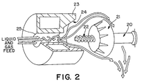
- Fig. 2 shows the essential elements of a Bematek mill.
- This mill is similar in structure and function to the Gaulin mill of Fig. 1 -- the main difference being the configurations of the rotor and the stator counterbore. It includes a rotary shaft (20) that carries a frustroconical rotor (21) that has a threaded leading end (22) and a stator (23) that has a central cylindrical opening (25) and a frustroconical counterbore (24) that is adapted to receive the rotor.
- the protein solution/gas mixture is fed into this mill via opening (25).
- the gas and solution are mixed as they pass by the threads on the shaft (22) and the mixture is emulsified and subjected to cavitation as it passes through the gap of the mill.
- the gap is defined by the space between the conical surfaces of the rotor and stator.
- Fig. 3 illustrates a Silverson mill.
- the structure of this mill is quite different from those of the mills of Figs 1 and 2.
- the depicted Silverson mill has a rotating shaft (30) that carries a paddle blade rotor (31).
- the rotor is received in a cup-shaped perforated screen stator (32).
- the stator is mounted on a housing (33) fitted with an inlet fitting (34).
- the inlet fitting (34) extends up into the housing (33) opening at the bottom center of the perforated screen stator (32).
- the housing has a central opening (not shown) that communicates with the inlet fitting and with an opening (also not shown) in the bottom of the stator.
- the solution/gas is fed via the inlet fitting into the bottom of the stator and is emulsified and cavitated between the flat faces (35) of the paddle rotor and the inner cylindrical surface of the stator.
- the "gap" of this mill can be defined as the space between the rotor (31) and stator (32), but the effect of the gap size on the process is influenced by the size of the perforations (36) of the stator.
- the product After passing through the mill the product may be cooled, typically to 10 - 20°C, and defoamed by settling or by adding biocompatible defoaming agents that do not adversely affect the microspheres.
- Microsphere size may be determined by a suitable particle counter, for example a Coulter Multisizer II (Coulter Electronics, Hialeah, Fl).
- the rotor speed, gap size and gas:liquid ratio are the principal process parameters which affect the characteristics (mean size, size distribution, and concentration of microspheres) of the microsphere product. Those parameters are adjusted empirically to provide a product having the desired characteristics. For any given product, its characteristics are defined clinically. For instance, putative specifications for perfluoropropane microspheres used for myocardial perfusions are: mean size, 4 microns; size distribution, 90% under 10 microns; concentration, 7 x 10 8 to 2 x 10 9 microspheres/mL.
- the protein solution is pre-heated before processing so that the process temperature can reach and maintain the incipient denaturation temperature.
- a Model 2 1/2" Bematek Colloid Mill (Fig. 2; Bematek Systems, Beverly MA), was piped so that the inlet port was connected to a heat exchanger. Gas impermeable tubing was used to make the soft connections between the heat exchanger hose barbs.
- the outlet port from the process head was connected to a stainless steel post-process chiller.
- T1 thermocouple was mounted in a Swagelok "Tee" between the pre-heat heat exchanger and the mill head to measure the feed temperature of the protein solution.
- a second "Tee” for introducing gas was also placed at the feed port.
- the T2 thermocouple was placed inside the exit from the process head, approximately 1 cm from the rotor and 2 cm from the shaft so that the temperature of the process could be accurately measured. In this way, the two temperatures can be measured independently, the feed temperature (T1) and the process temperature, (T2), and compared to determine the amount of heating of the solution during processing.
- U.S.P. albumin was diluted with normal saline to make up a 1% (w/v) solution.
- the denaturation temperature was determined experimentally, as described, to be 78°C. It was fed into the mill at 200 mL/min following degassing along with perfluoropropane at 100 mL/min (50% v/v). Differences between T1 and T2 of 10° to 15°C were noted. In order to obtain a process temperature of 77°C (1°C below denaturation temperature), the feed temperature was adjusted to a range of 62° to 67°C.
- T1 and T2 are differences between milling parameters, (choice of mill, mill settings, flow rate, gas:liquid ratio, etc.) in order to target the process temperature to avoid bulk denaturation of the protein while successfully encapsulating the gas microbubbles with a thin shell of denatured protein.
- the chiller-out temperature (T3) was also monitored, and for best results was targeted at 20°C.
- Microspheres containing various gases were produced as follows: 5% human albumin solution (USP) was deaerated under continuous vacuum for two hours. The vacuum was released by filling the evacuated vessel with the gas of interest. Insoluble gases utilized include sulfur hexafluoride, perfluoroethane, and perfluoropropane. Microspheres containing more soluble gases, air, nitrogen, oxygen and argon, were also produced. The use of argon was representative of a high molecular weight, but relatively soluble, gas.
- the albumin solution was adjusted to 68°C via an in-line heat exchanger and pumped at 100 mL/min into a 2 1/2" (63.5 mm) colloid mill (Greerco, Hudson, NH, model W250V or AF Gaulin, Everett, MA, model 2F).
- the specific gas at room temperature, was added to the liquid feed just upstream of the inlet port at a flow rate of 120-220 mL/min.
- the gap between the rotor and the stator was adjusted to 2/1000th inch (0.005 cm) and the albumin solution was milled continuously at about 7000 rpm at a process temperature of 73°C.
- Air, sulfur hexafluoride and perfluoroethane microspheres were prepared by both batch and continuous ultrasound cavitation processes.
- a solution of human albumin, 5%, USP was degassed under vacuum and stored under the specific gas.
- the continuous sonication process was performed as described by Cerny (USP 4,957,656), substituting the insoluble gases for air.
- the batch process was performed utilizing a 3/4" (1.9 mm) liquid processing horn (Sonics and Materials, Danbury CT). Gas was passed through the horn and into the albumin such that during the entire process air was excluded.
- the albumin was warmed to 73°C and sonicated for 5 sec at 20 KHz at 60 microns double amplitude, using a Branson piezoelectric converter and power source (Branson Ultrasonics, Danbury CT). The product was immediately transferred to a glass vial and sealed under gas.
- the product consisted of a thick, milky suspension of microspheres at concentrations of 1.4 x 10 8 to 1.0 x 10 9 microspheres/mL with a mean size of 2.5 to 3.3 microns.
- Albumin microspheres containing various gases were prepared as described in Example 2 or 5. Microscopic examination of the products revealed a monodisperse suspension of spherical microspheres. The microspheres were collapsed by application of high pressure in a syringe until the suspension clarified. In all cases, microscopic reexamination revealed the presence of hyaline, membranous shells from the collapsed microspheres.
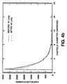
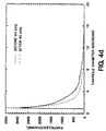
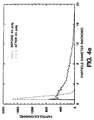
- Albumin microspheres containing various gases were prepared as described in Example 2. Ten mL of each suspension was placed in a 10 mL glass gas-tight syringe (Hamilton, Reno NV) fitted with a pressure gauge. All headspace was removed and the apparatus was sealed. A constant pressure of 40 psig (2.76 bar) was applied for 3 min. A Coulter Counter was then used to measure the sample particle concentration and distribution. Comparisons of the data ( Figures 4a-4e) before and after pressurization demonstrated a relative resistance of the insoluble gas microspheres to 40 psig (2.76 bar).
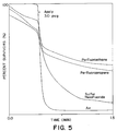
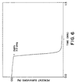
- Microspheres containing various gases were prepared as described in Example 2. Each sample of microspheres was diluted to an equal volume of encapsulated gas per mL of phosphate-buffered saline (0.15M), about a 1:60 dilution. The diluted suspension was subjected to instant static pressures of 0.5 psig (0.035 bar) to 7.5 psig (0.52 bar) in a sealed vessel with adequate head space. Figure 5 shows the effect of pressure on microsphere concentration. Microspheres containing the insoluble gases perfluoropropane, perfluoroethane and sulfur hexafluoride are much more pressure-resistant than air or high molecular weight argon-filled microspheres of the same concentration and size distributions ( Figure 6). Physiological pressures in the bloodstream range from a peripheral venous pressure of 1.5 psig (0.10 bar) to 2.5 psig (0.17 bar) in the myocardial wall.
- Albumin microspheres containing various gases were prepared as described in Example 2.
- Phosphate-buffered saline (PBS) was degassed by boiling just before use. 0.05 mL to 1.5 mL aliquots of the hot buffer were placed in 13 x 100 test tubes and allowed to cool 1 min to room temperature in a water bath. A constant volume of microspheres was added to each tube. After mixing, the final volume was brought to 3.0 mL with PBS and the microsphere concentration was determined.
- Figure 7 shows that improved survival in degassed solutions is obtained for the microspheres containing the insoluble gases perfluoropropane, perfluoroethane and sulfur hexafluoride.
- Microspheres containing air, sulfur hexafluoride or perfluoroethane were diluted into whole blood. Air-filled microspheres exhibited collapse. The insoluble gas-filled microspheres were shown to survive dilution in fresh whole blood.
- Microspheres prepared from various gases were prepared as described in Example 2. Microspheres were diluted into phosphate-buffered saline, as described in Example 8, and placed in a clear cell positioned on the stage of a microscope. The cell was connected to a nitrogen source that allowed for observing the effects of rapid application and release of physiological pressures on the microspheres.
- Sulfur hexafluoride microspheres also exhibited enhanced elasticity under applied physiological pressure relative to air-filled microspheres, but less elasticity relative to the perfluorocarbon microspheres.
- a 20 cc syringe barrel was fitted with a T-type thermocouple inserted through the tip and mounted onto a support stand.
- the syringe was filled to the 16 cc mark with Swiss Red Cross 5% human serum albumin.
- Gas perfluoropropane (C 3 F 8 ) or sulfur hexafluoride (SF 6 )) was introduced into the top of the syringe barrel and flowed over the surface of the liquid.
- a sonicating horn was lowered to the 10 cc mark, below the surface of the solution, and operated at 50% power until the temperature of the solution rose to 72.8 - 73°C; approximately 1 minute.
- the horn was immediately withdrawn to the meniscus ⁇ 1 mm and the power level increased to 65%. Sonication continued for 5 seconds, with an additional temperature increase of 1.2 - 2°C.
- the product was poured into a glass vial to capacity and sealed.
- Human serum albumin was diluted to a 1% w/v solution with sterile saline. The solution was heated to incipient denaturation, approximately 76°C. The system was closed to the external atmosphere and perfluoropropane or sulfur hexafluoride gas was introduced into the liquid flow (1:1) in place of air. The product was made continuously by flowing the gas/albumin mixture past the sonicator horn at approximately 100 ml liquid/min. The product was chilled upon exit from the sonication chamber by passage through a heat exchanger and collected as a bulk liquid suspension of microspheres. Handling and storage conditions were similar to that given for manually produced microspheres.
- Albumin microspheres containing perfluoropropane or sulfur hexafluoride gas were also produced in a closed system by milling a mixture of 1% human serum albumin and gas, similar to that described in Example 2.
- Albumin solution heated to a temperature sufficient to allow microsphere formation by the mechanical cavitation of a given mill, was mixed 1:1 (v:v) gas and introduced into a colloid mill.
- the liquid flow rate was dependent upon the capacity or size of the mill, typically 100 to 500 ml/min.
- a Silverson L4R mill and a Bematek 3" (76.2 mm) production colloid mill were used for this evaluation.
- the outflow from the mill was cooled by passage through a heat exchange system and the resulting albumin microsphere suspension was collected in bulk.
- the product was filled into glass vials, similar to the other processes.
- the percentage of perfluoropropane entrapped in duplicate lots of microspheres prepared as described in PROCESS METHODS was determined by gas chromatography on a Hewlett Packard 5890. A sample of the microsphere suspension was taken in a gas tight syringe. The gas was released from the microspheres using an anti-foam agent in ethanol and the entrapped gas was detected by thermal conductivity.
- Pressure resistance of albumin microspheres was evaluated by a method similar to that reported by Sintetica in European Patent Application 554,213. Microspheres were diluted in aerated phosphate buffered saline, to approximately 1 absorbance unit at 600 nm, in a 3 ml pressure cuvette. The neck was attached to a pressure source and the cuvette placed in a recording spectrophotometer. The pressure in the cuvette was increased linearly from 0 to 5 or 10 psig (0.35 to 0.69 bar) over 150 seconds, at which time the pressure was released.
- the pressure ramp was created by a proportioning solenoid valve (Honeywell) and a pressure transducer (Omega) that were placed between a 20 psi (1.38 bar) pressure source (N 2 tank) and a 5 liter stainless steel reservoir.
- the cuvette was connected to the steel reservoir through a digital pressure gauge.
- the reservoir, and cuvette was pressurized at a selected rate until the desired pressure was achieved.
- the optical density of the microsphere suspension was monitored as a function of time and pressure. The data was corrected for the natural flotation rate of microspheres in the cuvette.
- Albumin microspheres produced by the methods of manual sonication, continuous sonication and mechanical cavitation were analyzed for concentration, mean size, encapsulated gas volume and size distribution within 24 hours after manufacture. All measurements were performed in duplicate, as a minimum, and are presented as the average. The results of these measurements are given in Table 6. Gas Method Conc.
- microspheres made in the open system using manual sonication encapsulate much less of the gas used to form the microspheres than those made in the closed systems (continuous sonication and mechanical cavitation.)
- the microspheres made in the closed system were made in the absence of oxygen, as determined using an oxygen electrode.
- Microspheres made by all three methods were subjected to the same amount of exposure to the atmosphere during handling and sampling (which accounts for less than 100% perfluoropropane gas being measured in microspheres made using the two closed system procedures), thus there was oxygen (and other atmospheric gases) present during formation in the open system which diminished the efficiency of gas encapsulation.
- a suspension of gas-filled microspheres will decrease in optical density with increasing pressure due to a decrease in size and associated change in surface area.
- Shrinkage is due to two factors; reversible compression according to the gas laws, and irreversible loss of the gas core to the surrounding liquid due to increased solubility according to Henry's law.
- Upon the release of an applied pressure only that fraction of the volume loss due to compression is recovered, and which can be observed by an increase in optical density.
- the loss of entrapped gas to the surrounding solution does not reenter the microspheres upon depressurization, but is lost to the head space above the solution.
- Fig. 8 shows the result of imposing a linear pressure gradient up to 10 psi (0.69 bar) on 1 OD suspensions of albumin microspheres prepared with perfluoropropane gas by the manual sonication (open system) method as well as the continuous sonication and mechanical cavitation (closed system) methods. Both closed system methods yielded microspheres that exhibited compression with increasing pressure, with a total recovery of volume upon release of the pressure at the end of the gradient. Loss of entrapped gas to the surrounding solution was not observed.
- Albumin microspheres prepared in the open system (manual sonication method) exhibited greater compression with applied pressure and only a partial recovery of volume upon release of pressure due to the irreversible loss of the gas core, resulting in a 40% destruction of microspheres.
Abstract
Description
| BUNSEN COEFFICIENTS OF GASES IN WATER (1 atmosphere, mL/mL) | ||
| | 5° | 25°C |
| Carbon Dioxide | 1.383 | 0.824 |
| Argon | 0.047 | 0.033 |
| Oxygen | 0.043 | 0.031 |
| Nitrogen | 0.021 | 0.016 |
| Sulfur hexafluoride | 0.008 | 0.0054 |
| Perfluoromethane | 0.0082 | 0.00504 |
| Perfluoroethane | 0.0027 | 0.00138 |
| Perfluoropropane | 0.0016 | N/A |
| Perfluorobutane | 0.0007 | N/A |
| PROTEIN | CONCENTRATION | pH | SOLVENT | Tdenaturation |
| Human Serum Albumin, USP Swiss Red Cross (Bern, Switzerland) | 50 mg/mL | 6.9 | 0.9% NaCl, 4mM Sodium Caprylate, 4mM Tryptophanate | 75°C |
| Human Serum Albumin, USP Swiss Red Cross (Bern, | 10 mg/mL | 6.9 | 0.9% NaCl, 1 mM Sodium Caprylate, 1 mM Tryptophanate | 78°C |
| β-Lactoglobulin, Sigma (St. Louis, MO) | 25 mg/mL | 7.6 | USP Water | 90°C |
| αβ-Globin, Sigma (St. Louis, MO) | 25 mg/mL | 5.0 | USP Water | 90°C |
| Lysozyme Sigma (St. Louis, MO) | 100 mg/mL | 7.5 | 5 mM TRIS, | 31°C as determined immediately after addition of DTT |
| Human Gamma Globulin, acid pH method, Sigma (St. Louis, MO) | 40 mg/mL | 5.0 | 10 mM MES, pH 5.0 | 66°C |
| Human Gamma Globulin, alkaline pH method, Sigma (St. Louis, MO) | 40 mg/mL | 9.8 | 10 mM TRIS, pH 9.8 | 69°C |
| apo-Transferrin, Sigma (St. Louis, MO) | 20 mg/mL | 7.5 | 10 mM TRIS* | 71°C |
- Model #2 1/2
- - Bematek, Beverly, MA
- Model W250V
- - Greerco, Hudson, NH
- Model 2F
- - APV Gaulin, Everett, MA
- Model L4R
- - Silverson, Chesham, UK
- Model Polytron PT3000
- - Kinematica, Littaw, Switzerland
The chiller-out temperature (T3) was also monitored, and for best results was targeted at 20°C.
| Concentration (µspheres/mL) | Mean Size (microns) | |
| Perfluoropropane | 8.3 x 10⁸ | 3.8 |
| Perfluoroethane | 10.6 x 10⁸ | 4.0 |
| Sulfur hexafluoride | 8.4 x 10⁸ | 3.9 |
| Air | 9.2 x 10⁷ | 3.4 |
| Nitrogen | 5.4 x 10⁷ | 5.0 |
| Oxygen | 6.1 x 10⁷ | 3.9 |
| Argon | 4.1 x 10⁷ | 3.5 |
| Rotor Tip Speed (ft/min) | Gap (cm) | Concentration (µspheres/mL) | Mean Size (microns) |
| 3500 | 0.01 | 0.76 x 10⁸ | 13.4 |
| 4300 | 0.01 | 2.43 x 10⁸ | 9.6 |
| 4800 | 0.01 | 9.38 x 10⁸ | 3.8 |
| 9700 | 0.01 | 20.96 x 10⁸ | 4.3 |
| 5200 | 0.02 | 12.87 x 10⁸ | 5.0 |
| 7000 | 0.02 | 12.50 x 10⁸ | 3.4 |
| 8700 | 0.02 | 14.42 x 10⁸ | 3.0 |
| 9600 | 0.02 | 15.22 x 10⁸ | 2.9 |
| Gas/Liquid % (v/v) | Gap (cm) | Concentration (µspheres/mL) | Mean Size (microns) |
| 20 | 0.03 | 6.54 x 10⁸ | 3.6 |
| 50 | 0.03 | 7.51 x 10⁸ | 4.3 |
| 70 | 0.03 | 8.76 x 10⁸ | 5.0 |
| 100 | 0.03 | 8.66 x 10⁸ | 5.9 |
| Gas | Method | Conc. (10 8 /ml) | Mean Size (µm) | Vol (ml/ml) |
| SF6 | Manual Sonication | 8.7 | 3.2 | 0.046 |
| SF6 | Continuous Sonication | 12.7 | 2.7 | 0.034 |
| SF6 | Mechanical Cavitation | 10.0 | 3.8 | 0.054 |
| C3F8 | Manual Sonication | 13.4 | 2.8 | 0.033 |
| C3F8 | Manual Sonication | 17.7 | 2.8 | 0.056 |
| C3F8 | Continuous Sonication | 10.1 | 3.0 | 0.050 |
| C3F8 | Continuous Sonication | 6.7 | 4.3 | 0.127 |
| C3F8 | Mechanical Cavitation (Bematek Mill) | 31.0 | 3.0 | 0.23 |
| C3F8 | Mechanical Cavitation (Silverson Mill) | 6.9 | 5.0 | 0.34 |
| Method | %C 3 F 8 |
| Manual Sonication | 70.0 |
| Continuous Sonication | 89.5 |
| Mechanical Cavitation | 95.5 |
Claims (16)
- A method of making encapsulated gas microspheres useful as an ultrasonic imaging agent the method comprising:a. providing an aqueous solution of a heat-denaturable protein at a temperature necessary to achieve incipient denaturation temperature during subsequent mechanical emulsification;b. combining the solution with a gas;c. emulsifying the protein solution and gas mixture by mechanically shearing the mixture to form a suspension of gas microbubbles having a mean diameter in the range of about 0.1 to about 10 microns; andd. encapsulating the gas microbubbles to form microspheres by mechanically cavitating the suspension to cause the protein to become denatured and thereby deposited at the gas-solution interface.
- A method according to claim 1 wherein the temperature is achieved by heating the solution.
- A method according to claim 1 wherein the temperature is achieved by including additives in the solution that alter the denaturation temperature of the protein.
- A method according to any preceding claim wherein the protein is a naturally occurring protein.
- A method according to any preceding claim wherein the protein is human serum albumin.
- A method according to any of claims 1 to 3 wherein the protein is a synthetic protein.
- A method according to any preceding claim wherein the concentration of the protein in the solution is from 0.1 to 10% w/v.
- A method according to any preceding claim wherein the concentration of the protein in the solution is from 1 to 5% w/v.
- A method according to any preceding claim wherein the concentration of the protein in the solution is about 1% w/v.
- A method according to any preceding claim wherein the gas is insoluble.
- A method according to any preceding claim wherein the gas is sulphur hexafluoride, perfluoromethane, perfluoroethane, perfluoropropane or perfluorobutane.
- A method according to any of claims 1 to 9 wherein the gas is air.
- A method according to any preceding claim wherein the ratio of gas to protein solution is 5% to 200% v/v.
- A method according to any preceding claim wherein the ratio of gas to protein solution is 20% to 100% v/v.
- A method according to any preceding claim wherein steps (c) and (d) are effected by passing the mixture through a mill.
- A method according to any preceding claim wherein the incipient denaturation temperature is about 1°C to 5°C below the denaturation temperature of the protein.
Applications Claiming Priority (5)
| Application Number | Priority Date | Filing Date | Title |
|---|---|---|---|
| US8671793A | 1993-07-02 | 1993-07-02 | |
| US86717 | 1993-07-02 | ||
| US18765694A | 1994-01-26 | 1994-01-26 | |
| US187656 | 1994-01-26 | ||
| EP94304902A EP0633030B1 (en) | 1993-07-02 | 1994-07-04 | The preparation of protein encapsulated insoluble gas microspheres. |
Related Parent Applications (1)
| Application Number | Title | Priority Date | Filing Date |
|---|---|---|---|
| EP94304902A Division EP0633030B1 (en) | 1993-07-02 | 1994-07-04 | The preparation of protein encapsulated insoluble gas microspheres. |
Publications (2)
| Publication Number | Publication Date |
|---|---|
| EP0885615A1 true EP0885615A1 (en) | 1998-12-23 |
| EP0885615B1 EP0885615B1 (en) | 2003-03-26 |
Family
ID=26775071
Family Applications (2)
| Application Number | Title | Priority Date | Filing Date |
|---|---|---|---|
| EP98116817A Expired - Lifetime EP0885615B1 (en) | 1993-07-02 | 1994-07-04 | Protein encapsulated insoluble gas microsperes and their preparation and use as ultrasonic imaging agents |
| EP94304902A Expired - Lifetime EP0633030B1 (en) | 1993-07-02 | 1994-07-04 | The preparation of protein encapsulated insoluble gas microspheres. |
Family Applications After (1)
| Application Number | Title | Priority Date | Filing Date |
|---|---|---|---|
| EP94304902A Expired - Lifetime EP0633030B1 (en) | 1993-07-02 | 1994-07-04 | The preparation of protein encapsulated insoluble gas microspheres. |
Country Status (20)
| Country | Link |
|---|---|
| US (1) | US5552133A (en) |
| EP (2) | EP0885615B1 (en) |
| JP (3) | JP2905598B2 (en) |
| KR (1) | KR100218642B1 (en) |
| CN (1) | CN1129910A (en) |
| AT (1) | ATE179334T1 (en) |
| AU (1) | AU683485B2 (en) |
| BR (1) | BR9406993A (en) |
| CA (1) | CA2166459C (en) |
| CZ (1) | CZ350895A3 (en) |
| DE (2) | DE69418101T2 (en) |
| ES (2) | ES2134321T3 (en) |
| FI (1) | FI956312A (en) |
| HU (1) | HUT74827A (en) |
| IL (1) | IL110185A (en) |
| NO (1) | NO955351L (en) |
| NZ (1) | NZ268826A (en) |
| PL (1) | PL312387A1 (en) |
| TW (1) | TW283646B (en) |
| WO (1) | WO1995001187A1 (en) |
Cited By (2)
| Publication number | Priority date | Publication date | Assignee | Title |
|---|---|---|---|---|
| US8647696B2 (en) | 2008-12-12 | 2014-02-11 | The University Of Birmingham | Low fat food containing gas bubbles |
| US9486416B2 (en) | 2009-12-22 | 2016-11-08 | Evonik Corporation | Emulsion-based process for preparing microparticles and workhead assembly for use with same |
Families Citing this family (137)
| Publication number | Priority date | Publication date | Assignee | Title |
|---|---|---|---|---|
| US5773024A (en) | 1989-12-22 | 1998-06-30 | Imarx Pharmaceutical Corp. | Container with multi-phase composition for use in diagnostic and therapeutic applications |
| US6146657A (en) | 1989-12-22 | 2000-11-14 | Imarx Pharmaceutical Corp. | Gas-filled lipid spheres for use in diagnostic and therapeutic applications |
| US5776429A (en) | 1989-12-22 | 1998-07-07 | Imarx Pharmaceutical Corp. | Method of preparing gas-filled microspheres using a lyophilized lipids |
| US6001335A (en) | 1989-12-22 | 1999-12-14 | Imarx Pharmaceutical Corp. | Contrasting agents for ultrasonic imaging and methods for preparing the same |
| US5733572A (en) | 1989-12-22 | 1998-03-31 | Imarx Pharmaceutical Corp. | Gas and gaseous precursor filled microspheres as topical and subcutaneous delivery vehicles |
| US6551576B1 (en) | 1989-12-22 | 2003-04-22 | Bristol-Myers Squibb Medical Imaging, Inc. | Container with multi-phase composition for use in diagnostic and therapeutic applications |
| US5469854A (en) | 1989-12-22 | 1995-11-28 | Imarx Pharmaceutical Corp. | Methods of preparing gas-filled liposomes |
| US5305757A (en) | 1989-12-22 | 1994-04-26 | Unger Evan C | Gas filled liposomes and their use as ultrasonic contrast agents |
| US5542935A (en) | 1989-12-22 | 1996-08-06 | Imarx Pharmaceutical Corp. | Therapeutic delivery systems related applications |
| US5580575A (en) | 1989-12-22 | 1996-12-03 | Imarx Pharmaceutical Corp. | Therapeutic drug delivery systems |
| US5656211A (en) | 1989-12-22 | 1997-08-12 | Imarx Pharmaceutical Corp. | Apparatus and method for making gas-filled vesicles of optimal size |
| US6088613A (en) | 1989-12-22 | 2000-07-11 | Imarx Pharmaceutical Corp. | Method of magnetic resonance focused surgical and therapeutic ultrasound |
| US5922304A (en) | 1989-12-22 | 1999-07-13 | Imarx Pharmaceutical Corp. | Gaseous precursor filled microspheres as magnetic resonance imaging contrast agents |
| US5585112A (en) | 1989-12-22 | 1996-12-17 | Imarx Pharmaceutical Corp. | Method of preparing gas and gaseous precursor-filled microspheres |
| US7083778B2 (en) * | 1991-05-03 | 2006-08-01 | Bracco International B.V. | Ultrasound contrast agents and methods of making and using them |
| US6989141B2 (en) * | 1990-05-18 | 2006-01-24 | Bracco International B.V. | Ultrasound contrast agents and methods of making and using them |
| US20040208826A1 (en) * | 1990-04-02 | 2004-10-21 | Bracco International B.V. | Ultrasound contrast agents and methods of making and using them |
| US5445813A (en) * | 1992-11-02 | 1995-08-29 | Bracco International B.V. | Stable microbubble suspensions as enhancement agents for ultrasound echography |
| US6613306B1 (en) | 1990-04-02 | 2003-09-02 | Bracco International B.V. | Ultrasound contrast agents and methods of making and using them |
| US20010024638A1 (en) * | 1992-11-02 | 2001-09-27 | Michel Schneider | Stable microbubble suspensions as enhancement agents for ultrasound echography and dry formulations thereof |
| US5578292A (en) | 1991-11-20 | 1996-11-26 | Bracco International B.V. | Long-lasting aqueous dispersions or suspensions of pressure-resistant gas-filled microvesicles and methods for the preparation thereof |
| USRE39146E1 (en) | 1990-04-02 | 2006-06-27 | Bracco International B.V. | Long-lasting aqueous dispersions or suspensions of pressure-resistant gas-filled microvesicles and methods for the preparation thereof |
| IN172208B (en) | 1990-04-02 | 1993-05-01 | Sint Sa | |
| AU636481B2 (en) * | 1990-05-18 | 1993-04-29 | Bracco International B.V. | Polymeric gas or air filled microballoons usable as suspensions in liquid carriers for ultrasonic echography |
| US20030194376A1 (en) * | 1990-05-18 | 2003-10-16 | Bracco International B.V. | Ultrasound contrast agents and methods of making and using them |
| US5874062A (en) | 1991-04-05 | 1999-02-23 | Imarx Pharmaceutical Corp. | Methods of computed tomography using perfluorocarbon gaseous filled microspheres as contrast agents |
| US5205290A (en) | 1991-04-05 | 1993-04-27 | Unger Evan C | Low density microspheres and their use as contrast agents for computed tomography |
| MX9205298A (en) | 1991-09-17 | 1993-05-01 | Steven Carl Quay | GASEOUS ULTRASOUND CONTRASTING MEDIA AND METHOD FOR SELECTING GASES TO BE USED AS ULTRASOUND CONTRASTING MEDIA |
| US6875420B1 (en) | 1991-09-17 | 2005-04-05 | Amersham Health As | Method of ultrasound imaging |
| US6723303B1 (en) | 1991-09-17 | 2004-04-20 | Amersham Health, As | Ultrasound contrast agents including protein stabilized microspheres of perfluoropropane, perfluorobutane or perfluoropentane |
| US5409688A (en) * | 1991-09-17 | 1995-04-25 | Sonus Pharmaceuticals, Inc. | Gaseous ultrasound contrast media |
| IL104084A (en) * | 1992-01-24 | 1996-09-12 | Bracco Int Bv | Long-lasting aqueous suspensions of pressure-resistant gas-filled microvesicles their preparation and contrast agents consisting of them |
| IL108416A (en) | 1993-01-25 | 1998-10-30 | Sonus Pharma Inc | Phase shift colloids as ultrasound contrast agents |
| CN1068230C (en) * | 1993-01-25 | 2001-07-11 | 索纳斯药品有限公司 | Phase shift colloids as ultrasound contrast agents |
| US5362478A (en) * | 1993-03-26 | 1994-11-08 | Vivorx Pharmaceuticals, Inc. | Magnetic resonance imaging with fluorocarbons encapsulated in a cross-linked polymeric shell |
| US5701899A (en) * | 1993-05-12 | 1997-12-30 | The Board Of Regents Of The University Of Nebraska | Perfluorobutane ultrasound contrast agent and methods for its manufacture and use |
| US5695740A (en) * | 1993-05-12 | 1997-12-09 | The Board Of Regents Of The University Of Nebraska | Perfluorocarbon ultrasound contrast agent comprising microbubbles containing a filmogenic protein and a saccharide |
| EP0711179B2 (en) | 1993-07-30 | 2010-09-01 | IMCOR Pharmaceutical Co. | Stabilized microbubble compositions for ultrasound |
| US5798091A (en) | 1993-07-30 | 1998-08-25 | Alliance Pharmaceutical Corp. | Stabilized gas emulsion containing phospholipid for ultrasound contrast enhancement |
| PT682530E (en) * | 1993-12-15 | 2003-06-30 | Bracco Research Sa | UTEIS GAS MIXTURES AS CONTRAST MEANS FOR ULTRASSONS |
| DE4406474A1 (en) * | 1994-02-23 | 1995-08-24 | Schering Ag | Gas-containing microparticles, agents containing them, their use in ultrasound diagnostics, and methods for producing the particles and agents |
| US5736121A (en) | 1994-05-23 | 1998-04-07 | Imarx Pharmaceutical Corp. | Stabilized homogenous suspensions as computed tomography contrast agents |
| US5965109A (en) * | 1994-08-02 | 1999-10-12 | Molecular Biosystems, Inc. | Process for making insoluble gas-filled microspheres containing a liquid hydrophobic barrier |
| US5730955A (en) * | 1994-08-02 | 1998-03-24 | Molecular Biosystems, Inc. | Process for making gas-filled microspheres containing a liquid hydrophobic barrier |
| US5540909A (en) * | 1994-09-28 | 1996-07-30 | Alliance Pharmaceutical Corp. | Harmonic ultrasound imaging with microbubbles |
| US6743779B1 (en) | 1994-11-29 | 2004-06-01 | Imarx Pharmaceutical Corp. | Methods for delivering compounds into a cell |
| US5830430A (en) | 1995-02-21 | 1998-11-03 | Imarx Pharmaceutical Corp. | Cationic lipids and the use thereof |
| US5997898A (en) | 1995-06-06 | 1999-12-07 | Imarx Pharmaceutical Corp. | Stabilized compositions of fluorinated amphiphiles for methods of therapeutic delivery |
| US6521211B1 (en) | 1995-06-07 | 2003-02-18 | Bristol-Myers Squibb Medical Imaging, Inc. | Methods of imaging and treatment with targeted compositions |
| US6033645A (en) | 1996-06-19 | 2000-03-07 | Unger; Evan C. | Methods for diagnostic imaging by regulating the administration rate of a contrast agent |
| US6231834B1 (en) | 1995-06-07 | 2001-05-15 | Imarx Pharmaceutical Corp. | Methods for ultrasound imaging involving the use of a contrast agent and multiple images and processing of same |
| US6139819A (en) | 1995-06-07 | 2000-10-31 | Imarx Pharmaceutical Corp. | Targeted contrast agents for diagnostic and therapeutic use |
| US5897851A (en) * | 1995-06-07 | 1999-04-27 | Sonus Pharmaceuticals, Inc. | Nucleation and activation of a liquid-in-liquid emulsion for use in ultrasound imaging |
| US5674469A (en) * | 1995-06-07 | 1997-10-07 | Molecular Biosystems, Inc. | Gas-exchange method of making gas-filled microspheres |
| US5648098A (en) * | 1995-10-17 | 1997-07-15 | The Board Of Regents Of The University Of Nebraska | Thrombolytic agents and methods of treatment for thrombosis |
| US6245747B1 (en) | 1996-03-12 | 2001-06-12 | The Board Of Regents Of The University Of Nebraska | Targeted site specific antisense oligodeoxynucleotide delivery method |
| JP2001507207A (en) | 1996-05-01 | 2001-06-05 | イマアーレクス・フアーマシユーチカル・コーポレーシヨン | Methods for delivering compounds to cells |
| US5976501A (en) * | 1996-06-07 | 1999-11-02 | Molecular Biosystems, Inc. | Use of pressure resistant protein microspheres encapsulating gases as ultrasonic imaging agents for vascular perfusion |
| CN1042700C (en) * | 1996-06-25 | 1999-03-31 | 谢峰 | Dextran albumin acoustical contrast medium containing perfluocarbon and its producing process |
| US5849727A (en) | 1996-06-28 | 1998-12-15 | Board Of Regents Of The University Of Nebraska | Compositions and methods for altering the biodistribution of biological agents |
| US6414139B1 (en) | 1996-09-03 | 2002-07-02 | Imarx Therapeutics, Inc. | Silicon amphiphilic compounds and the use thereof |
| CA2263568C (en) | 1996-09-11 | 2008-12-02 | Imarx Pharmaceutical Corp. | Methods for diagnostic imaging using a contrast agent and a renal vasodilator |
| US5846517A (en) | 1996-09-11 | 1998-12-08 | Imarx Pharmaceutical Corp. | Methods for diagnostic imaging using a renal contrast agent and a vasodilator |
| US6083484A (en) | 1996-10-17 | 2000-07-04 | Molecular Biosystems, Inc. | Microparticles stabilized by polynuclear chromium complexes and their use as ultrasound contrast agents |
| US6120751A (en) | 1997-03-21 | 2000-09-19 | Imarx Pharmaceutical Corp. | Charged lipids and uses for the same |
| US6537246B1 (en) | 1997-06-18 | 2003-03-25 | Imarx Therapeutics, Inc. | Oxygen delivery agents and uses for the same |
| US6143276A (en) | 1997-03-21 | 2000-11-07 | Imarx Pharmaceutical Corp. | Methods for delivering bioactive agents to regions of elevated temperatures |
| US6090800A (en) | 1997-05-06 | 2000-07-18 | Imarx Pharmaceutical Corp. | Lipid soluble steroid prodrugs |
| US6416740B1 (en) | 1997-05-13 | 2002-07-09 | Bristol-Myers Squibb Medical Imaging, Inc. | Acoustically active drug delivery systems |
| AU7702798A (en) | 1997-05-30 | 1998-12-30 | Alliance Pharmaceutical Corporation | Methods and apparatus for monitoring and quantifying the movement of fluid |
| EP1007181A1 (en) | 1997-07-04 | 2000-06-14 | Nycomed Imaging As | Process for the selection of particles of a preselected size from a particulate pharmaceutical product |
| JP4808842B2 (en) * | 1997-08-18 | 2011-11-02 | ジーイー・ヘルスケア・アクスイェ・セルスカプ | Vesicle preparation method |
| GB9717476D0 (en) * | 1997-08-18 | 1997-10-22 | Nycomed Imaging As | Process |
| US6548047B1 (en) | 1997-09-15 | 2003-04-15 | Bristol-Myers Squibb Medical Imaging, Inc. | Thermal preactivation of gaseous precursor filled compositions |
| US7654728B2 (en) * | 1997-10-24 | 2010-02-02 | Revalesio Corporation | System and method for therapeutic application of dissolved oxygen |
| US7128278B2 (en) | 1997-10-24 | 2006-10-31 | Microdiffusion, Inc. | System and method for irritating with aerated water |
| US6386751B1 (en) | 1997-10-24 | 2002-05-14 | Diffusion Dynamics, Inc. | Diffuser/emulsifier |
| US20110075507A1 (en) * | 1997-10-24 | 2011-03-31 | Revalesio Corporation | Diffuser/emulsifier |
| US6702949B2 (en) | 1997-10-24 | 2004-03-09 | Microdiffusion, Inc. | Diffuser/emulsifier for aquaculture applications |
| US6123923A (en) | 1997-12-18 | 2000-09-26 | Imarx Pharmaceutical Corp. | Optoacoustic contrast agents and methods for their use |
| US20010003580A1 (en) | 1998-01-14 | 2001-06-14 | Poh K. Hui | Preparation of a lipid blend and a phospholipid suspension containing the lipid blend |
| US20010021372A1 (en) * | 1998-08-18 | 2001-09-13 | Tore Omtveit | Apparatus having partially gold-plated surface |
| ATE243947T1 (en) * | 1998-12-23 | 2003-07-15 | Unilever Nv | WATER-IN-OIL EMULSIONS CONTAINING FLOWING GAS BUBBLES |
| US6444192B1 (en) | 1999-02-05 | 2002-09-03 | The Regents Of The University Of California | Diagnostic imaging of lymph structures |
| US20050150618A1 (en) * | 2000-05-17 | 2005-07-14 | Bijan Kazem | Methods of processing lignocellulosic pulp with cavitation |
| US6627784B2 (en) * | 2000-05-17 | 2003-09-30 | Hydro Dynamics, Inc. | Highly efficient method of mixing dissimilar fluids using mechanically induced cavitation |
| CA2410887C (en) | 2000-06-02 | 2012-07-24 | Bracco Research Usa | Compounds for targeting endothelial cells, compositions containing the same and methods for their use |
| US6962071B2 (en) | 2001-04-06 | 2005-11-08 | Bracco Research S.A. | Method for improved measurement of local physical parameters in a fluid-filled cavity |
| US7087212B2 (en) | 2001-08-17 | 2006-08-08 | Mallinckrodt, Inc | Multicomponent assemblies having enhanced binding properties for diagnosis and therapy |
| US7261876B2 (en) | 2002-03-01 | 2007-08-28 | Bracco International Bv | Multivalent constructs for therapeutic and diagnostic applications |
| CA2513044A1 (en) | 2002-03-01 | 2004-08-05 | Dyax Corp. | Kdr and vegf/kdr binding peptides and their use in diagnosis and therapy |
| US7794693B2 (en) | 2002-03-01 | 2010-09-14 | Bracco International B.V. | Targeting vector-phospholipid conjugates |
| US7211240B2 (en) | 2002-03-01 | 2007-05-01 | Bracco International B.V. | Multivalent constructs for therapeutic and diagnostic applications |
| US8623822B2 (en) | 2002-03-01 | 2014-01-07 | Bracco Suisse Sa | KDR and VEGF/KDR binding peptides and their use in diagnosis and therapy |
| ES2398393T3 (en) | 2002-03-01 | 2013-03-15 | Dyax Corp. | KDR and VEGF / KDR binding peptides and their use in diagnosis and therapy |
| US20040126400A1 (en) * | 2002-05-03 | 2004-07-01 | Iversen Patrick L. | Delivery of therapeutic compounds via microparticles or microbubbles |
| US6823820B2 (en) | 2002-12-03 | 2004-11-30 | Christian Helmut Thoma | Apparatus for heating fluids |
| ES2396368T3 (en) | 2003-03-03 | 2013-02-21 | Dyax Corporation | Peptides that specifically bind to the HGF receptor (CMET) and uses thereof |
| CA2526333C (en) * | 2003-05-19 | 2011-12-06 | Hydro Dynamics, Inc. | Method and apparatus for conducting a chemical reaction in the presence of cavitation and an electrical current |
| US6910448B2 (en) | 2003-07-07 | 2005-06-28 | Christian Thoma | Apparatus and method for heating fluids |
| WO2005021050A1 (en) * | 2003-08-22 | 2005-03-10 | Hydro Dynamics, Inc. | Method and apparatus for irradiating fluids |
| US8076117B2 (en) * | 2004-03-18 | 2011-12-13 | Mayo Foundation For Medical Education And Research | Microbial biofilm removal methods and systems |
| US7316501B2 (en) * | 2004-05-20 | 2008-01-08 | Christian Thoma | Apparatus and method for mixing dissimilar fluids |
| US8012457B2 (en) | 2004-06-04 | 2011-09-06 | Acusphere, Inc. | Ultrasound contrast agent dosage formulation |
| CA2662765A1 (en) * | 2006-09-05 | 2008-03-13 | Cerion Technology, Inc. | Cerium dioxide nanoparticle-containing fuel additive |
| US8883865B2 (en) | 2006-09-05 | 2014-11-11 | Cerion Technology, Inc. | Cerium-containing nanoparticles |
| US10435639B2 (en) | 2006-09-05 | 2019-10-08 | Cerion, Llc | Fuel additive containing lattice engineered cerium dioxide nanoparticles |
| US8784897B2 (en) | 2006-10-25 | 2014-07-22 | Revalesio Corporation | Methods of therapeutic treatment of eyes |
| US8445546B2 (en) | 2006-10-25 | 2013-05-21 | Revalesio Corporation | Electrokinetically-altered fluids comprising charge-stabilized gas-containing nanostructures |
| AU2007349224B2 (en) | 2006-10-25 | 2014-04-03 | Revalesio Corporation | Methods of wound care and treatment |
| US8609148B2 (en) | 2006-10-25 | 2013-12-17 | Revalesio Corporation | Methods of therapeutic treatment of eyes |
| AU2007308840C1 (en) | 2006-10-25 | 2014-09-25 | Revalesio Corporation | Methods of therapeutic treatment of eyes and other human tissues using an oxygen-enriched solution |
| AU2007308838B2 (en) | 2006-10-25 | 2014-03-13 | Revalesio Corporation | Mixing device and output fluids of same |
| US8784898B2 (en) | 2006-10-25 | 2014-07-22 | Revalesio Corporation | Methods of wound care and treatment |
| US8465642B2 (en) * | 2007-05-04 | 2013-06-18 | Hydro Dynamics, Inc. | Method and apparatus for separating impurities from a liquid stream by electrically generated gas bubbles |
| US20090036856A1 (en) * | 2007-07-31 | 2009-02-05 | Kimberly-Clark Worldwide, Inc. | Triggerable self-generating liquid foam barrier/interceptor |
| US10125359B2 (en) | 2007-10-25 | 2018-11-13 | Revalesio Corporation | Compositions and methods for treating inflammation |
| US9745567B2 (en) | 2008-04-28 | 2017-08-29 | Revalesio Corporation | Compositions and methods for treating multiple sclerosis |
| US9523090B2 (en) | 2007-10-25 | 2016-12-20 | Revalesio Corporation | Compositions and methods for treating inflammation |
| US8430968B2 (en) | 2008-01-22 | 2013-04-30 | Hydro Dynamics, Inc. | Method of extracting starches and sugar from biological material using controlled cavitation |
| MX2010011856A (en) | 2008-05-01 | 2011-02-15 | Revalesio Corp | Compositions and methods for treating digestive disorders. |
| US8846063B2 (en) * | 2008-12-16 | 2014-09-30 | Kimberly-Clark Worldwide, Inc. | Personal care composition containing a volatile and a terpene alcohol |
| US8815292B2 (en) | 2009-04-27 | 2014-08-26 | Revalesio Corporation | Compositions and methods for treating insulin resistance and diabetes mellitus |
| KR20130114581A (en) | 2010-05-07 | 2013-10-18 | 레발레시오 코퍼레이션 | Compositions and methods for enhancing physiological performance and recovery time |
| CN101926821B (en) * | 2010-07-12 | 2012-01-25 | 四川大学华西医院 | Drug composition for targeted release of trace elements and preparation method and application thereof |
| CN102302507B (en) * | 2010-07-12 | 2014-02-05 | 四川大学华西医院 | Medicinal composition for directional controlled release of trace elements and preparation method and application |
| JP2013533320A (en) | 2010-08-12 | 2013-08-22 | レバレジオ コーポレイション | Compositions and methods for treating tauopathy |
| CZ303992B6 (en) * | 2010-11-04 | 2013-08-07 | Student Science, S.R.O. | Nanofibrous carriers with photoaffinity-bound microspheres and process for preparing thereof |
| CN103534014B (en) * | 2011-05-27 | 2016-04-13 | M技术株式会社 | Fine bubble generating apparatus, micro-bubble method for generation and employ its gas liquid reaction method |
| KR20120140290A (en) * | 2011-06-21 | 2012-12-31 | 주식회사 퍼시픽시스템 | Method of manufacturing bubble for accelerating functional material and device or transferring functional material |
| CN104349827A (en) * | 2012-04-30 | 2015-02-11 | 通用电气医疗集团股份有限公司 | Method for filling container with foamable composition |
| US10350314B2 (en) * | 2012-06-26 | 2019-07-16 | Ge Healthcare As | Preparation of composition comprising gas microbubbles |
| WO2015058037A1 (en) | 2013-10-17 | 2015-04-23 | Cerion, Llc | Malic acid stabilized nanoceria particles |
| CN105233308B (en) * | 2015-11-02 | 2018-10-30 | 中国科学院深圳先进技术研究院 | A kind of acoustic contrast agent and the preparation method and application thereof for HIFU |
| EP3419742B1 (en) | 2016-02-25 | 2022-12-14 | King Abdullah University Of Science And Technology | Acoustically excited encapsulated microbubbles and mitigation of biofouling |
| JP7104709B2 (en) | 2016-10-11 | 2022-07-21 | トーマス・ジェファーソン・ユニバーシティ | Non-invasive method for pressure measurement |
| CN107519502B (en) * | 2017-08-20 | 2019-01-15 | 湖南康润药业有限公司 | The preparation method and encapsulated microbubble of encapsulated microbubble |
Citations (5)
| Publication number | Priority date | Publication date | Assignee | Title |
|---|---|---|---|---|
| EP0224934A2 (en) * | 1985-12-05 | 1987-06-10 | Steven B. Feinstein | Contrast agent, process for its preparation and its use for ultrasonic imaging |
| EP0324938A1 (en) * | 1987-12-29 | 1989-07-26 | Molecular Biosystems, Inc. | Concentrated stabilized microbubble-type ultrasonic imaging agent and method of production |
| EP0359246A2 (en) * | 1988-09-14 | 1990-03-21 | Molecular Biosystems, Inc. | Continuous sonication method for preparing protein encapsulated microbubbles |
| WO1992005806A1 (en) * | 1990-10-05 | 1992-04-16 | Sintetica S.A. | Method for the preparation of stable suspensions of hollow gas-filled microspheres suitable for ultrasonic echography |
| WO1993005819A1 (en) * | 1991-09-17 | 1993-04-01 | Sonus Pharmaceuticals, Inc | Gaseous ultrasound contrast media and method for selecting gases for use as ultrasound contrast media |
Family Cites Families (5)
| Publication number | Priority date | Publication date | Assignee | Title |
|---|---|---|---|---|
| US4572203A (en) * | 1983-01-27 | 1986-02-25 | Feinstein Steven B | Contact agents for ultrasonic imaging |
| AU636481B2 (en) * | 1990-05-18 | 1993-04-29 | Bracco International B.V. | Polymeric gas or air filled microballoons usable as suspensions in liquid carriers for ultrasonic echography |
| GB9107628D0 (en) * | 1991-04-10 | 1991-05-29 | Moonbrook Limited | Preparation of diagnostic agents |
| US5409688A (en) * | 1991-09-17 | 1995-04-25 | Sonus Pharmaceuticals, Inc. | Gaseous ultrasound contrast media |
| IL104084A (en) * | 1992-01-24 | 1996-09-12 | Bracco Int Bv | Long-lasting aqueous suspensions of pressure-resistant gas-filled microvesicles their preparation and contrast agents consisting of them |
-
1994
- 1994-07-01 KR KR1019960700002A patent/KR100218642B1/en not_active IP Right Cessation
- 1994-07-01 AU AU72184/94A patent/AU683485B2/en not_active Ceased
- 1994-07-01 PL PL94312387A patent/PL312387A1/en unknown
- 1994-07-01 NZ NZ268826A patent/NZ268826A/en not_active IP Right Cessation
- 1994-07-01 BR BR9406993A patent/BR9406993A/en not_active Application Discontinuation
- 1994-07-01 WO PCT/US1994/007533 patent/WO1995001187A1/en not_active Application Discontinuation
- 1994-07-01 CA CA002166459A patent/CA2166459C/en not_active Expired - Fee Related
- 1994-07-01 IL IL11018594A patent/IL110185A/en not_active IP Right Cessation
- 1994-07-01 CZ CZ953508A patent/CZ350895A3/en unknown
- 1994-07-01 JP JP7503679A patent/JP2905598B2/en not_active Expired - Lifetime
- 1994-07-01 HU HU9503977A patent/HUT74827A/en unknown
- 1994-07-01 CN CN94193227A patent/CN1129910A/en active Pending
- 1994-07-04 ES ES94304902T patent/ES2134321T3/en not_active Expired - Lifetime
- 1994-07-04 DE DE69418101T patent/DE69418101T2/en not_active Expired - Lifetime
- 1994-07-04 AT AT94304902T patent/ATE179334T1/en not_active IP Right Cessation
- 1994-07-04 DE DE69432370T patent/DE69432370T2/en not_active Expired - Lifetime
- 1994-07-04 EP EP98116817A patent/EP0885615B1/en not_active Expired - Lifetime
- 1994-07-04 EP EP94304902A patent/EP0633030B1/en not_active Expired - Lifetime
- 1994-07-04 ES ES98116817T patent/ES2195247T3/en not_active Expired - Lifetime
- 1994-08-01 TW TW083107084A patent/TW283646B/zh active
- 1994-08-12 US US08/290,022 patent/US5552133A/en not_active Expired - Lifetime
-
1995
- 1995-12-29 NO NO955351A patent/NO955351L/en unknown
- 1995-12-29 FI FI956312A patent/FI956312A/en not_active Application Discontinuation
-
1998
- 1998-09-11 JP JP25901998A patent/JP3285544B2/en not_active Expired - Lifetime
-
2001
- 2001-11-20 JP JP2001355407A patent/JP3809368B2/en not_active Expired - Lifetime
Patent Citations (5)
| Publication number | Priority date | Publication date | Assignee | Title |
|---|---|---|---|---|
| EP0224934A2 (en) * | 1985-12-05 | 1987-06-10 | Steven B. Feinstein | Contrast agent, process for its preparation and its use for ultrasonic imaging |
| EP0324938A1 (en) * | 1987-12-29 | 1989-07-26 | Molecular Biosystems, Inc. | Concentrated stabilized microbubble-type ultrasonic imaging agent and method of production |
| EP0359246A2 (en) * | 1988-09-14 | 1990-03-21 | Molecular Biosystems, Inc. | Continuous sonication method for preparing protein encapsulated microbubbles |
| WO1992005806A1 (en) * | 1990-10-05 | 1992-04-16 | Sintetica S.A. | Method for the preparation of stable suspensions of hollow gas-filled microspheres suitable for ultrasonic echography |
| WO1993005819A1 (en) * | 1991-09-17 | 1993-04-01 | Sonus Pharmaceuticals, Inc | Gaseous ultrasound contrast media and method for selecting gases for use as ultrasound contrast media |
Cited By (2)
| Publication number | Priority date | Publication date | Assignee | Title |
|---|---|---|---|---|
| US8647696B2 (en) | 2008-12-12 | 2014-02-11 | The University Of Birmingham | Low fat food containing gas bubbles |
| US9486416B2 (en) | 2009-12-22 | 2016-11-08 | Evonik Corporation | Emulsion-based process for preparing microparticles and workhead assembly for use with same |
Also Published As
Similar Documents
| Publication | Publication Date | Title |
|---|---|---|
| EP0633030B1 (en) | The preparation of protein encapsulated insoluble gas microspheres. | |
| US5855865A (en) | Method for making encapsulated gas microspheres from heat denatured protein in the absence of oxygen gas | |
| US4957656A (en) | Continuous sonication method for preparing protein encapsulated microbubbles | |
| AU703652B2 (en) | Gas-filled microspheres with fluorine-containing shells | |
| EP0743860B2 (en) | Preparation of hollow microcapsules | |
| US5606973A (en) | Liquid core microdroplets for ultrasound imaging | |
| EP0907380B1 (en) | Pressure resistant protein microspheres as ultrasonic imaging agents | |
| EP0324938A1 (en) | Concentrated stabilized microbubble-type ultrasonic imaging agent and method of production | |
| US5730955A (en) | Process for making gas-filled microspheres containing a liquid hydrophobic barrier | |
| Wheatley et al. | Structural studies on stabilized microbubbles: development of a novel contrast agent for diagnostic ultrasound | |
| EP0927047A2 (en) | Use of hollow microcapsules | |
| AU722742B2 (en) | Methods for making encapsulated microspheres from heat denatured protein using mechanical cavitation | |
| CA2373869A1 (en) | Multi-stage method for producing gas-filled microcapsules |
Legal Events
| Date | Code | Title | Description |
|---|---|---|---|
| PUAI | Public reference made under article 153(3) epc to a published international application that has entered the european phase |
Free format text: ORIGINAL CODE: 0009012 |
|
| AC | Divisional application: reference to earlier application |
Ref document number: 633030 Country of ref document: EP |
|
| AK | Designated contracting states |
Kind code of ref document: A1 Designated state(s): DE ES FR GB IE IT NL |
|
| RIN1 | Information on inventor provided before grant (corrected) |
Inventor name: LOHRMANN, ROLF Inventor name: HAMILTON, KENNETH Inventor name: HULLE, CARL Inventor name: JABLONSKI, EDWARD G. Inventor name: PODELL, SHEILA BENAY Inventor name: LAMBERT, KAREL J. |
|
| 17P | Request for examination filed |
Effective date: 19990621 |
|
| 17Q | First examination report despatched |
Effective date: 19991018 |
|
| GRAG | Despatch of communication of intention to grant |
Free format text: ORIGINAL CODE: EPIDOS AGRA |
|
| GRAG | Despatch of communication of intention to grant |
Free format text: ORIGINAL CODE: EPIDOS AGRA |
|
| GRAH | Despatch of communication of intention to grant a patent |
Free format text: ORIGINAL CODE: EPIDOS IGRA |
|
| GRAG | Despatch of communication of intention to grant |
Free format text: ORIGINAL CODE: EPIDOS AGRA |
|
| GRAH | Despatch of communication of intention to grant a patent |
Free format text: ORIGINAL CODE: EPIDOS IGRA |
|
| GRAG | Despatch of communication of intention to grant |
Free format text: ORIGINAL CODE: EPIDOS AGRA |
|
| GRAH | Despatch of communication of intention to grant a patent |
Free format text: ORIGINAL CODE: EPIDOS IGRA |
|
| GRAH | Despatch of communication of intention to grant a patent |
Free format text: ORIGINAL CODE: EPIDOS IGRA |
|
| GRAA | (expected) grant |
Free format text: ORIGINAL CODE: 0009210 |
|
| AC | Divisional application: reference to earlier application |
Ref document number: 0633030 Country of ref document: EP Kind code of ref document: P |
|
| AK | Designated contracting states |
Designated state(s): DE ES FR GB IE IT NL |
|
| PG25 | Lapsed in a contracting state [announced via postgrant information from national office to epo] |
Ref country code: NL Free format text: LAPSE BECAUSE OF FAILURE TO SUBMIT A TRANSLATION OF THE DESCRIPTION OR TO PAY THE FEE WITHIN THE PRESCRIBED TIME-LIMIT Effective date: 20030326 |
|
| REG | Reference to a national code |
Ref country code: GB Ref legal event code: FG4D |
|
| REF | Corresponds to: |
Ref document number: 69432370 Country of ref document: DE Date of ref document: 20030430 Kind code of ref document: P |
|
| REG | Reference to a national code |
Ref country code: IE Ref legal event code: FG4D |
|
| PG25 | Lapsed in a contracting state [announced via postgrant information from national office to epo] |
Ref country code: IE Free format text: LAPSE BECAUSE OF NON-PAYMENT OF DUE FEES Effective date: 20030704 |
|
| NLV1 | Nl: lapsed or annulled due to failure to fulfill the requirements of art. 29p and 29m of the patents act | ||
| ET | Fr: translation filed | ||
| PLBE | No opposition filed within time limit |
Free format text: ORIGINAL CODE: 0009261 |
|
| STAA | Information on the status of an ep patent application or granted ep patent |
Free format text: STATUS: NO OPPOSITION FILED WITHIN TIME LIMIT |
|
| 26N | No opposition filed |
Effective date: 20031230 |
|
| REG | Reference to a national code |
Ref country code: IE Ref legal event code: MM4A |
|
| PGFP | Annual fee paid to national office [announced via postgrant information from national office to epo] |
Ref country code: DE Payment date: 20130729 Year of fee payment: 20 Ref country code: ES Payment date: 20130726 Year of fee payment: 20 |
|
| PGFP | Annual fee paid to national office [announced via postgrant information from national office to epo] |
Ref country code: FR Payment date: 20130717 Year of fee payment: 20 Ref country code: GB Payment date: 20130729 Year of fee payment: 20 |
|
| PGFP | Annual fee paid to national office [announced via postgrant information from national office to epo] |
Ref country code: IT Payment date: 20130724 Year of fee payment: 20 |
|
| REG | Reference to a national code |
Ref country code: DE Ref legal event code: R071 Ref document number: 69432370 Country of ref document: DE |
|
| REG | Reference to a national code |
Ref country code: GB Ref legal event code: PE20 Expiry date: 20140703 |
|
| REG | Reference to a national code |
Ref country code: ES Ref legal event code: FD2A Effective date: 20140911 |
|
| PG25 | Lapsed in a contracting state [announced via postgrant information from national office to epo] |
Ref country code: DE Free format text: LAPSE BECAUSE OF EXPIRATION OF PROTECTION Effective date: 20140705 |
|
| PG25 | Lapsed in a contracting state [announced via postgrant information from national office to epo] |
Ref country code: GB Free format text: LAPSE BECAUSE OF EXPIRATION OF PROTECTION Effective date: 20140703 Ref country code: ES Free format text: LAPSE BECAUSE OF EXPIRATION OF PROTECTION Effective date: 20140705 |